The Utility of Mammi-Pet in Multifocal Breast Cancer
Received: 01-Dec-2016 / Accepted Date: 06-Dec-2016 / Published Date: 16-Dec-2016 DOI: 10.4172/2572-4118.1000i101
35927Clinical Image Description
We present the case of a 65 year-old woman referred from the breast cancer prevention programme by the appearance of a suspicious lesion in the mammography. There were not personal or familiar antecedents of breast cancer in the clinical history and was mother of two child. The patient was asymptomatic and the physical examination revealed a tumor of 4 cm in the upper outer quadrant of the left breast. She also presented a palpable lymph node in the left axilla.
The ultrasound image described a bad defined nodule of 25 × 24 × 20 mm with and adjacent nodule of 12 × 9 × 11 mm in the upper outer quadrant of left breast, one nodule of 7 × 7 × 7 mm in the axillary tail, and one of 4 × 3 × 4 mm near the nipple. All of them had malignant characteristics and were classified as BIRADS 5. Core biopsy was performed of all the nodules and the lymph node. The result was of ductal invasive carcinoma in the biopsy of the big nodule in the upper outer quadrant. The result of the biopsy of the tumour near the nipple was of carcinoma with doubtful invasion. The minor nodule in the axillary tail was benign and metastasis of carcinoma was found in the lymph node biopsy. The immunohistochemistry results were: ER++, PR-, Cerb B2 -, Ki 67 10%.
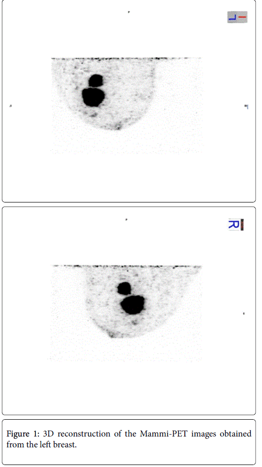
MRI was performed and described a round nodule of irregular shape in the outer quadrant of left breast with a malignant pattern. Also described two more suspicious nodules in the same location of 15 mm and 7 mm. The sum up of the three lesions presented and extension of 6 cm. The study was completed with a PET-CT and a Mammo-dedicated PET (Mammi-PET). The Mammi-PET showed an hipermetabolic lesion (SUVmax 7.8) in the upper outer quadrant of left breast. It also described another hypercaptation focus (SUVmax 7.8) near the first one, and another little focus of activity near the nipple (Figure 1).
A radical modified left mastectomy was performed with radical axillary lymphadenectomy of levels I, II and III of Berg. The anatomopathological results revealed a ductal invasive carcinoma of 2.3 × 2.5 × 3 cm, Grade II with the same immunohistochemistry as obtained with the core biopsy. The second nodule described in the Mammi-PET was also malignant with the same characteristics as the first one. One focus of in situ carcinoma of 0.4 × 0.2 cm was found near the nipple. In this lesion the immunohistochemistry was: ER+++, PR++, Cerb B2-, Ki-67 8%. The results of the lymphadenectomy showed affection of 4/11 lymph nodes, all of them located at level I of Berg.
The aim of this report is to show the correlation of the Mammi-PET images with the real histological findings. In this case the PET described two foci of high metabolic activity (SUV max 7.8) corresponding to the invasive carcinoma and another focus of less capitation which was finally in relation with an in situ carcinoma with different immunohistochemistry.
Citation: Gomez AA, Jurado RS, Diana CF (2016) The Utility of Mammi-Pet in Multifocal Breast Cancer. Breast Can Curr Res 1:i101. DOI: 10.4172/2572-4118.1000i101
Copyright: © 2016 Gomez AA, et al. This is an open-access article distributed under the terms of the Creative Commons Attribution License, which permits unrestricted use, distribution, and reproduction in any medium, provided the original author and source are credited.
Select your language of interest to view the total content in your interested language
Share This Article
Recommended Journals
Open Access Journals
Article Tools
Article Usage
- Total views: 5338
- [From(publication date): 12-2016 - Aug 30, 2025]
- Breakdown by view type
- HTML page views: 4436
- PDF downloads: 902

