Thrombosed Left Circumflex Artery Aneurysm presenting with Syncope
Received: 11-Oct-2021 / Accepted Date: 20-Oct-2021 / Published Date: 27-Oct-2021 DOI: 10.4172/2167-7964.1000345
Image Article
A 79-year-old man with history of coronary artery bypass graft (CABG), atrial fibrillation and recent abdominal aortic aneurysm repair presented after a sudden loss of consciousness. Examination revealed a blood pressure of 130/95 mm Hg, heart rate 84 beats per minute and respiratory rate 18 per minute without any distress. On physical exam he had bilateral riles, elevated jugular venous distension, and bilateral pitting edema. Labs were significant for Troponin T increased from 0.060 to 0.120 ng/mL (Normal <=0.010 ng/mL) and Pro BNP of 3,008 pg/mL (Normal range: 1-450 pg. /mL). Clinically patient’s presentation was consistent with acute decompensated heart failure.
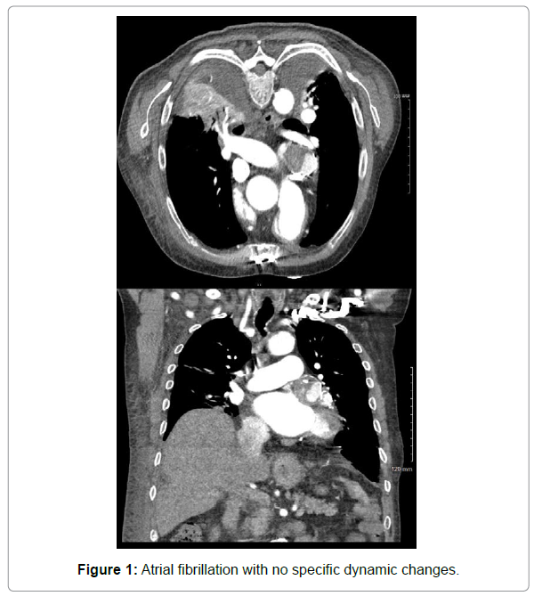
EKG was obtained showed atrial fibrillation with no specific dynamic changes. Echocardiogram revealed reduced ejection fraction and left ventricular diastolic dysfunction, bicuspid aortic valve and moderately dilated aortic root and mild dilation of the ascending aorta. Further imaging of the thoracic aorta was recommended. Contrast tomography angiography of the chest revealed coronary artery aneurysm of the left circumflex artery with mural thrombus measuring 3 cm x 3.8 cm (Figure 1). Surgical intervention was deemed risky given his overall condition. To our knowledge there has been no previously reported cases of thrombosis aneurysm in the left circumflex contributing to symptoms of heart failure or syncope as all other causes of syncope have been ruled out.
Left Circumflex aneurysm thrombosis
Left circumflex artery aneurysm is an extremely rare clinical condition which requires careful evaluation of the coronary anatomy [1]. They are seen in 1.1% to 4.9% of patients undergoing coronary angiography and in about 0.02-0.04% of the general population [2]. They are commonly located in the right coronary artery. The techniques for diagnosing include non-invasive and invasive methods, such as echocardiography, CT, magnetic resonance imaging and coronary angiography. There have been no clinical trials to determine the best therapy for these patients with thrombus formation. The pathophysiology is still unclear, and the optimal treatment remains debatable. In some cases, surgical intervention is preferred. There is lack of consensus regarding the optimal management of coronary artery aneurysm; however, guideline directed medical therapy is preferred and dual antiplatelet therapy is considered if thrombosis/ embolism is a concern [3].
References
- Gupta, Vishal (2008) Giant Left Circumflex Coronary Artery Aneurysm with Arteriovenous Fistula to the Coronary Sinus. Circulation 118:2304-2304.
- Genç B, Taştan A, Abacılar AF, Akpınar MB, Uyar S (2016) Thrombosis left circumflex artery aneurysm presenting with myocardial infarction. Asian Cardiovascular Thorac Ann 24:39-41.
- Bath, Anandbir (2019) Coronary Artery Aneurysm Presenting as STEMI.†BMJ Case Reports 12:6.
Citation: Lakhdar S, Buttar C, Nyabera A, Munira MS (2021) Thrombosed Left Circumflex Artery Aneurysm presenting with Syncope. OMICS J Radiol 10: 345. DOI: 10.4172/2167-7964.1000345
Copyright: © 2021 Lakhdar S, et al. This is an open-access article distributed under the terms of the Creative Commons Attribution License, which permits unrestricted use, distribution, and reproduction in any medium, provided the original author and source are credited.
Select your language of interest to view the total content in your interested language
Share This Article
Open Access Journals
Article Tools
Article Usage
- Total views: 2704
- [From(publication date): 0-2021 - Dec 08, 2025]
- Breakdown by view type
- HTML page views: 1994
- PDF downloads: 710

