Whole body computed tomography scan of the polytrauma patient at Kira Hospital in Bujumbura. About 17 cases
Received: 02-Nov-2023 / Manuscript No. roa-23-119893 / Editor assigned: 06-Nov-2023 / PreQC No. roa-23-119893 / Reviewed: 20-Nov-2023 / QC No. roa-23-119893 / Revised: 24-Nov-2023 / Manuscript No. roa-23-119893 / Published Date: 30-Nov-2023
Abstract
Aim: To determine the epidemiological and tomodensitometric aspects of multiple trauma patients who have benefited from a whole body CT scan at Kira Hospital in Bujumbura.
Patients and Methods: This is a descriptive retrospective study focusing on patients who underwent a whole-body CT scan for multiple traumas over a 20-month period from January 2016 to August 2017. The analysis focused on the following data: age, sex, multiple trauma circumstances, whole body scan protocol and CT results.
Results: This study involved 17 patients including 13 men (76.47%) and 4 women (23.53%), aged between 18 and 82 years. Their average age was 43.7 years. A road traffic accident was the main cause of multiple traumas (82,35%). During the whole body CT scan, traumatic lesions were found in 94.1% of patients. The most affected region was the thorax with 64.7% of cases, with a predominance of pleural lesions (52.94%). Abdomino-pelvic lesions were discovered in 47.1% of cases and intraperitoneal effusions represented 29.41% of cases. In the cranioencephalic stage, lesions were found in 29.41% of patients with a predominance of bone lesions (29.41%). Cervical lesions were less represented with a single case (5.88%).
Conclusion: A polytrauma patient presents with lesional polymorphism, with lesions predominantly on the thoracic level. The whole body computed tomography examination takes pride of place in establishing a complete and exhaustive lesion assessment.
Keywords
Polytrauma; Whole body CT scan
Introduction
A multiple trauma patient is a patient suffering from a violent trauma likely to have caused multiple injuries and / or threatening the life or function [1-3]. A precise and exhaustive lesional assessment is necessary for a good management of the polytrauma patient. Indeed, some polytrauma patients die from preventable causes. Among these patients, there are surgical indications not identified in time and unrecognized lesions [4-6]. The imaging of the multiple trauma patient is therefore one of the linchpins of the initial hospital treatment. The role of imaging in this area has been significantly modified by the advent of multi-detector CT scanners. They allow rapid volume acquisition, the exploration of different anatomical segments in thin sections, optimization of vascular exploration times and multiplanar reconstructions. Whole-body CT scan can detect lesions that are often unrecognized with standard radiography and ultrasound, with great precision in a single acquisition [7]. Several studies have noted a benefit from performing a complete systematic CT scan of patients with suspected multiple traumas, with a markedly reduced number of unnoticed lesions [8-10]. The objective of this work is to analyze the epidemiological and lesion profile found in multiple trauma patients who have benefited from a whole body CT scan at Kira Hospital in Bujumbura.
Patients and Methods
This is a descriptive retrospective study of patients who underwent a whole body CT scan for multiple traumas. It took place over a period of 20 months, from January 2016 to August 2017, in the Radiodiagnostic and Medical Imaging Department of Kira Hospital in Bujumbura, with two CT scanners of 16 and 64 detectors. The analysis focused on the following data: age, sex, circumstances of multiple trauma, whole body scan protocol, and scan results, region by anatomical region (cranioencephalic, cervical, thoracic and abdominopelvic). The fate of these patients during hospitalization has not been studied. Data analysis and entry was performed using Microsoft Word and Excel 2019 software.
Results
Out of 1518 computed tomography scans performed during the period of our study, 30 patients had undergone a computed tomography scan for suspected multiple traumas. Whole-body computed tomography was performed in 17 patients (56.7% of multiple trauma patients) among these 30 patients. There was a predominance of patients aged between 21 and 40 years (7 cases or 41.17%). Their average age was 43.76 with extremes of 18 and 82. Among them, 13 patients were male (76.47%), ie a sex ratio of 3.25. The circumstances in which the polytrauma occurred were road accidents in 14 patients (82.3%) and the other 3 patients (17.6%) had suffered an assault with a firearm.
Before the tomodensitometric examination, only 3 patients had a notification of procedures performed: 1 case of emergency splenectomy, 1 case of chest drainage from a hemothorax and 1 case of transfusion for hemodynamic instability. Cranioencephalic and cervical tomodensitometric examination had been performed in all patients without injection of contrast medium. On the thoracoabdominal- pelvic level, it was performed with and without injection of iodine contrast in 12 patients (70.58%) and without injection of iodine contrast in 5 patients.
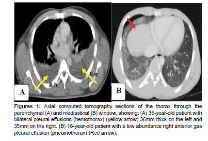
Of the 17 patients, lesions were found in 16 patients (94.11%). The site of these lesions was mainly thoracic (11 patients or 64.7% of cases), abdomino-pelvic in 8 patients (47.1% of cases), cranioencephalic in 5 patients (29.4% of cases). The cervical stage was affected in one single patient (5.9% of cases). In the thorax, there were 11 patients (64.7%) who presented lesions (6 cases of hemothorax or 35.3% of cases, 5 cases of rib fracture or 29.4% of cases, 4 cases of pulmonary contusions or 23.5%, 3 cases of pneumothorax or 17.6% and 3 cases of thoracic vertebra fracture or 17.6% (Figure 1A and 1B).
Figure 1: Axial computed tomography sections of the thorax through the parenchymal (A) and mediastinal (B) window, showing: (A) 35-year-old patient with bilateral pleural effusions (hemothorax) (yellow arrow) 36mm thick on the left and 35mm on the right. (B) 18-year-old patient with a low abundance right anterior gas pleural effusion (pneumothorax) (Red arrow).
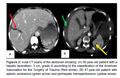
Traumatic lesions in the abdomino-pelvic area were observed in eight patients (47.1%). Those are five cases of hemoperitoneum (29.4%), four cases of pelvic fracture, three cases of splenic involvement, two cases of pneumoperitoneum linked to gastric perforation for one and intestinal for the other. Only one case of hepatic laceration was noted (Figure 2A and 2B).
Figure 2: Axial CT scans of the abdomen showing: (A) 60-year-old patient with a hepatic laceration> 3 cm, grade III according to the classification of the American Association for the Surgery of Trauma (Red arrow). (B) 47-year-old patient with splenic contusions (green arrow) and perihepatic hemoperitoneum (yellow arrow).
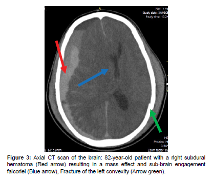
On the cranioencephalic level, 5 patients (31.2%) presented lesions with 4 cases of fracture of the facial mass (23.5%) and 2 cases of fracture of the vault (11.7%). There were 2 cases of subdural hematoma associated with subdural hematoma in the brain in one of the patients (Figure 3) and a subarachnoid hemorrhage in another patient. There was also 1 case of cerebral hematoma and 1 case of cerebellar contusion.
One single case of cervical lesion was noted associating a fracture of C7 and D1 with an antelisthesis of C7 on D1. Overall, a lesion association was observed in 12 patients (75%) with at least 2 lesions on different anatomical stages. Four patients had a single lesion (23.5%).
Discussion
In our study, CT scan was performed with and without injection of contrast product in 70.58% of patients. As Dreizin, Munera and Blunt [11] pointed out in their study, the injection of contrast product makes it possible to detect active bleeding requiring rapid management in a specialized setting. Most lesions in our polytrauma patients predominated on the thoracic level (64.7% of cases). Some authors had made this observation, in particular Babaud et al. [12] with 30.38% of cases, Ahvenjarvi et al. [13] with 44% of cases, Khov et al. [10] with 62% of cases and Maurin et al. [14] with 66% of cases. Other authors such as Sampson et al [15] and Bingol et al. [16] had noted in their studies a predominance of cranioencephalic lesions on the other hand. This could be explained, in our series, by the fact that whole body computed tomography is not done routinely in all patients suspected of polytrauma because of the relatively high cost (around the equivalent of US $ 300). Thus, in some patients, tomodensitometric exploration is carried out segment by segment according to clinical signs but also according to the financial means available for the patient.
On the thoracic level, the lesions were dominated by hemothorax (35.3%), rib fractures (29.4%) and pulmonary parenchymal contusion (23.5%). Unlike our study where pneumothorax (17.6%) was the less noted thoracic lesion, certain data in the literature place parenchymal contusions first among thoracic lesions with 27.6% of cases for Bingol et al. [16] and 33% of cases for Sampson et al. [15]. Pulmonary contusion poses a problem of definition, which explains the variations found to estimate its frequency (from 10 to 100% depending on the studies). The tomodensitometric examination remains very reliable in chest trauma. It helps in the diagnosis of pulmonary contusions, anterior pneumothorax and in the study of the mediastinum [17]. Since the thorax contains vital structures, especially the heart and aorta, the diagnostic delay risks compromising the vital prognosis in the event of its traumatic impairment. Computed tomography is very sensitive in the diagnosis of ruptured aorta [13, 18]. In our series, no case of aortic rupture was noted.
Traumatic abdominal-pelvic involvement was marked by hemoperitoneum (29.4%) following full organ involvement as also noted in the study by Bingol et al. [16], where hemoperitoneum represented 23.1% of cases. Splenic and hepatic lesions were lesions of the solid organs found in 11.76% and 5.88% of cases, respectively. Several studies reported a predominance of splenic involvement during abdominal trauma. This is the case of Bingol et al. [16] with 16.9% of cases, Ahvenjarvi et al. [13] with 15% of cases and Salim et al. [19] with 2% of the cases. However, other studies have placed the liver in the first rank of the intra-abdominal solid organs damaged during abdominal trauma, such as Frampas et al. [20] with 10.3% of cases and Sampson et al. [15] with 6% of cases. The intestine was the least affected organ in our study with a single case of intestinal perforation. Intestinal involvement is reported in all studies to be the least represented. Intestinal perforation is reported in 3.8% and 4% of the cases respectively in the studies performed by Bingol et al. [16] and Salim et al. [19].
At the cranio-encephalic level, cranio-encephalic lesions represented 31.2% of the population with 23.5% of cases for fractures of the facial mass. Ahvenjarvi et al. [13] and Bingol et al. [16] had reported an attack of the facial mass respectively in 8% and 26% of the population. In our series, the subdural hematoma was the haematic collection noted in 2 patients and there was only one case of subarachnoid hemorrhage. Authors such as Salim et al. [19] and Bingol et al. [16] reported a predominance of subarachnoid hematoma (HSA) with 7.8% and 11.7% of cases, respectively. The encephalic parenchymal lesions consisted of cerebellar contusions, cerebral intraparenchymal hematoma with sub-falcor involvement. Several studies report a predominance of hemorrhagic contusions, such as reported in the study of Salim et al. [19] with 7.7% of the cases and the study of Ahvenjarvi et al. [13] with 14% of the cases. Our sample size is too small to clarify the importance of each type of head injury occurring in the context of multiple traumas.
In our study, a lesion association was observed in 12 patients (75%), with at least 2 lesions on different anatomical stages. The existence of several lesions in different anatomical regions is one of the factors of poor prognosis as shown in the German study performed by Regel et al. [21]. In fact, in this study, in a sample of 3,406 polytrauma patients between 1972 and 1991, mortality varied from 14.3 to 26.3% depending on whether one or two regions were affected. It was then about 27-30% if three, four and five anatomical regions were involved.
Conclusion
Whole-body computed tomography has a special place in performing the lesion assessment in multiple trauma patients. These patients are mostly young adult males involved in road accidents. Whole body computed tomography remains inaccessible for some of these patients. There was at least one lesion in most of our patients with a clear predominance of lesions of the thoracic floor, followed by abdomino-pelvic lesions. It must be performed as early as possible to improve the survival of patients with multiple traumas.
References
- Mathieu L, Desfemmes F, Jancovici R (2006) Prise en charge chirurgicale du polytraumatisé en situation précaire. 143: 349-354.
- Albanese J (2002) Le polytraumatisé. Springer Science & Business Media.
- Butcher N, Balogh ZJ (2009) The definition of polytrauma: the need for international consensus. Injury 40: S12-S22.
- Novak V, Kostić A, Berilažić L, Milošević P (2016) Severity of brain injury-influence on treatment outcome in trauma patients. 55: 27-31.
- Sanddal TL, Esposito TJ, Whitney JR, Hartford D, Taillac PP, et al. (2011) Analysis of preventable trauma deaths and opportunities for trauma care improvement in Utah. 70: 970-977.
- Sharma B, Gupta M, Harish D, Singh V (2005) Missed diagnoses in trauma patients vis-à-vis significance of autopsy. 36: 976-983.
- Savry C, Dy L, Quinio P (2002) Prise en charge initiale d’un patient polytraumatisé aux urgences. Réanimation. 11: 486-492.
- Rieger M, Czermak B, El Attal R, Sumann G, Jaschke W, et al. (2009) Initial clinical experience with a 64-MDCT whole-body scanner in an emergency department: better time management and diagnostic quality? 66: 648-657.
- Agostini C, Durieux M, Milot L, Kamaoui I, Floccard B, et al. (2008) Emergency-Value of double reading of whole body CT in polytrauma patients.89: 325-330.
- Khov M, Vialle R, Chan M, Hannequin J, Jouhanneaud A, et al. (2007) DIV-WP-2 Prise en charge radiologique du polytraumatisme. 88: 1541.
- Dreizin D, Munera F (2012) Blunt polytrauma: evaluation with 64-section whole-body CT angiography. Radiographics. 32: 609-631.
- Babaud J, Ridereau-Zins C, Bouhours G, Lebigot J, Bertrais S, et al. (2012) Intérêt des critères de Vittel pour l’indication d’un scanner corps entier chez un patient traumatisé grave. Journal de Radiologie diagnostique et interventionnelle 93: 399-407.
- Ahvenjärvi L, Mattila L, Ojala R, Tervonen O (2005) Value of multidetector computed tomography in assessing blunt multitrauma patients. Acta Radiologica 46: 177-183.
- Maurin O, Prunet B, De Régloix S, Delort G, Lacroix G, et al. (2012) Épidémiologie des traumatisés graves accueillis à l’hôpital d’instruction des armées Sainte-Anne. Recueil prospectif sur six mois. Med Armées 40: 333-337.
- Sampson M, Colquhoun K, Hennessy N (2006) Computed tomography whole body imaging in multi-trauma: 7 years experience. Clin Radiol 61: 365-369.
- Bingol O, Ayrık C, Kose A, Bozkurt S, Narcı H, et al. (2015) Retrospective analysis of whole-body multislice computed tomography findings taken in trauma patients. Turk J Emerg Med 15: 116-121.
- Bléry M, Kraiem A, Edouard A, Iffenecker C, Rocher L, et al. (1997) Approche diagnostique du polytraumatisé en urgence. Feuillets de radiologie 37: 103-116.
- Wurmb TE, Frühwald P, Hopfner W, Roewer N, Brederlau J (2007) Whole-body multislice computed tomography as the primary and sole diagnostic tool in patients with blunt trauma: searching for its appropriate indication. Am J Emerg Med 25: 1057-1062.
- Salim A, Sangthong B, Martin M, Brown C, Plurad D, et al. (2006) Whole body imaging in blunt multisystem trauma patients without obvious signs of injury: results of a prospective study. Arch Surg 141: 468-475.
- Frampas E, Regenet N, Meurette G, Metairie S, Léauté F, et al. (2006) DIG55 Place et apports de l’imagerie dans la prise en charge des traumatismes abdominaux. Journal de Radiologie 87: 1469.
- Regel G, Lobenhoffer P, Grotz M, Pape H, Lehmann U, et al. (1995) Treatment results of patients with multiple trauma: an analysis of 3406 cases treated between 1972 and 1991 at a German Level I Trauma Center. J Trauma Acute Care Surg 38: 70-78.
Indexed at, Google Scholar, Crossref
Indexed at, Google Scholar, Crossref
Indexed at, Google Scholar, Crossref
Indexed at, Google Scholar, Crossref
Indexed at, Google Scholar, Crossref
Indexed at, Google Scholar, Crossref
Indexed at, Google Scholar, Crossref
Indexed at, Google Scholar, Crossref
Indexed at, Google Scholar, Crossref
Indexed at, Google Scholar, Crossref
Indexed at, Google Scholar, Crossref
Indexed at, Google Scholar, Crossref
Citation: Manirakiza S, Mbonicura JC, Murekatete C, Niyondiko JC, BarasukanaP, et al. (2023) Whole Body Computed Tomography Scan of the Polytrauma Patientat Kira Hospital in Bujumbura: About 17 Cases. OMICS J Radiol 12: 506.
Copyright: © 2023 Manirakiza S, et al. This is an open-access article distributedunder the terms of the Creative Commons Attribution License, which permitsunrestricted use, distribution, and reproduction in any medium, provided theoriginal author and source are credited.
Select your language of interest to view the total content in your interested language
Share This Article
Open Access Journals
Article Usage
- Total views: 2047
- [From(publication date): 0-2023 - Nov 27, 2025]
- Breakdown by view type
- HTML page views: 1709
- PDF downloads: 338


