A Pediatric Case of Gorham Disease with Extensive Maxillofacial Involvement
Received: 17-Jan-2013 / Accepted Date: 26-Jul-2013 / Published Date: 02-Aug-2013 DOI: 10.4172/2161-119X.1000139
Abstract
Objectives: To present a rare pediatric case of Gorham disease with craniofacial involvement. Study design: Case report and literature review.
Methods: Literature review of craniofacial Gorham disease in pediatrics and discussion of a representative case within our health system.
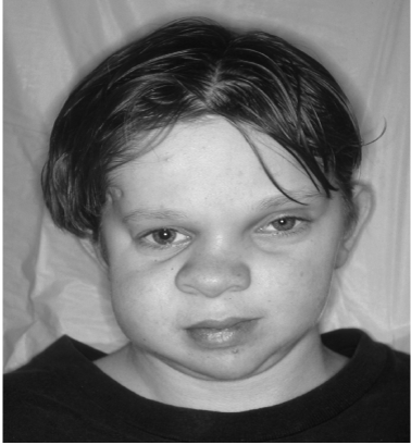
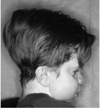
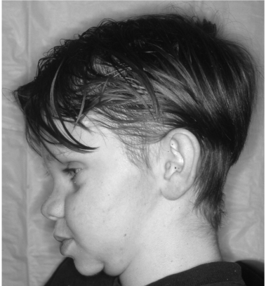
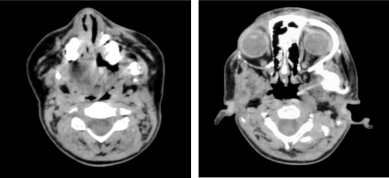
Results: A nine year old male presented after mandibular dental trauma. Massive maxillofacial osteolysis ensued. Photographs and radiologic images demonstrate the dysmorphic facial features of this case of Gorham disease.
Conclusion: To the best of our knowledge, our case represents the fourth case in the literature to document Gorham disease in a male pediatric patient with mandibular involvement.
Keywords: Gorham disease, Vanishing bone disease, Massive osteolysis
254004Introduction
Few cases of Gorham Disease (GD) have been reported in the literature. It has been called vanishing bone disease, massive osteolysis, phantom bone disease, and is also known as Gorham-Stout syndrome. GD was first reported by Jackson in 1838 in a patient description of a boneless arm due to massive osteolysis [1]. GD often presents as a pathologic fracture more commonly in males in the second and third decades of life. No hereditary correlation or racial predilection is known. This disease frequently affects the long bones, pelvic girdle, shoulder girdles, vertebrate, and ribs. Since 1928, forty-two cases involving the maxillofacial region have been documented. Of those, only three have involved the mandible of a male patient younger than ten years of age [2]. We report the case of a nine year old male suffering from massive osteolysis with involvement of the mandible, maxilla, sphenoid, temporal bone, and infratemporal fossa.
Case Report
RG is a 9 year old male who suffered mild superficial trauma to the face after falling. He did not sustain any bony fractures but 3 months later, his right mandibular molar teeth were noted to be loose and inflamed requiring extraction. Pathologic examination of a soft tissue biopsy revealed sulfur granules and the patient was treated with periodic debridement and antibiotics for presumed Actinomycosis osteomyelitis. The patient’s mandible underwent continued osteolysis despite aggressive intravenous and oral antibiotics. Eighteen months later, contralateral right mandibular involvement was noted. Five months later, the left maxillary molars were found to be loose, and defects in the hard palate as well as nasal speech were present.
At the age of 13, a Magnetic Resonance Imaging (MRI) scan of the head and skull base revealed contiguous spread of the osteolysis to include the clivus, petrous mastoid, carotid canal, jugular fossa, right pterygoid plates, sphenoid body and occipital bone with erosion of the floor and anterior wall of the right middle cranial fossa. A Computerized Tomography (CT) scan the following month showed dehiscence of the right middle cranial fossa floor onto the right infratemporal fossa. Further analysis of the CT scan showed that the patient’s right cochlea was at risk of this progressive osteolysis. At the age of 14 the osteolysis ensued and an orbital rim biopsy revealed massive osteolysis without concern for malignancy. After 5 years of progressive osteolysis without evidence of identified etiology our patient was diagnosed with GD. By this time, the patient had developed a quite abnormal facies (Figures 1-3). Two years later, the patient developed cervical spine involvement and underwent spinal fusion.
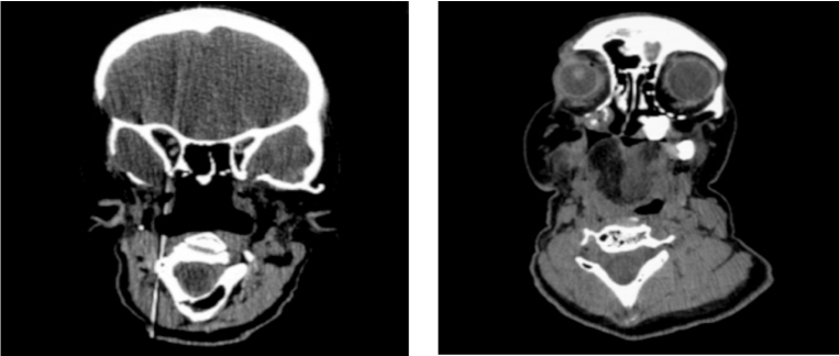
By the age of 16, the patient had developed near complete osteolysis of the mandible, demineralization of the occipital bone, tympanic and mastoid portions of the temporal bone, greater wing of the right sphenoid bone, hypoglossal canal, and bilateral maxillary involvement (Figure 4). Seven years later, CT scan showed dramatic osteolytic defects of the right parietal, sphenoid, and right temporal bones as well as the posterior cervical spine (Figure 5). The disease progressed to involve the clavicle and ribs. In addition to the patient’s spinal fusion and external beam radiation therapy, he was also treated with intravenous calcitonin and bisphosphonates. Although these treatment modalities may have slowed the progression of our patient’s disease, they were not curative.
Discussion
GD is defined by osteolysis that can progress to complete dissolution of the involved bone [3]. The disease is usually monocentric, with contiguous involvement of adjacent bones [4]. It can involve multiple skeletal regions, but rarely involves the facial skeleton. It usually affects patients in their second and third decades of life with an average age at diagnosis of 33 years old [2]. Our case represents a pediatric patient with extensive maxillofacial and skull base involvement. Both the age of the patient and location of disease are exceptionally rare manifestations of GD.
To the best of our knowledge, since 1928, there have only been three other cases of GD in the literature involving the mandible of a male patient younger than ten years of age. Regeneration following the osteolysis is extremely rare [2]. GD seems to progress through two phases. The first phase consists of active bone resorption and contiguous spread to adjacent bone. Clinically, this may manifest as pathologic fractures, and may be the etiology of difficulties with speech, mastication, and a prominent facial deformity, all expressed in our patient [5]. Latent spread of GD represents the second phase of massive osteolysis [5]. During this quiescent phase, the haversian structure of the bone undergoes fibrosis [2]. The duration of these stages is unpredictable and may last months to years. The lack of new bone formation is a hallmark of GD [6].
The diagnosis of GD is based on clinical, histologic, and molecular features. Diagnosis is difficult and requires exclusion of neoplastic, inflammatory, infectious, and endocrinologic disease. The etiology of the massive osteolysis is still unknown. The lack of a standard treatment regimen for patients suffering from GD correlates with its widely disputed pathogenesis. The range of treatment consists of either surgery and radiation therapy, or medical therapy. Medical therapy is controversial as there is a lack of definitive evidence for its benefit. Early irradiation of the affected region has been observed to induce remission in some patients [2,5]. Close observation for spinal instability is critical as these patients commonly require surgical stabilization. Medical therapy has been used in refractory cases and consists of bisphosphonates, calcitonin, calcium, vitamin D, and alpha- 2b interferon therapy [2]. Surgical treatment of disease involving the skull base and maxillo-facial regions may not be an option given the potential post-surgical morbidity. According to Escande, treatment of maxillofacial GD is local resection of the involved bone in an attempt to stop progression of the disease [2]. Although GD is an insidious progressive disease, spontaneous regression has been described [2]. Involvement of the cervical and thoracic spine resulting in persistent chylothorax is the usual cause of death in GD patients [7]. Due to the rarity of pediatric GD, there is limited information on specific treatment options in pediatric patients.
The radiographic findings of GD can be quite extensive without confinement to anatomical boundaries. Johnson and McClure divided the radiographic features of Gorham’s disease into early intraosseous and later extraosseous stages [3,8]. Early disease portrays patchy osteoporosis with multiple intramedullary and subcortical radiolucencies [8]. In the later phase, disruption of the cortex leads to destruction and aggressive resorption of the bone. CT and MRI scanning are the imaging modalities of choice for following the progression of GD [8].
Conclusion
GD is a complex disorder hallmarked by progressive osteolysis of unknown etiology that rarely manifests in the maxillo-facial region. This case report emphasizes GD as a rare differential for lesions of the facial skeleton. To the best of our knowledge, this report represents the fourth known case of GD in the mandible of a male pediatric patient younger than ten years of age.
Acknowledgements
This research was presented at the Combined Otolaryngological Spring Meeting in Las Vegas, Nevada from April 28 to May 2, 2010.
Conflict of Interest
None of the authors have any conflicts of interest or financial disclosures.
References
- Frankel DG, Lewin JS, and Cohen B (1997) Massive Osteolysis of the Skull Base. Pediatric Radiology 27: 265-267.
- Lo CP, Chen CY, Chin SC, Juan CJ, Hsueh CJ, et al. (2004) Disappearing Calvarium in Gorham Disease: MR Imaging Characteristics with Pathologic Correlation. AJNR Am J Neuroradiol 25: 415-418.
- Bouloux GF, Walker DM, and McKellar G (1999) Massive Osteolysis of the Mandible Report of a Case with Multifocal Bone Loss. Oral Surg Oral Med Oral Pathol Oral Radiol Endod. 87: 357-361.
- Benhalima H, Lazrak A, Boulaich M, Mezahi M, Kzadri M, et.al. (2001) Massive Osteolysis of the Maxillo-Facial Bones: Case Report and Review of the Literature. Odonto-Stomatologie Tropicale 96: 35-40.
- Dominguez R and Washowich TL (1994) Gorham’s Disease or Vanishing Bone Disease: Plain Film, CT, and MRI Findings of Two Cases. Pediatr Radiol 24: 316-318.
- Pfleger A, Schwinger W, Maier A, Tauss J, Popper HH, et.al. (2006) Gorham-Stout Syndrome in a Male Adolescent-Case Report and Review of the Literature. J Pediatr Hematol Oncol 28: 231-233.
- Dunbar SF, Rosenberg A, Mankin H, Rosenthal D, Suit HD (1993) Gorham’s Massive Osteolysis: The Role of Radiation Therapy and a Review of the Literature. Int J Radiat Oncol Biol Phys 26: 491-497.
Citation: Toma MS, Al-khudari S, Schweitzer VG (2013) A Pediatric Case of Gorham Disease with Extensive Maxillofacial Involvement. Otolaryngology 3:139. DOI: 10.4172/2161-119X.1000139
Copyright: © 2013 Toma MS, et al. This is an open-access article distributed under the terms of the Creative Commons Attribution License, which permits unrestricted use, distribution, and reproduction in any medium, provided the original author and source are credited.
Select your language of interest to view the total content in your interested language
Share This Article
Recommended Journals
Open Access Journals
Article Tools
Article Usage
- Total views: 18309
- [From(publication date): 9-2013 - Dec 06, 2025]
- Breakdown by view type
- HTML page views: 13439
- PDF downloads: 4870

