Research Article Open Access
Decolorization of Triphenylmethane Dyes by Six White-Rot Fungi Isolated from Nature
| Kavita Vasdev* | |
| Department of Microbiology, Gargi College, New Delhi, India | |
| Corresponding Author : | Dr. Kavita Vasdev Associate Professor Department of Microbiology Gargi College, New Delhi, India E-mail: vasdevkavi@yahoo.com |
| Received September 09, 2011; Accepted October 27, 2011; Published October 29, 2011 | |
| Citation: Vasdev K (2011) Decolorization of Triphenylmethane Dyes by Six White- Rot Fungi Isolated from Nature. J Bioremed Biodegrad 2:128. doi:10.4172/2155-6199.1000128 | |
| Copyright: © 2011 Vasdev K. This is an open-access article distributed under the terms of the Creative Commons Attribution License, which permits unrestricted use, distribution, and reproduction in any medium, provided the original author and source are credited. | |
Related article at Pubmed Pubmed  Scholar Google Scholar Google |
|
Visit for more related articles at Journal of Bioremediation & Biodegradation
Abstract
The ability to decolorize three Triphenyl methane dyes (crystal violet, Bromophenol blue and Malachite green) by six white rot fungi, isolated from nature, was evaluated in liquid media. All six-fungi showed high decolorization capacity and were able to decolorize all three dyes within 72 hours. All six fungal strains not only decolorized these dyes, they also showed varied levels of laccase production during decolorization. Their growth was not affected much by presence of dyes in the medium. Three of these fungi were found to have capacity to decolorize as high as 6g/L concentration of these dyes. Out of the three dyes tested here, Malachite green was decolorized fastest. The dyes were not just being decolorized; they were actually being degraded as was evident from their UV-Visible Spectra. Furthermore, the fungi degraded these dyes without accumulating any phenolic compounds.
| Introduction |
| Industrial dyes are released into environment as waste water by industry. More than 10,000 synthetic dyes commercially available are used in textile, dyeing, printing and other industrial applications. These are chemically diverse and divided into azo, anthraquinone, heterocyclic polymers and triphenylmethane dyes. Most of these are stable against light, temperature and biodegradation and have therefore accumulated in the environment as recalcitrant compounds [1]. |
| Conventional waste water treatment is not efficient to remove recalcitrant dyestuffs from effluents [2]. Several physical and chemical methods are effective but have high operating costs and limited applicability. Although, abiotic means of reduction of azo and other dyes exist but they require highly expensive catalysts and reagents. A number of biotechnological approaches have been suggested by recent research as of potential interest towards combating this pollution source in an ecoefficient manner, including the use of bacteria or fungi, often in combination with physicochemical process [3-6]. In recent years, the biological decolorization method has been considered as effective, specific, less energy intensive and environmentally benign, since it results in partial or complete bioconversion of pollutants to stable nontoxic end products [7]. |
| By far the single class of microorganisms most efficient in breaking down synthetic dyes is white rot fungi. These fungi are a class of microorganisms that are known to produce efficient enzymes capable of decomposing dyes under aerobic conditions. They produce various oxidoreductases that degrade lignin and a very wide range of pollutants; such as polychlorinated biphenyls (PCB) polycyclic aromatic compounds (PAH), pesticides, synthetic dyes, Industrial dyes [8,9]. Owing to extracellular, non-specific, free radical based ligninolytic systems of WRF; they can completely eliminate a variety of xenobiotics, including synthetic dyes and industrial dyes giving rise to non-toxic compounds [7]. Triphenylmethane dyes released by textile industries into aquatic systems leads to pollution of aqueous habitats. These dyes are found in soil and river sediments as a result of improper waste water treatment [10]. The decolorization of Triphenylmethane dyes by various species of Cyathus has already been reported by Vasdev et al. [10]. Since then there have been a number of reports on decolorization of triphenylmethane dyes by fungi [1,11], as well as by bacteria [12,13]. Most of these reports are limited to describing the decolorization, involvement of ligninolytic system with decolorization, effect of dyes on growth of organisms and extent of decolorization. |
| This is the first most comprehensive study, that describes, not just the ability of fungi to decolorize the dyes, but also studies their degradation by UV-visible spectrum, linking the decolorization with ligninolytic system and growth of fungi but also describes the ability of these fungi to detoxify the environment, while degrading the dyes i.e. by testing the concentration of toxic phenolic compounds produced in the culture, as a result of degradation of these dyes. |
| Materials and Methods |
| Chemicals |
| 10mM Guiacol prepared in 0.1M citrate phosphate buffer, Crystal violet (C.I.42555), Malachite green (C.I. 42000), Bromophenol blue, Malt extract broth medium were purchased from Hi media laboratories India. All chemicals and media components used were of analytical grade. |
| Isolation of micro-organism |
| Fungi were isolated from nature. The mushrooms were collected from the Gargi campus, during rainy season. All mushrooms were surface sterilized before culturing, with 0.1 % HgCl2, then with distilled water, then with 70% ethyl alcohol and finally 2-3 washes with distilled water. Mycelium was aseptically removed from fruit bodies and inoculated on to 2% MEA (Malt extract medium) plates, containing streptomycin. These newly isolated fungi from natural sources were- KV9, KV10, KV11, KV12, KVA5, AKCH These fungal strains were grown on 2% Malt extract agar (MEA) media, containing Malt extract 20g/L, Ca(NO3)2.4H2O 0.5g/L, MgSO4.7H2O 0.5 g/L, KH2PO4 0.5 g/L, Agar 20 g/L, pH 5.4 and incubated at 28°C. |
| Pure cultures of these strains were maintained by repeated sub culturing on MEA. These strains were kept at 4°C till use. All strains were examined microscopically to see basidiomycete's characteristics and named alphabetically. |
| Agar plate screening for laccase |
| The fungal isolates were screened for laccase production by growing them on 2 % MEA media. After 8 days of growth, plates were flooded with 10mM Guiacol and incubated at 28°C overnight. The production of an intense brown colour under and around the fungal colony in media after flooding with Guiacol indicated presence of laccase activity [14]. |
| Culture conditions |
| All white rot fungi (WRF) were grown in 250ml Erlenmeyer flask containing 50ml of sterile 2% Malt extract broth (MEB) containing, Malt extract 20 g/L, Ca(NO3)2.4H2O 0.5 g/L, MgSO4.7H2O 0.5 g/L, KH2PO4 0.5 g/L, pH 5.4 at 28°C. Each flask was inoculated with two 8mm diameter fungal discs, obtained from the periphery of the actively growing fungal cultures in MEA plates and incubated at 28°C. After 8 days of growth, in liquid media, 1ml of Crystal violet, Malachite green and Bromophenol blue (1g/L) was added to each flask, containing respective grown fungal culture. The time of addition of dyes in grown culture flask was considered to be day zero. Three fungi were tested for decolorization of Malachite green at a higher concentration of 6g/L. |
| Decolorization assays |
| Decolorization of Crystal violet, Bromophenol blue and Malachite green in liquid media, was measured in culture filtrates (three replicate flasks), after removing 1ml of culture filtrate from each aseptically. The change in absorbance was monitored spectrophotometrically at their maximum wavelength (590nm for Crystal violet, 620nm for Malachite green and 595nm for Bromophenol blue).The uninoculated Malt extract broth with respective dye served as abiotic control. The decolorization efficiency was determined using the following equation: |
| Decolorization (%) = [{(Aλ initial – Aλ final / Aλ initial} X 100] |
| Where, Aλ initial= Absorbance of dye in uninoculated control, Aλ final= Absorbance of dye in inoculated culture flasks. |
| Estimation of total phenolics |
| The total Phenolics were estimated in the culture filtrates, after complete decolorization of Malachite green, according to Singleton et al. [15]. The reaction mixture containing 3 ml distilled water, 50µl cultures filtrate and 250µl of Folin-Ciocalteu reagent was incubated at 30°C, for 1 minute. After incubation, 750µl Na2 CO3 (10%) was added to the reaction mixture followed by incubation at 30°C for 60 minutes in dark. The amount of phenolics was determined by measuring absorbance at 760 nm against reaction blank. |
| UV-VIS characterization |
| The dye degradation under static cultivation conditions, in the culture filtrates of all six fungi with all three dyes, were monitored by following the change in the UV-VIS spectra in range of 400-800nm and comparing it with UV-VIS spectrum of the Native dyes, using a UVVIS spectrophotometer. |
| Enzyme assays |
| The 250ml Erlenmeyer flasks with 50ml of MEB were inoculated in same way as for decolorization assay. Enzyme activity was measured in culture filtrates from replicate flasks, obtained after mycelia removal by filtration through filter paper. Laccase activity was determined spectrophotometrically by monitoring increase in absorbance at 470nm due to oxidation of guiacol, according to Vasdev & Kuhad [16]. Heat killed enzyme was used as control. The change in 0.01/ ml/min was defined as 1 U of laccase activity. |
| Determination of biomass production |
| Biomass production in liquid media was evaluated by determining the dry mass of mycelia .Mycelia were harvested from their cultivation liquid by filtration through whatman filter paper no.1, dried at 80°C for 48 hours and weighed. |
| Results |
| Screening white rot fungi for laccase production |
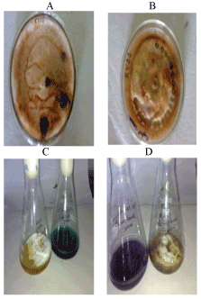
| All newly isolated white rot fungi from natural sources were basidiomycetes (namely KV9, KV10, KV11, KV12, KVA5, and AKCH). All the strains grew well on 2% Malt extract agar medium. The fungal isolates, when tested microscopically, showed the presence of clamp connections which is the characteristic feature of basidiomycetous fungi. These fungal strains grown for 8 days at 28°C, when flooded with 10mM Guaicol, produced intense brown color around the colonies, indicating the production of laccase (Figure 1a,b). |
| Decolorization in liquid media |
| Decolorization of Triphenylmethane dyes (Crystal violet, Malachite green and Bromophenol blue) was studied with six white-rot fungi, at effective concentration of 0.3g/L, were found to be capable of decolorization, though to different extents. Among the dyes decolorized by these six fungi, malachite green was decolorized fastest (table 1). |
| All fungal strains were found to be highly efficient, showing more than 80% decolorization of Malachite green within first 48 hours from the time of dye addition (Table 2). |
| Presence of dyes in the medium had no effect on the growth of these fungi, except KVAS, indicating, these fungi were able to tolerate the dyes and decolorize them to completion. At the same time, none of the cultures showed any adsorption of dye to the mycelium (Figure 1c,d). |
| Three out of the six fungal strains namely KV10, KV11 and AKCH were found capable of decolorizing as high as 6g/L concentration of Malachite green. |
| UV-VIS characterization |
| All the six fungal strains not only decolorized these triphenylmethane dyes, but also degraded them which is evident from the visible spectra 400-800nm of each of the dyes being completely degraded by fungal strains and that of native dyes in uninoculated control (Figure 2,3 shown as supplementary). All six fungal strains, not only decolorized and degraded triphenylmethane dyes, but also carried out these phenomenon in a non-toxic manner. This is evident from the concentration of phenolics present in the culture filtrates, after complete decolorization (Figure 3 shown as supplementary). |
| Estimation of total phenolics |
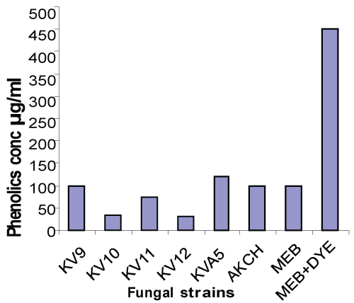
| The concentration of phenolics accumulated by these fungal cultures was found to be nearly same as that present in uninoculated ME Broth without dye i.e. less than 80µg/ml, which is much less than those present in ME Broth with dyes indicating complete detoxification (Figure 4). |
| Laccase enzyme |
| The production of laccase by all fungal strains was studied under the same conditions as in the decolorization experiments. The malt extract broths without the dyes, inoculated by respective fungi were used as controls. The results showed in table 3 that the laccase productions were affected very slightly by the kind of dye added. There was correlation between laccase production and dye decolorization. The two fungi (KV10 and KV12) which showed fastest dye decolorization, also showed highest laccase production, while AKCH which was less efficient showed low laccase activity (Table 3). |
| Discussion |
| This study of the ability of white rot fungi to decolorize different triphenylmethane dyes (Crystal violet, Malachite green and Bromophenol blue) in liquid medium revealed, a high decolorization capacity as well as high efficiency of decolorization of these fungi. Although higher dye concentrations have been reported to have a toxic effect on fungi, it was found that even a concentration of 6g/L of malachite green, was tolerated by fungal strain KV10 & KV11 and did not limit the decolorization capacity. |
| Three out of six fungal strains KV10, KV11, AKCH showed the ability to decolorize up to 6 g/L, Malachite green, which is much higher than 2g/L reported for D.squalens [17]. Contrary to the reports mentioned by Nyanhongo et al. [18] and in their own study laccases from Trametes hirsuta and from Trametes modesta, showed, that highly substituted triphenylmethane dyes require a longer time to be decolorized or could not be decolorized completely, in our study all six strains of fungi decolorized triphenylmethane dyes to 100% in 72 hours at concentration of 0.3g/L malachite green. Five out of six fungi tested have shown faster decolorization rates, more than 80% decolorization within first 48 hours, which is much faster than reported elsewhere Vasdev et al. [10], reported decolorization time of 96 hours for various species of Cyathus, while Yesilada et al. [19] reported decolorization time of 72 hours for 62 % decolorization of Crystal violet by Phanerochaete chrysosporium ME446, while Knapp et al. [20] reported 216 hours decolorization time for Phanerochaete chrysosporium NCIM1197. |
| Dye decolorization carried out by fungi did not show any effect on their growth at concentration till 0.3g/L. Among the three triphenylmethane dyes tested, Malachite green was decolorized fastest, which is in accordance with the observations of R.K. Sani & U.L. Banerjee [12]. |
| The decolorization process requires the destruction of chromophores. Small structural differences also affect the decolorization process. Malachite green has two dimethyl groups in two side chains whereas crystal violet is having three dimethyl groups in three side chains. It may be the reason that decolorization of more substituted triphenylmethane dyes took longer time. |
| In the present study, not only decolorization of Triphenylmethane dyes were studied, but the Biodegradation of these dyes was also studied by taking their absorption spectrum with UV-Visible spectrophotometer. UV-Visible spectra of the three dyes showed their biodegradation by all six fungal cultures. The native dyes showed absorbance peak at their wavelength maxima, Crystal violet at 590nm, Malachite green at 620nm and Bromophenol blue at 595nm, while in case of dyes being degraded by fungi, there was complete disappearance of peaks in the spectrum, along with exponential fall, indicating complete decolorization and degradation, as has also been reported by Franciscon Elisangela et al. [13], in case of biodegradation of textile dyes by S. arlette. |
| Biodegradation of triphenylmethane dyes by six white rot fungi was also being carried out in a non-toxic, ecofriendly manner. During decolorization, all six fungi brought down the level of Phenolics from 450µg/ml in presence of native dye in MEB broth, to less than 80µg/ ml which is the concentration of Phenolics present in the malt extract broth in the absence of any dye. Therefore these fungi degraded as well as detoxified these dyes completely, without accumulating any phenolics compound, leaving the environment clean. |
| As all these fungi were found to be laccase producers and showed correlation between dye decolorization and laccase production. With fastest decolorizers KV10 & KV12 showing highest laccase level, while AKCH, which, showed least laccase production, during decolorization, was the slowest decolorizer. So, these fungi also showed involvement of ligninolytic system in decolorization, which is in accordance with the observations of Vasdev K et al., Eichelerova I et al., Nyanhongo et al., Sani & Banerjee [10,17,18,12]. |
| Conclusion |
| The results from the present study indicates the six fungal isolates; with high efficiency, faster rate of decolorization, tolerance to high concentration of dyes and their ability to degrade them completely in a nontoxic, ecofriendly manner, could be potential targets for future biotechnological applications, particularly in decolorization of textile dyes, effluents and other xenobiotic compounds. |
| Since the dyes are being decolorized by extracellular laccase, these fungi can be used for overproduction of laccase enzyme and could be a potential source for further studies on production and regulation of lignolytic enzymes. |
| Acknowledgements |
| Author gratefully acknowledges University Grants Commission, New Delhi for providing funds for this work through Major Research project no F No. 33- 216/2007(SR). |
References
|
Tables and Figures at a glance
| Table 1 | Table 2 | Table 3 |
Figures at a glance
 |
 |
| Figure 1 | Figure 4 |
Relevant Topics
- Anaerobic Biodegradation
- Biodegradable Balloons
- Biodegradable Confetti
- Biodegradable Diapers
- Biodegradable Plastics
- Biodegradable Sunscreen
- Biodegradation
- Bioremediation Bacteria
- Bioremediation Oil Spills
- Bioremediation Plants
- Bioremediation Products
- Ex Situ Bioremediation
- Heavy Metal Bioremediation
- In Situ Bioremediation
- Mycoremediation
- Non Biodegradable
- Phytoremediation
- Sewage Water Treatment
- Soil Bioremediation
- Types of Upwelling
- Waste Degredation
- Xenobiotics
Recommended Journals
Article Tools
Article Usage
- Total views: 15934
- [From(publication date):
November-2011 - Nov 22, 2025] - Breakdown by view type
- HTML page views : 10980
- PDF downloads : 4954
