A comparative study of functional outcome of distal tibia extra articular fractures managed with intramedullary nailing and plating
Received: 27-Feb-2022 / Manuscript No. crfa-22-55686 / Editor assigned: 01-Mar-2022 / PreQC No. crfa-22-55686 (PQ) / Reviewed: 15-Mar-2022 / QC No. crfa-22-55686 / Revised: 21-Mar-2022 / Manuscript No. crfa-22-55686 (R) / Published Date: 28-Mar-2022 DOI: 10.4172/2329-910X.1000336
Abstract
Introduction: The mechanism of injury and the prognosis of dis-placed, extra-articular fractures of the distal tibia is different to that for Pilon fractures, though ideal form of fixation for displaced, extra-articular fractures of the distal tibia remains controversial. In the many tertiary care centres, open reduction and internal fixation with locking-plates and intramedullary nailing are the two most common forms of treatment. Both of these techniques provide reliable fixation, but both are associated with specific complications. There is little information regarding the functional recovery following either procedure.
Material and Methods: We performed a randomised interventional prospective study for 18 months from January 2020 to June 2021 to determine the functional outcome of 48 patients managed with either a locking-plate (n = 24) or an intramedullary nailing (n = 24). Both groups were monitored with Olerud and Monrad ankle score (OMAS), visual analog score for pain assessment, physiotherapy was started in both group depending on the assessment of fixation and initially toe touch weight bearing was started on visibility of radiological callus and then later progressed to full weight bearing, patients were followed for minimum 9 months and relevant statistical tests were applied.
Results: amongst the two groups we had an average time to union of 16.5 weeks in the interlocking group while plating group had an average time of 19.23 weeks which was significant, also the average time required for the weight bearing was 4.5 weeks in nailing group compared to the 8.6 weeks in the plating group which was also significantly lower, there was lower incidence of the complications in the nailing group compared to the plating group.
Conclusion: our study demonstrates that both methods can be used in management of distal tibia extra articular fractures. Closed reduction and internal fixation with intramedullary interlocking nail has advantage of reduced time to union, early mobilisation and lesser incidence of complications compared to open reduction and internal fixation with plating.
Keywords: Pilon fractures; Distal tibia; Diaphyseal fractures
Introduction
Fractures of distal tibia are among the most challenging injuries faced by the surgeon. The common concern among these fractures is associated soft tissue injury component and if not treated properly may result in serious complications and disability [1]. High energy motor vehicle accidents constitute the lead because especially in the middle aged adult male because of their more outdoor activity .The incidence of distal tibia fractures in most series is 0.6% and it constitutes to about 10-13% of all tibial fractures [2]. Stable fixation becomes a difficult task as it is devoid of muscular attachments. The mechanism of injury and the prognosis of dis-placed, extra-articular fractures of the distal tibia are different to that for Pilon fractures. Their proximity to the ankle, however, makes the surgical treatment more complicated than the treatment of diaphyseal fractures of the tibia [3,4]. Locked intramedullary nailing is considered the treatment of choice for diaphyseal fractures but there are concerns about the stability of the fixation, break-age of the nail and locking screws, risk of propagation of the fracture into the ankle joint, and unsatisfactory alignment in fractures involving the metaphysis [5]. Distal tibia metaphyseal fractures can be managed with open reduction and plate fixation. This approach often necessitates extensive soft tissue dissection and devitalisation, creating an environment, less favourable for fracture healing and more prone to infection and postoperative ankle stiffness [6]. As a result other methods such as intramedullary nailing, percutaneous plating have become the standard treatment for distal tibia fractures. Fracture fixation with intramedullary nails was developed in an effort to limit these potential operative complications [7,8]. The use of intramedullary nails obviates the need for extensive surgical dissection, spares the extraosseous blood supply, and allows the device to function in a load-sharing manner. However, intramedullary management of distal tibia metaphyseal fractures is accompanied by its own complications, including malalignment, hardware failure, and the risk of fracture propagation into the ankle joint [9]. Locked plate designs act as fixed-angle devices whose stability is provided by the axial and angular stability at the screw-plate interface instead of relying on the frictional force between the plate and bone, which is thought to preserve the periosteal blood supply around the fracture site. Locked plates are indicated for fracture management in osteoporotic bone and in periarticular fracture patterns, making them a feasible treatment option for distal tibia metaphyseal fractures [10,11]. Due to absence of defined criteria in the literature for the surgical treatment to extra articular distal tibia fractures, this study is conducted to compare the treatment results of intramedullary nailing and locking plate technique in terms of rate of healing, functional outcome and complications.
Material And Methods
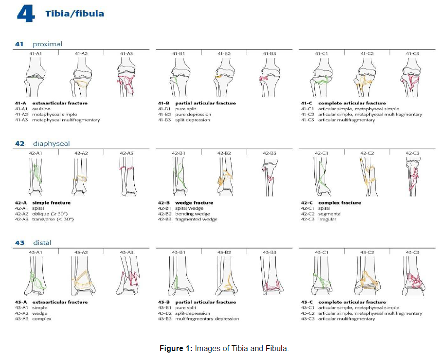
This is a prospective interventional study included 48 patients aged 21 to 68 years and were diagnosed as having a proximal tibial fracture with or without diaphyseal involvement. The fractures were stabilized with two method first the single lateral insertion of locking compression plate (LCP-) proximal lateral tibia (-PLT), using the percutaneous plate osteosynthesis technique and secondly using closed reduction and intermedullary interlocking nailing system at a tertiary trauma care centre from Jan 2019 to July 2020 (Figure 1).
Inclusion Criteria
1. Age more than 18 years and less than 75 years and willing for surgery.
2. Patients with competent neurological and vascular status of the affected limb and patients who meet the medical standards for routine elective surgery.
3. Patients with fracture meeting the AO criteria (41-A), duration of injury < 2 weeks.
Exclusion Criteria
1. Patient with open fractures, intra articular extension, pathological fractures, poor medical health or who did not give consent were excluded.
2. Patients with immature skeleton, segmental fracture of tibia, old injury were excluded.
Surgical techniques for MIPO plating
Patients were operated under spinal anaesthesia in supine position on a standard radiolucent table. The key concept of the approaches to the distal tibia is preserving the soft tissue envelope and the blood supply in the fractured metaphyseal area. Generally, the plate is inserted from distal to proximal through a tunnel between periosteum and intact overlying tissue. The medial approach is commonly used for the MIPO technique. A straight skin incision 3–5 cm is performed on the medial aspect of the distal tibia. The incision stops distally at the tip of the medial malleolus. The greater saphenous vein and saphenous nerve are held anteriorly with a blunt retractor. Separate stab incisions are usually sufficient for the insertion of the proximal screws in the diaphysis. It may be necessary to perform further stab incisions at the level of the fracture to percutaneously apply reduction forceps or insert a separate lag screw for better reduction especially in oblique or spiral fracture patterns at the meta diaphysis. In this periosteal space tunnelling towards the diaphysis can easily be achieved by using the blunt tip of the plate or an epiperiosteal tunnelling instrument. Reduction is achieved with manual traction and manipulation. Anatomically precontoured plate is inserted above the epiperiosteal space from distal to proximal on the anteromedial aspect of the tibia. First the plate is adjusted to the periarticular part of the tibia. It is important that the plate is in the correct position in relation to the joint space. After insertion of plate and achieving the reduction, the plate is temporarily fixed to bone with K wires. Then the plate is fixed to proximal fragment with one locking screw under image intensifier guidance through a small stab incision. The screw is then used as a hinge to rotate the plate clockwise or anticlockwise as required for accurate placement. Distal fragment fixation is done with combination of locking and cortical screws. The whole part was finally confirmed with IITV. The wounds are all closed in layers. Sterile dressings were applied over the wounds. The limb is wrapped in a compressive dressing.
Surgical technique for intramedullary nailing
A vertical patellar tendon splitting incision over skin extending from centre of the inferior pole of patella to the tibial tuberosity was made about 3 cms long. The patellar tendon was split vertically in its middle and retracted to reach the proximal part of tibial tuberosity. Next step was to determine the point of insertion. Essential for the success of the procedure is the correct choice of the insertion point. As a rule, the insertion point should be slightly distal to the tibial plateau, just medial to lateral tibial spine on a true AP view and exactly in line with the medullary canal on lateral view. If the insertion point is too distal, there is danger of fracturing the distal cortex of the main proximal fragment. On the other hand, insertion far too proximally bears the risk of opening the knee joint, patella comes in way of the zig or removal of nail may be difficult. After selecting the point of insertion curved bone awl was used to breach the proximal tibial cortex in, so that from perpendicular position its handle comes to be parallel to the shaft. In the metaphyseal cancellous bone, an entry portal was created, making sure it was in line with the centre of the medullary canal. After widening the medullary canal with a curved awl, a guide wire of size 3mms diameter x 950mms length was passed into the medullary canal of the proximal fragment. Reduction of the fracture fragments under image intensifier by maintaining longitudinal traction in line of the tibia was done. Accurate closed reduction of the fracture was verified under image intensifier before insertion of the guide wire in the distal tibial metaphysis. After reduction, the tip of the guide wire was passed till it enters the subchondral bone of distal tibia. In both AP and lateral views, the guide wire should lie in the centre of the tibial plafond. Reaming was initiated with hand reamer of size 8 mm, and then with one millimetre increment till the scratching sound of the isthmus was felt. Exact length of the nail was measured from the length of the guide wire remaining inside the medullary canal from the entry point. Size of the nail was assessed as one millimetre less than the diameter of the last reamer. Then a properly selected and assembled nail was passed into the medullary canal over the guide wire. Distal locking was always done first. It was done under image intensifier control by free hand technique. Cases in which the distal fragment was large enough to accommodate two mediolateral screws, two mediolateral screws were passed, and cases in which distal fragment was too small one mediolateral and one anteroposterior screw were passed. This was followed by proximal locking with the help of the zig using 4.9 mm interlocking bolts of appropriate length both static and dynamic. Screw positions were confirmed under C-arm image intensifier. After this, zig was removed, and stability was checked by performing flexion and extension at knee and ankle joint. Then all incisions were closed in layers. Sterile dressing was applied over the wound.
Post-operative care:
Patients were monitored for vascularity, swelling, discoloration, and movement for first 48 hrs, check dress was done on day 3 and then later Sutures will be removed on 14th day. The patients were placed in a well-padded posterior splint with the ankle neutral to prevent an equines deformity, during this period limb elevation and active toe movements were encouraged. Non weight bearing mobilization was started with walker which progressed to partial weight bearing after four weeks at least, Full weightbearing was advised once considerable callus was visualised radiologically. Active range of movements of knee and ankle were initiated as soon as the patient’s skin condition and pain permitted.
Follow up: Plain radiograph was carried out post operative, after two weeks, one month, two month, four month, and six month of surgery. Evaluation of clinical and radiological outcomes was made.
Result
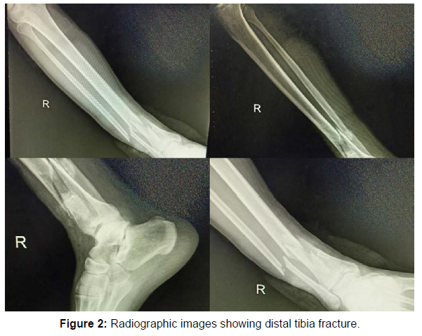
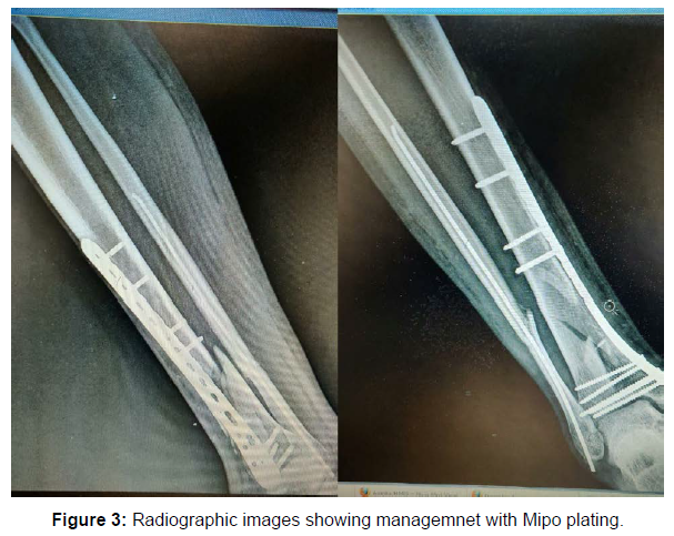
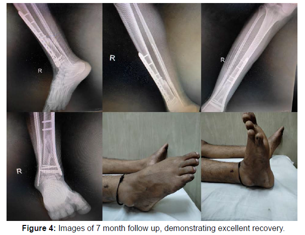
In our study, out of total 48 patients, the eldest patient in our study was 68 year old whereas youngest patient was 21 year old. The mean age of all patients was 41.31+13.35 year. However, 56.2% of our patients were from 31-50 age groups. The male gender was predominantly forming the sample size whereas females in the sample size. 32 patients had fracture due to road traffic accident, 8 had due to fall from height, 1 due to assault and 7 patients due to slip and fall. 2 patients had associated femur fracture and 30 patients did not have any associated injury. Of the total 24 patients were managed with locking plate and 24 patients were managed with intramedullary nail. There are disadvantages of these locking plate are costly, removal of plate is difficult, Plate cannot be contoured according to special need Locking head screws gives false feeling of the hold, Lag effect cannot be given, in proximal tibial locking plates the peroneal nerve is at risk when used through the lateral approach. 2 Patients had superficial infection and 1 had deep infection. In the present study, the range of knee flexion was 100 to 146 degrees, with a mean flexion of 136 degrees. Out of 48 patient 40 patient had no extension lag only 8 patient had extension lag more than 10 degree who had showed poor compliance to pain and physiotherapy Other than infection other complication included, delayed union (3) varus deformity(0), post traumatic arthritis in 2 patients. No complications like Compartment syndrome, DVT, Iatrogenic foot drop and Avascular necrosis. Out of 48 patients 22 patients had radiological union at 12 weeks with average time for union was 16 weeks. Majority of our patients 80% started walking in 18-20 weeks (Tables 1-6 and Figures 2-4).
| Male/Female | 35/13 |
|---|---|
| Age (Range and Mean) | 41.31+13.35 years (21 to 68 years) |
| Right/left side | 29/19 |
| Fracture type 43-A1 | 10 |
| Fracture type 43-A2 | 21 |
| Fracture type 43-A3 | 10 |
| Proximal fracture extension | 7 |
| Closed/Open injury | 48/0 |
Table 1: Demographics and Fracture type
| Motor Vehicle crash | 12 | 25% |
|---|---|---|
| Fall | 8 | 16.66% |
| Isolated Fracture | 24 | 50% |
| Multiple Fracture | 3 | 6.25% |
| Polytrauma | 1 | 2.08% |
| TOTAL | 48 | 100% |
Table 2: Mechanism of Injury
| Plating group n=24 | Nailing group n=24 | |
|---|---|---|
| 8.2±2.3 | 5.2±1.3 | |
| Days post-surgery (Hospital stay) | 6.6±2.3 | 4.6±1.3 |
| Surgical time in minutes | 110±20 | 95±20 |
| Estimated blood loss | 40.5±20.5 | 30.5±10.5 |
| Fluoroscopy time in minutes | 20.3±6.3 | 10.3±6.3 |
| Plate size (9/10/11 hole) | (28/10/10) | Intra-medullary nail used |
| Time to union in | 5.2±1.3 | 3.2±1.3 |
| Immobilization in days | 20.5±10.6 | 5.5±2.6 |
Table 3: Surgical Details
| Parameters to be compared | Plating group n=24 | Nailing group n=24 |
|---|---|---|
| Radiographic Healing Time(months) | 5.1±1.1 | 3.2±1.6 |
| Delayed Union | 3 cases | 2 cases |
| Non-Union | 1 case | Nil |
| Valgus Malunion | 2 (7 deg. & 8 deg.) | Nil |
| Varus Malunion | 0 | 0 |
| Secondary Loss of Reduction | 0 | 0 |
| Superficial wound infection | 4 | 0 |
| Implant removal | 0 | 0 |
Table 4: Post-operative Complications and Outcomes
| Parameter | Degree | Score |
|---|---|---|
| Pain | None | 25 |
| While walking on uneven surface | 20 | |
| While walking on even surface outdoors | 10 | |
| While walking indoors | 5 | |
| Constant and severe | 0 | |
| Stiffness | None | 10 |
| Stiffness | 0 | |
| Swelling | None | 10 |
| Only evenings | 5 | |
| Constant and severe | 0 | |
| Stair climbing | No problems | 10 |
| Impaired | 5 | |
| Impossible | 0 | |
| Running | Possible | 5 |
| Impossible | 0 | |
| Jumping | Possible | 5 |
| Impossible | 0 | |
| Squating | No problem | 5 |
| Impossible | 0 | |
| Supports | None | 10 |
| Taping, wrapping | 5 | |
| stick or crutches | 0 | |
| Work activities of daily life | Same as before injury | 20 |
| Loss of tempo | 15 | |
| Change to a job/part-time work | 10 | |
| Severely impaired work capacity | 0 |
Table 5: Olerud and Molander’s clinical Criteria for outcome assessment
| Range | Results | 1 Month follow-up | 2 Month follow-up |
|---|---|---|---|
| 100 | Excellent | 10 | 40 |
| 80-100 | Good | 30 | 8 |
| 60-80 | Fair | 8 | 0 |
| <60 | Poor | 0 | 0 |
Table 6: Outcome analysis
Discussion
Extra articular distal tibial fracture which are presented to the orthopaedician, often poses a challenge to the surgeon as status of soft tissue and degree of comminution itself complicates the plan of management [12,13]. The goal of operative treatment is to obtain anatomical alignment of the joint surface while providing enough stability to allow early motion. This should be accomplished using techniques that minimize osseous and soft tissue devascularization in the hopes of decreasing the complications resulting from treatment [14,15]. With the development of minimally invasive surgery, percutaneous plating has challenged interlocking nailing as locked plate designs act as fixed-angle devices whose stability is provided by the axial and angular stability at the screw-plate interface instead of relying on the frictional force between the plate and bone, which is thought to preserve the periosteal blood supply around the fracture site [16,17]. In present series, 48 cases of extrarticular distal tibia fractures were treated primarily over a period of two years with follow up ranging from 12 months to 22 months. We evaluated our results and compared them with the result of various studies in the literature. In present study, the average duration of surgery in interlocking group was 57.53 min and the average duration of surgery in plating group was 69.33 min which was comparable to the studies done by Guo JJ, et al. [18]. In present study, fracture of fibula was seen in 7 cases in ILN group whereas in 8 cases in plating group. Fixation of fibula was done in 04 cases in ILN group whereas in 7 patients in plating group which was comparable to Guo JJ, et al. They concluded that fixation of associated fibula fracture reduced the incidence of non-union in distal tibial fractures. In present study, in the interlocking group average time to weight bearing was 9.53 weeks whereas in the plating group average time to full weight bearing was 13.06 weeks which was comparable to studies done by Redfern DJ, et al. [14] and Guo JJ, et al. [18]. In present study, average time for union was 17.43 weeks in interlocking group compared to 21.40 weeks in plating group which was comparable to the studies done by Redfern DJ, et al. [14] and Bahari S, et al. [13]. In our study implant irritation and ankle stiffness were the most common complications in plating group (26.66%), superficial infection in two patients (13.33%) of plating group. These results were comparable to the studies done by Bahari S. et al. [13], Lau TW, et al. [8], Guo JJ, et al. [18].
Conclusion
Minimally invasive plate osteosynthesis by locking plate have shown a reliable method of fixation for distal tibial fractures. This procedures preserving most of the osseous vascularity, fracture hematoma which provide biological repair so there is lesser incidence of delayed union and non-union .This technique can be used in fractures where locked nailing cannot be done like distal tibial fractures with small distal metaphyseal fragments and comminuted fractures. The decision to fix the fibula was based on intraoperative reduction of tibia fracture. If significant malalignment was still persisting after fixation of tibia, only then the decision to fix fibula fracture was made. Thus we do not recommend fibular fixation routinely because the essential benefit of closed MIPO in the avoidance of soft tissue dissection might be compromised in this way and also reduces strain over the tibial fracture, which heightens the potential for delayed healing or nonunion but to support this , larger trial are needed. Implant prominence and its related complications because of mismatching of the implant contouring and supra malleolar anatomy especially in thin built patients. Meticulous soft tissue handling is imperative. Our results have shown that both closed intramedullary nailing and locking plating can be used safely to treat distal metaphyseal fractures of the tibia. Closed nailing has the advantage of shortened operating time, early weight bearing, decreased wound problems, early union of the fracture, decreased implant related problems and overall reduced morbidity, hence we prefer closed intramedullary interlocking nailing in treatment of distal tibia fractures. For fractures of the distal tibia and fibula, the proportion of patients with mal-alignment was significantly greater without fixation of fibula after intramedullary nailing or locking plate fixation. Thus, we recommend fibular fixation whenever intramedullary nailing or locking plate fixation is used in distal tibiofibular fractures A Randomized Controlled Trial, possibly triple blinded or at least double blinded in nature, involving many patients with long term follow-up is clearly needed to bring the differences between the two techniques.
Funding
Nil
Conflicts of Interest
Nil
References
- Stannard J, Schmidt A, Kregor P (2007) Surgical treatment of orthopaedic trauma (1st edn). pp:767-797.
- Schatzker J, Tile M (2005) The rationale of operative fracture care (3rd edn). Springer pp: 475-476.
- Bucholz RW, Court-Brown CM, Heckman JD, Tornet P (2015) Rockwood and Green's Fractures in adults. New York [u.a.]: Lippincott. 98
- Borrelli J, Prickett W, Song E, Becker D, Ricci W (2002) Extraosseous blood supply of the tibia and the effects of different plating techniques: a human cadaveric study. J Orthop Trauma. 16(10):691-5.
- Bedi A, Toan T, Karunakar M (2006) Surgical Treatment of Nonarticular Distal Tibia Fractures. J Am Acad Orthop Surg 14(7): 406-16.
- Prasad PNVSV, Nemade A, Anjum R, Joshi N (2019) Extra-articular distal tibial fractures, is interlocking nailing an option? A prospective study of 147 cases. Chin J Traumatol 22:103-107.
- Ghera S, Santori F, Calderaro M, Giorgini T (2004) Minimally Invasive Plate Osteosynthesis in Distal TibialFractures: Pitfalls and Surgical Guidelines. Trauma Orthop 27(9):903.
- Lau T, Leung F, Chan F, Chow S (2008) Wound complication of minimally invasive plate osteosynthesis in distal tibia fractures. Int Orthop 32(5): 697-703.
- Nork S, Schwartz A, Agel J, Holt S, Schrick J,et al. (2005) Intramedullary nailing of distal metaphyseal tibial fractures. J Bone Joint Surg Am 87(6):1213-21.
- Johner R, Wruhs O (1983) Classification of tibial shaft fractures and correlation with results after rigid internal fixation. Clin Orthop 178: 7-25.
- Gautier E, Sommer C (2003) Guidelines for the clinical application of the LCP. Inj Int J Care Inj 34:S-B63-SB76.
- Wagner M (2003) General principles for the clinical use of the LCP. Inj Int J Care Inj 34:S-B31-S-B42.
- Bahari S, Lenehan B, Khan H, McElwain JP (2007) Minimally invasive percutaneous plate fixation of distal tibia fractures. Acta Orthop Belg 73(5):635-40
- Redfern DJ, Syed SU, Davies SJ (2004) Fractures of the distal tibia: minimally invasive plate osteosynthesis. Injury 35(6):615-20.
- Sitnik AA, Beletsky AV (2013) Minimally invasive percutaneous plate fixation of tibia fractures: results in 80 patients. Clin Orthop Relat Res 471(9):2783-9.
- Paluvadi SV, Lal H, Mittal D, Vidyarthi K (2014) Management of fractures of the distal third tibia by minimally invasive plate osteosynthesis-A prospective series of 50 patients. J Clin Orthop Trauma 5(3): 129-136.
- Kumar D, Ram GG, Vijayaraghavan PV (2014) Minimally Invasive Plate Versus Intramedullary Interlocking Nailing In Distal Third Tibia Fractures. IOSR-J Dent Med Sci 13:15-17.
- Guo JJ, Tang N, Yang HL, Tang TS (2010) A prospective, randomised trial comparing closed intramedullary nailing with percutaneous plating in the treatment of distal metaphyseal fractures of the tibia. J Bone Joint Surg Br 92: 984-8.
Indexed at, Google Scholar, Crossref
Indexed at, Google Scholar, Crossref
Indexed at, Google Scholar, Crossref
Indexed at, Google Scholar, Crossref
Indexed at, Google Scholar, Crossref
Indexed at, Google Scholar, Crossref
Indexed at, Google Scholar, Crossref
Indexed at, Google Scholar, Crossref
Indexed at, Google Scholar, Crossref
Indexed at, Google Scholar, Crossref
Citation: Mahajan NP, Patil T, Mhatre JA, Kamble M, Sarkunde P, et al. (2022) A comparative study of functional outcome of distal tibia extra articular fractures managed with intramedullary nailing and plating. Clin Res Foot Ankle, 10: 336. DOI: 10.4172/2329-910X.1000336
Copyright: © 2022 Mahajan NP, et al. This is an open-access article distributed under the terms of the Creative Commons Attribution License, which permitsunrestricted use, distribution, and reproduction in any medium, provided the original author and source are credited.
Select your language of interest to view the total content in your interested language
Share This Article
Recommended Journals
Open Access Journals
Article Tools
Article Usage
- Total views: 3478
- [From(publication date): 0-2022 - Dec 16, 2025]
- Breakdown by view type
- HTML page views: 2740
- PDF downloads: 738


