Clinical images Open Access
A Rare Case of Herniated Renal Cyst Through Grynfeltt’s Triangle
Antonio Pesce* and Stefano PuleoDepartment of Medical and Surgical Sciences and Advanced Technologies “G.F. Ingrassia”, University of Catania, Via S. Sofia 84, 95123 Catania, Italy
- *Corresponding Author:
- Antonio Pesce, MD
Via Taranto n° 14 Catania 95125 – Catania, Italy
Tel: +39 3286680943
Fax: +39-95-3782912
E-mail: nino.fish@hotmail.it
Received date: May 23, 2015 Accepted date: June 19, 2015 Published date: June 31, 2015
Citation: Pesce A and Puleo S (2015) A Rare Case of Herniated Renal Cyst Through Grynfeltt’s Triangle.J Gastrointest Dig Syst 5:304. doi:10.4172/2161-069X.1000304
Copyright: © 2015 Pesce A, et al. This is an open-access article distributed under the terms of the Creative Commons Attribution License, which permits unrestricted use, distribution, and reproduction in any medium, provided the original author and source are credited.
Visit for more related articles at Journal of Gastrointestinal & Digestive System
Clinical Image
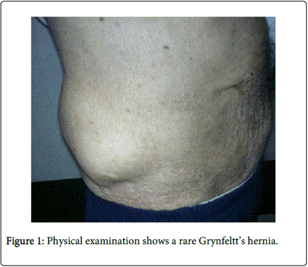
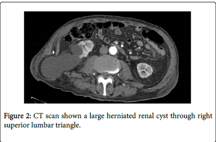
Lumbar hernias represent a rare entity among the abdominal wall defects. They are rare defects involving two weak areas of the postero-lateral abdominal wall: the superior lumbar triangle of Grynfeltt-Lesshaft, which is the most common site, and the inferior lumbar triangle of Petit. Antomically Grynfeltt’s triangle is bounded: above by the twelfth rib, medially by the sacrospinalis muscle, laterally by the posterior border of the internal oblique muscle. This rare hernia can be classified as congenital (approximately 20%), generally associated with other malformations, or acquired (around 80%), presenting in adults spontaneously or secondary to trauma or surgical incisions. The hernial sac content is generally characterized by retroperitoneal fat. We present a case of a 80 years old man who presented right lower back pain associated with a palpable mass for 5 years. The past medical history revealed a not well defined surgical procedure for a right renal trauma. The diagnosis of incisional lumbar hernia was made by physical examination (Figure 1). Radiological investigations such as abdominal ultrasonography and CT scan were performed in order to confirm the clinical suspicion. CT scan showed a herniated renal cyst of 9 cm diameter through right superior lumbar triangle (Figure 2). The patient underwent a small lumbotomy and a polypropylene mesh was placed.
--Relevant Topics
- Constipation
- Digestive Enzymes
- Endoscopy
- Epigastric Pain
- Gall Bladder
- Gastric Cancer
- Gastrointestinal Bleeding
- Gastrointestinal Hormones
- Gastrointestinal Infections
- Gastrointestinal Inflammation
- Gastrointestinal Pathology
- Gastrointestinal Pharmacology
- Gastrointestinal Radiology
- Gastrointestinal Surgery
- Gastrointestinal Tuberculosis
- GIST Sarcoma
- Intestinal Blockage
- Pancreas
- Salivary Glands
- Stomach Bloating
- Stomach Cramps
- Stomach Disorders
- Stomach Ulcer
Recommended Journals
Article Tools
Article Usage
- Total views: 15667
- [From(publication date):
August-2015 - Aug 29, 2025] - Breakdown by view type
- HTML page views : 11055
- PDF downloads : 4612


