A Rare Case of Metastatic Retinoblastoma with Cervical Mass Presenting as a Massive Swelling in Child
Received: 04-Oct-2021 / Accepted Date: 18-Oct-2021 / Published Date: 25-Oct-2021
Abstract
Retinoblastoma (Rb) is a common intraocular malignancy in childhood, due to mutation of RB1 gene, approximately one-third of these tumor are bilateral retinoblastoma, which are more heritable and usually found in younger children.
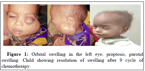
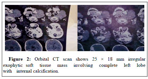
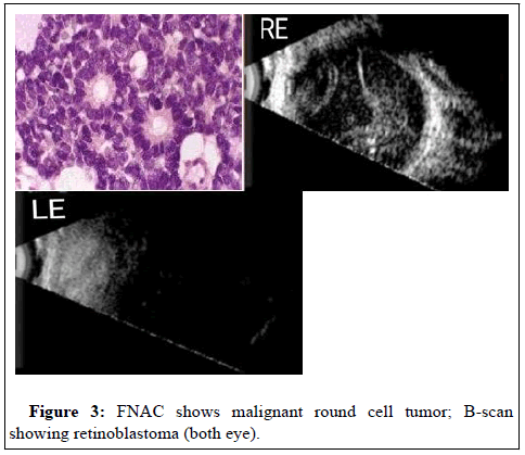
A 4 year old boy presented with swelling of left eye with proptosis and swelling extending up to cervical and parotid region, necrosed conjunctiva and cornea. Ocular examination included visual acuity, slit lamp examination. Detailed fundus evaluation after total mydriasis (right eye) under general anesthesia was done. Investigations like BScan; Computed Tomography/MRI imaging of orbit and brain was done. General examination to look for lymphadenopathy, hepatomegaly and bony swellings was done. Metastatic work-up included peripheral smear, bone marrow aspiration, CSF analysis, X-ray chest and USG abdomen. B-scan Ultrasonography indicated a mass under the retina in both eye (left>right). Orbital Computed Tomography scan was showing orbital soft mass with calcification.
Keywords: Retinoblastoma; Proptosis; Orbital; Calcification
Introduction
Retinoblastoma is a childhood ocular malignancy generally presents with leukocoria, diminution of vision, strabismus, proptosis or even developmental delay. Retinoblastoma is the most frequent malignant intraocular tumor of childhood. It occurs with an estimated frequency of 1 in 15,000 children. Prior to 20th century, retinoblastoma was a uniformly fatal disease. Today, survival is better than 95% in developed countries. However, late presentation and delayed diagnosis are still problems in the developing world, resulting in lower survival rates. The role of hereditary in retinoblastoma was not appreciated until the 19th century, because the disease was poorly defined and there were practically no survivors. During the 20th century, more scientific studies were conducted regarding the hereditary patterns of retinoblastoma. Although, these general observations served to explain some of the clinical facts, they were unable to offer any basis for the genetic abnormality affecting the siblings and parents of children affected with retinoblastoma [1]. The “two-hit hypothesis” was based on the observation that there were two distinct forms of retinoblastoma seen in the clinic. Children with the family history of retinoblastoma often had bilateral multifocal retinoblastoma. In contrast, the children with unilateral retinoblastoma rarely had any family history of the disease. They inherited a defective copy of this tumor suppressor gene and were likely to have retinal cells that sustained mutations in the second allele, leading to multifocal bilateral retinoblastoma.
Pawius described retinoblastoma as early as in 1597 referred to the tumor as fungus hematodes and suggested enucleation as the primary mode of management [2]. Flexner (1891) and Wintersteiner (1897) described the histology of retinoblastoma. The rosettes were named after them [3]. The term retinoblastoma was coined by Verhoeff (1922) [4]. Von Graffe (1884) advocated enucleation of eye with long optic stump. Hilgartner (1903) first used external beam radiotherapy and Kupfer (1953) used chemotherapy for retinoblastoma. Reese- Ellsworth (1958) proposed a classification of retinoblastoma [5].
Tumor originated from retinoblasts and the American Ophthalmological Society officially accepted the term retinoblastoma in 1962. Retinoblastoma arises from the malignant transformation of primitive retinal cells due to mutation in the Rb gene in chromosome 13q14 [6]. Genetic counseling is important aspect in the management of retinoblastoma. These landmark studies on the genetics ofretinoblastoma susceptibility have had a major impact on our understanding of tumor suppressor genes and have formed the framework for much of our current knowledge of genetic lesions associated with tumorigenesis.
Case Report
A 4 year old boy presented with left orbital swelling with proptosis and swelling extending up to cervical region and parotid region, necrosed conjunctiva and cornea. On indirect ophthalmoscopy right eye shows friable mass infero-temporal to disc. B-scan Ultra sonography shows large mass under the retina in left eye involving cervical region and small size mass under the retina, right eye. Orbital CT scan shows 25 × 18 mm irregular exophytic soft tissue mass involving complete left lobe with internal calcification extending to anterior chamber and outside the posterior margin of globe. Right globe shows 6-7 mm nodular soft tissue masses with calcification and 50 × 60 mm soft tissue left cervical region. FNAC from left eye shows necrotic material and from left side of face shows features of malignant round cell tumor. There was no family history. There was no evidence hepatomegaly found. He has an elder male sibling with no obvious illness. The patient is showing significant improvement in swelling after treatment with chemotherapy (Figures 1-3).
Discussion
Retinoblastoma is the most common intraocular malignancy in children. The average age of diagnosis of Rb is 18 months with 90% cases being diagnosed in children less than 5 years of age without any racial or sex predilection. In developing countries due to poverty, low socioeconomic statuts, illiteracy causing a delay in the presentation, diagnosis and treatment. Rb commonly presents with cat’s eye reflection or leukocoria in the pupil. Studies in India also had leucocoria as most common presenting feature. In all the studies more than 50% of patients had leucocoria. Strabismus was not the presenting sign in our study but was one of the significant features in studies by Abramson et al. and Honavar et al. [7,8]. Rb patients with extra-ocular or metastatic disease are known to have a poor prognosis, however chemoreduction (systemic, intraarterial, intravitreal) and radiation, enucleation have been quite encouraging. Regular follow-up is mandatory also because of higher risk of other malignancies.
However, retinoblastoma is still a deadly cancer worldwide, with an estimated death rate of more than 40%. Most of the deats from retinoblastoma occur in Asia and Africa. Successful treatment requires early diagnosis as well as availability of modern treatment modalities and the major goal should be to improve the delivery of ocular health care in non-industrialized countries. Patients of extraocular and metastatic Rb is rare and have had a poor prognosis but recent approach of combining chemotherapy with radiotherapy has shown better outcome as seen with my patient.
Conclusion
Retinoblastoma has excellent prognosis for survival, eye salvage and vision salvage with current treatment modalities. It requires integrated approach involving adequate screening, prompt referral and a team of ophthalmologist and pediatric oncologist at a comprehensive tertiary care centre.
Our study shows that patients with retinoblastoma seek treatment at late stage of disease due to lack of awareness and delayed access to tertiary health care. This highlights the need for improved awareness among public and primary health care providers so that diagnosis can be made at an early stage.
References
- Halperin EC, Brady LW, Wazer DE, Perez CA (2013) Perez & Brady's principles and practice of radiation oncology. Lippincott Williams & Wilkins.
- Abramson DH (1990) Retinoblastoma incidence in the United States. Arch Ophthalmol 108: 1514.
- Flexner S (1891) A peculiar glioma (neuroepithelioma?) of the retina. Bull Johns Hopkins Hosp 2: 115-119.
- Verhoeff FH, Jackson E (1926) Minutes of the proceedings. In62nd annual meeting. Trans Am Ophthalmol Soc 24: 38-43.
- Albert DM (1987) Historic review of retinoblastoma. Ophthalmology 94: 654-662.
- Sparkes RS, Murphree AL, Lingua RW (1983) Gene for hereditary retinoblastoma assigned to human chromosome 13 by linkage to esterase D. Science 219: 971-973.
- Abramson DH, Frank CM, Susman M, Whalen MP, Dunkel IJ, et al. (1998) Presenting signs of Retinoblastoma. Pediatrics 132: 505-508.
- Reddy VP, Honavar SG, Shome D, Shah K, Naik MN, et al. (2007) Demographics, clinical profile, management, and outcome of retinoblastoma in a tertiary care center in southern India. J Clin Oncol 25: 20011.
Citation: Kumari A, Kumar S(2021) A Rare Case of Metastatic Retinoblastoma with Cervical Mass Presenting as a Massive Swelling in Child. Diagn Pathol Open S7: 025.
Copyright: © 2021 Kumari A, et al. This is an open-access article distributed under the terms of the Creative Commons Attribution License, which permits unrestricted use, distribution, and reproduction in any medium, provided the original author and source are credited.
Select your language of interest to view the total content in your interested language
Share This Article
Open Access Journals
Article Usage
- Total views: 2268
- [From(publication date): 0-2021 - Dec 08, 2025]
- Breakdown by view type
- HTML page views: 1638
- PDF downloads: 630


