A Review of an Implantable Middle Ear Microphone: New Floating Piezoelectric Microphone
Received: 11-May-2018 / Accepted Date: 23-Jun-2017 / Published Date: 30-Jun-2017 DOI: 10.4172/2161-119X.1000312
255148Introduction
An increasing number of (those with severe and profound sensorineural hearing loss, SSHL / PSHL) have benefited from cochlear implants (CIs). The exterior components of CIs, including the speech processing system, exterior coil, power supply component, and microphone (sensor), can be attract attention and inconvenient, may embarrass patients, can limit patients’ social activities, can cause the inevitability of silent sleep (24/7 h) and complicate patients’ daily activities, for example, during bathing or while participating in swimming or other sports [1,2]. To eliminate these disadvantages, totally implantable cochlear implants (TICIs) must be studied. The most essential challenge is the implantable microphone (sensor), which needs to be collect acoustic signals and convert them into electrical signals.
Research on implantable microphones has made progress. The most representative designs are as follows: the TICA from Implex, Munich, Germany [3]; two products, the TIKI [1] and Carina [4], from Cochlear, Sydney, Australia; and the Esteem [5,6] from Envoy, Saint Paul, MN, USA. The above designs have different advantages and disadvantages, and further research is ongoing.
Beginning in early 2001, our team in the Department of Otology and Skull Base Surgery at the Eye, Ear, Nose and Throat Hospital of Fudan University has concentrated on researching miniature piezoelectric microphones for totally implantable middle ear systems. In 2004, we designed and built a piezoelectric microphone that can be connected to the head of the malleus of a cat in order to collect acoustic vibration signals.
By 2009, the piezoelectric microphone had been improved many times and was constructed from piezoelectric bimorph material cut into a linear shape [7]. The transducer was 6 mm long, 2 mm wide and 0.2 mm thick, and weighed 20 mg. A T-shaped titanium rod clamped to the piezoelectric microphone was connected to the head of a cat malleus. The other end of the piezoelectric microphone, as the signal output end, was connected to the amplifier. The piezoelectric microphone was coupled to the head of the malleus to convert the eardrum-malleus mechanical vibrations activated by tonal stimuli into electric signals. An audible acoustic signal that showed a nearly flat frequency response curve (FRC) was detected [7]. However, during the surgical procedure, the cat’s incus had to be removed because of the structure of the piezoelectric microphone. Biocompatibility was another important issue to be considered.
The floating piezoelectric microphone (FPM) prototype was developed in 2012 [8]. Based on micro-electro-mechanics systems (MEMS) technology, the FPM was composed of one piece of piezoelectric ceramic bimorph element (PCBE, 4.5 mm × 1.0 mm × 0.3 mm) and one preamplifier chip (LMV 1032, 1.18 mm 1.18 mm × 0.35 mm, National Semiconductor Co, Ltd., USA). It was then sealed inside a thin titanium shell (Ti6Al4V standard grade, 0.10 mm thick) and shaped into a single cantilever structure [8]. The FPM was 5.0 mm × 1.5 mm × 1.2 mm in size and weighed 38.4 mg. The FPM was supplied with 2 V of direct current voltage via 2 wires, and it exported signals to the data acquisition (DAQ) equipment via the other wire. During testing in a sound-proof box, the sound source, which supplied pure tones from 0.25 to 8.0 kHz, was placed at a distance of 1 m from the location of the cat implanted with the prototype. The average sensitivity of the piezoelectric transducer was -35.83 dB rms ref 1 V at 1000 Hz. The following characteristics were found: 1) though the method of connecting the FPM to the ossicular chain needs to be improved, the ossicular chain was kept intact in this study due to the new design of the FPM. 2) When implanted into a cat’s tympanic cavity, the FPM produces an inefficient output at low frequencies, especially those lower than 1.5 kHz, but the FRC remained flat compared to our previous research at frequencies above 2.0 kHz. Different coupling site might contribute to the above difference. 3) The FPM and the HiFi Hy-M30 microphone were tested simultaneously. In the high-frequency test, the FPM displayed a flatter FRC than the HiFi Hy-M30 microphone. However, the FPM showed less sensitivity compared to the HiFi Hy-M30 microphone. The reason for this might be that the acoustic vibrations of the tympanic membrane and ossicles at low frequencies was influenced by an overloading mass [9-11]. Additionally, an unsteady junction between the FPM and the ossicles may cause the above results. These problems were improved in the following studies.
The next year, further studies were performed to explore the feasibility of connecting the FPM to the ossicular chain of a human cadaver without destroying the middle ear structure [12]. The displacement FRC was studied using finite element (FE) analysis. In vitro experiments were also performed to investigate the amplitude of the FRC and to explore a feasible connection position on the ossicular chains of human cadavers.
To find a suitable implant position, a finite element (FE) analysis was performed. A 3D geometric model of the human middle ear was built using micro-computerized tomography (micro-CT) and Mimics. The geometric model was then used to establish the FE model and to perform the FE analysis. Two locations for the FPM were chosen: 1) the long process of the incus and 2) the manubrium of the malleus. On the basis of a 3D reconstruction, the model-derived FRC of the FPM’s displacement in the ossicular chain had two characteristics: 1) it was flat at low frequencies, 2) decreasing as the frequency increased. The results also showed that the displacement and the FRC were similar.
The FPM was implanted in the fresh human temporal bone (implanted in tympanic cavity without contacting any structure, or implanted at the ossicular chain) and tested. The FPM collected the vibration of the ossicular chain more effectively than that of the FPM in the tympanic cavity. The average sensitivity of the FPM is -44.22 dB rms ref 1 V at 1000 Hz in the long process of the incus, -53.33 dB rms ref 1 V at 1000 Hz in the malleus and -108.59 dB rms ref 1 V at 1000 Hz in the tympanic cavity. The in vivo experiments also showed a similar trend with that of the model-derived FRC of the FPM displacement: flat at low frequencies; decreasing at high frequencies.
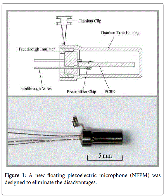
However, the FPM still lacked biocompatibility and needed a better coupling mechanism. A new floating piezoelectric microphone (NFPM) was designed to eliminate these disadvantages (Figure 1); a description of itwas first published in 2016 [13]. Based on the design of FPM, the NFPM included a titanium clip (1.38 mm in diameter), a thin titanium tube housing (0.1 mm in thickness), and was shaped like a cantilever structure. The tube feedthrough assembly included three feedthrough wires, a tube feedthrough insulator, and a tube feedthrough flanger. The wires, which were insulated platinum and 0.06 mm in diameter, were attached to the PCBE and preamplifier using a spot-welder. The PCBE size was reduced to 1.0 mm × 0.3 mm × 4.0 mm; the NFPM measured 5.91 mm × 2.4 mm × 2.02 mm and weighed 67.0 mg.
The NFPM’s advantages included its improved biocompatibility and its titanium clip, which should make surgical operations convenient and ensure a tight connection. With an acoustic stimulation strength of 90 dB SPL ref 20 lPa, the average sensitivity of the NFPM was -56.58 dB rms ref 1 V at 1000 Hz in the long process of the incus and -92.94 dB rms ref 1 V at 1000 Hz in the tympanic cavity. The FRC of the NFPM was flatter than that of the FPM. Although NFPM has the advantages above, there is still room for improvement. The average sensitivity should be further improved. The location of the wires could be more suitable (for example, move to the side of titanium tube, and the three wires might be combined into one) for the middle ear structure. The better design of the titanium clip (for example, the clip of a kind if stapes prosthesis named Piston might be a better design) will make operation more convenient.
At present, the NFPM is connected to a CI system, and it can be used as a signal source for the CI. At the same time, the research involving the NFPM has entered the small-scale clinical test phase, and the results are as expected.
As an implantable microphone, the NFPM is placed in a hermetically sealed and biocompatible titanium shell, and it converts the vibrations of the ossicular chain into electrical signals. In the future, with further researches of the signal process and power supply, the NFPM would work with an implantable speech processor and stimulate electrodes of CI.
Acknowledgement
The study was supported by National Natural Science Foundation of China (81420108010, 81271084 to F.C, 81500785 to N.G, 81000413, 81570920 and 81370022 to D.R.), the Major State Basic Research Development Program of China (973 Program) ( 2011CB504506) to F.C, and Innovation Project of Shanghai Municipal Science and Technology Commission (11411952300) to F.C.
Competing Financial Interests
The authors declare no competing interests.
References
- Briggs RJ, Eder HC, Seligman PM, Cowan RS, Plant KL, et al. (2008) Initial clinical experience with a totally implantable cochlear implant research device. Otol Neurotol 29: 114-119.
- Cohen N (2007) The totally implantable cochlear implant. Ear Hear 28: 100S-101S.
- Leysieffer H, Baumann JW, Mayer R, Müller D, Müller G, et al. (1998) A totally implantable hearing aid for inner ear deafness: TICA LZ 3001. HNO 46: 853-863.
- Haynes DS, Young JA, Wanna GB, Glasscock ME 3rd (2009) Middle ear implantable hearing devices: An overview. Trends Amplif 13: 206-214.
- Marzo SJ, Sappington JM, Shohet JA (2014) The envoy esteem implantable hearing system. Otolaryngol Clin North Am 47: 941-952.
- Kraus EM, Shohet JA, Catalano PJ (2011) Envoy esteem totally implantable hearing system: Phase 2 trial, 1 year hearing results. Otolaryngol Head Neck Surg 145: 100-109.
- Chi FL, Wu Y, Yan QB, Shen YH, Jiang Y, et al. (2009) Sensitivity and fidelity of a novel piezoelectric middle ear transducer. ORL J Otorhinolaryngol Relat Spec 71: 216-220.
- Kang HY, Na G, Chi FL, Jin K, Jin TZ, et al. (2012) Feasible pickup from intact ossicular chain with floating piezoelectric microphone. Biomed Eng Online 11: 10.
- Gyo K, Yanagihara N, Araki H (1984) Sound pickup utilizing an implantable piezoelectric ceramic bimorph element: Application to the cochlear implant. Am J Otol 5: 273-276.
- Tonndorf J (1988) Sensorineural and pseudosensorineural hearing losses. ORL J Otorhinolaryngol Relat Spec 50: 79-83.
- Needham AJ, Jiang D, Bibas A et al. (2005) The effects of mass loading the ossicles with a floating mass transducer on middle ear transfer function. Otol Neurotol 26: 218-224.
- Gao N, Chen YZ, Chi FL, Zhang TY, Xu HD, et al. (2013) The frequency response of a floating piezoelectric microphone for the implantable middle ear microphone. Laryngoscope 123: 1506-1513.
- Jia XH, Gao N, et al. (2016) A new floating piezoelectric microphone for the implantable middle ear microphone in experimental studies. Acta Otolaryngol 136: 1248-1254.
Citation: Xu XD, Chi FL (2017) A Review of an Implantable Middle Ear Microphone: New Floating Piezoelectric Microphone. Otolaryngol (Sunnyvale) 7:312. DOI: 10.4172/2161-119X.1000312
Copyright: © 2017 Xu XD, et al. This is an open-access article distributed under the terms of the Creative Commons Attribution License, which permits unrestricted use, distribution and reproduction in any medium, provided the original author and source are credited.
Select your language of interest to view the total content in your interested language
Share This Article
Recommended Journals
Open Access Journals
Article Tools
Article Usage
- Total views: 3824
- [From(publication date): 0-2017 - Jul 16, 2025]
- Breakdown by view type
- HTML page views: 2911
- PDF downloads: 913

