Case Report Open Access
"A Unique Case of Amelogenesis Imperfecta"
Lakshman Balaji*, Revathy V, Suganthi, Geethalakshmi and Prem Kumar SDepartment of Pediatric and Preventive Dentistry, Tamil Nadu Government Dental College and Hospital, Chennai, India
- *Corresponding Author:
- Dr. Lakshman B
House surgeon
Tamil Nadu Government Dental College and Hospital
Chennai, India, Plot no 32
Door no 3,7th Main Road, 5th Avenue
Dhandeeswaram, Velachery, Chennai-600 042, India
Tel: +91 9840392988
E-mail: blakgau@gmail.com
Received Date: May 18, 2016; Accepted Date: May 23, 2016; Published Date: May 27, 2016
Citation: Lakshman B, Revathy V, Suganthi, Geethalakshmi, Prem Kumar S (2016) A Unique Case of Amelogenesis Imperfecta. Pediatr Dent Care 1:109. doi:10.4172/pdc.1000109
Copyright: © 2016 Lakshman B, et al. This is an open-access article distributed under the terms of the Creative Commons Attribution License, which permits unrestricted use, distribution, and reproduction in any medium, provided the original author and source are credited.
Visit for more related articles at Neonatal and Pediatric Medicine
Abstract
Amelogenesis imperfecta is a developmental disturbance in the structure of the tooth enamel of genetic origin. Though it is a rare hereditary defect, it is not an uncommon entity and is encountered in clinical practice with varying degrees of frequency. Amelogenesis imperfecta, for the most part, is widely considered as a disease, which affects the enamel of all the teeth uniformly. We present here a case of amelogenesis imperfecta in which relative sparing of the maxillary and mandibular anteriors is seen. This report should serve to remind clinicians of the fact that overly simple descriptions of the distribution of the disease are unwarranted, and that it can present inhighly variablepatterns.
Keywords
Amelogenesis imperfect; Enamel hypoplasia; Sparing; Relative; Anteriors; Unaffected
Introduction
Amelogenesis imperfecta (AI) encompasses a group of genomic conditions, which affects the structural and clinical appearance of the enamel in all or nearly all the teeth in an equal manner [1]. AI may be associated with morphological or biochemical changes in other parts of the body while isolated AI occurs in the absence of a systemic disorder. The clinical presentation is diverse and depends entirely on the severity and type of the disease. Both primary and permanent teeth may be affected. Teeth may show a large variety of structural changes that deviate from the normal - In the first type of AI, the hypoplastic type - small, yellowish teeth or squarish teeth may be seen, with smooth, rough, thin or entirely absent enamel. Presence of generalized pitting or localized rows or bands of pitting may be observed. In the second type, the hypocalcified type, the enamel is soft and easily lost from the coronal portion, but it may be spared in the cervical region owing to its better calcification in that area. Anterior open bites may be seen. In the hypomaturation type, teeth are normal in shape but are mottled in appearance. A white-brown-yellow pigmentation of enamel is often noted. The enamel is soft enough to be punctured by a dental explorer and tends to chip from the underlying dentin. A zone of white opaque enamel may be seen on the incisal or occlusal one third of the crown, which is referred to as a snow capped appearance [2]. The fourth type of AI - the combination of hypomaturation-hypoplastic pattern and taurodontism was included in the classification by Witkop [3]. While there is a huge variability in appearance of the teeth affected with this disease, there is a general consensus that all or most of the teeth are affected in a similar manner [4].
Case Report
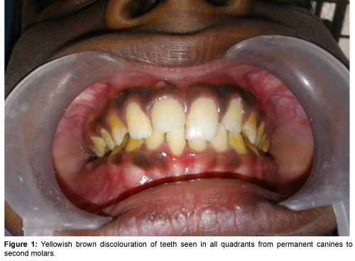
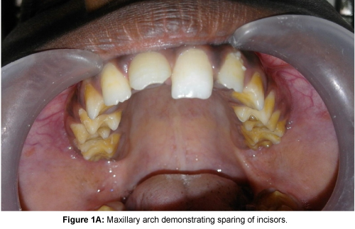
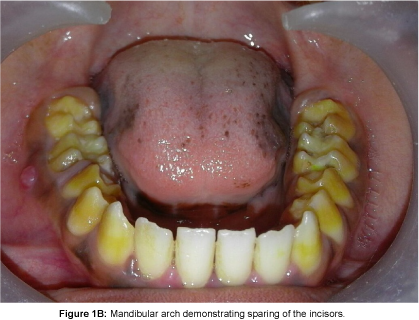
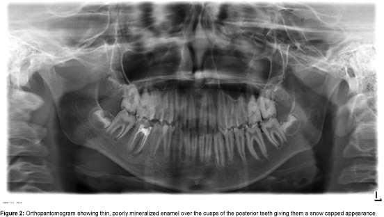
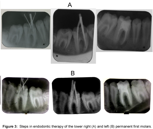
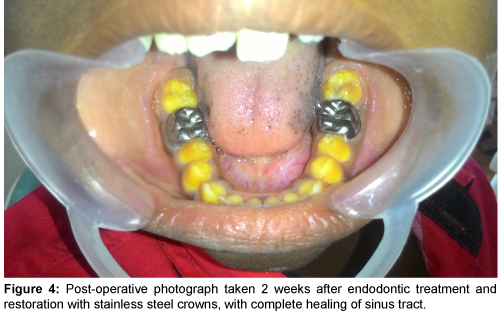
A 12-year-old male patient reported to the Department of Pediatric and Preventive Dentistry with the chief complaint of yellowish brown discolouration of the upper and lower back teeth. In addition to this primary complaint, the patient also reported repeated episodes of spontaneous pain and swelling in the lower right and left back tooth region. History revealed that the discolouration was present from the time of eruption of the permanent teeth. The patient’s mother affirmed that the primary teeth were unaffected and that there was no positive family history; medical history was noncontributory. On intraoral examination, a yellowish brown discolouration of the teeth was seen from the permanent canine to second molar region in all the quadrants (Figure 1). The discolouration was seen in the lingual surfaces of the teeth and the middle third of the buccal surfaces; however, this iscolouration did not involve the cervical third of the teeth. The surfaces of the teeth were rough, but the shape and size of the teeth appeared to be normal. On palpation, no softness or chipping of enamel was noted. None of the usual complications of amelogenesis imperfecta, viz. sensitivity, anterior open bite, attrition of teeth were present in this patient. Large carious lesions were seen in the occlusal surfaces of the left and right mandibular permanent first molars, with a sinus tract opening into the attached gingiva in relation to lower right first permanent molar. On examination of the periodontium, generalized plaque and calculus accumulation was seen, with a generalized chronic gingivitis. The striking feature was that the upper and lower central and lateral incisors on both arches seemed to be completely spared of the affliction (Figures 1A and 1B). These teeth had a normal appearance without any discolouration except for a fracture in the right maxillary central incisor involving the enamel and dentin (Figure 1A). An orthopantomogram revealed thin caps of enamel present on the posterior teeth with snowcapped appearance of the posterior teeth (Figure 2). Based on the history, the clinical features, and the radiographic findings, a provisional diagnosis of amelogenesis imperfecta was made with chronic apical periodontitis in the lower right and left first permanent molars. As to the type of AI, it was postulated that this case was a combination of the hypomaturative and the hypocalcified patterns. A treatment plan consisting of oral prophylaxis, endodontic management of lower first permanent molars and composite restoration of the fractured central incisor was framed. After the completion of endodontic treatment (Figure 3), stainless steel crowns were placed on the lower right and left first permanent molars. Follow up after 2 weeks revealed healing of the sinus tract which was associated with the lower right first permanent molar (Figure 4). The patient was advised to report for placement of full crown restorations in the other posterior teeth.
Discussion
AI is a diverse collection of inherited diseases that exhibit defects in the quality and quantity of enamel in the absence of systemic manifestations. As it is entirely a disease of the ectoderm (from which enamel arises), the Mesodermal components like dentin and pulp are entirely normal [5]. The estimated frequency of AI varies widely. Some studies estimate the incidence to be as high as 1 in 718 while others arrive at an incidence value as low as 1 in 14,000 [6]. AI may be inherited as an autosomal dominant, autosomal recessive or an X linked recessive disease. It arises from mutations that occur in one or more than one of four genes that are known to play a role in enamel formation viz., amelogenin (AMELX), enamelin (ENAM), enamelysin (MMP20) and kallikrein (KLK4). The amelogenin gene on the X chromosome is implicated in genetic defects in X-linked AI, but the molecular basis is unclear in this and other forms of the disease [7]. During amelogenesis, the enamel changes from a protein rich soft tissue to a hard tissue entirely devoid of protein - this change is dependent on certain molecular and cellular activities, which in turn, depends upon the genes mentioned above. Any mutation in the genes will result in aberrations in the enamel. The hypoplastic type of AI shows reduced thickness of enamel or entirely absent enamel, which is of normal radiopacity. A square shaped crown and low or absent cusps are also seen on radiographs. The hypocalcified type shows the chief feature of enamel chipping away after the tooth erupts into function. Even an explorer point may be able to penetrate into the enamel under pressure. Radiographically, enamel is of normal thickness but less dense than dentin. The hypomaturative type predominantly manifests as a yellowish to yellowish brown mottling of the enamel. The enamel is of decreased thickness and less dense than dentin. A snowcapped appearance is seen due to presence of white opaque enamel on the tips of the cusps. The present case interestingly shows some of the features of the hypomaturative and hypocalcified types in the absence of any systemic disease. This case is considered as a rare clinical presentation because the maxillary and mandibular anteriors are apparently unaffected in the patient. AI is usually thought of as affecting all the teeth uniformly, in a more or less equal manner. In this patient, the sparing of the anteriors has been advantageous as his appearance has not been compromised, which in itself is a rarity, as most patients having AI routinely cite the poor aesthetics as the chief complaint which prompted them to seek treatment.
For most patients, AI has severe psychosocial effects. Dental care for patients suffering from this disease is challenging and requires a good cooperation and understanding between the dentist, the patient, and the patient’s family. The treatment plan should not only relieve the patient’s chief complaint, but should also anticipate future problems. An interdisciplinary approach must be utilized to tackle the disease. The importance of maintaining oral hygiene cannot be stressed enough for these patients as the rough enamel surfaces tend to have a tendency for plaque accumulation and sensitivity [8]. Treatment is a combination of prevention, such as initiating use of fluoride mouthwashes, dentifrices and therapeutic modalities, including esthetic adhesive restorations, all ceramic crowns, and porcelain fused to metal crowns, inlays and onlays, and over dentures.
Conclusion
Amelogenesis imperfecta need not necessarily affect all the teeth uniformly as previously believed. The purpose of this case report is to record that relative sparing of teeth is yet another feature that dentists must be ready to look for amidst the already variable patterns of the disease.
Acknowledgement
This work is attributed to Tamil Nadu Government Dental College and Hospital, Chennai.
References
- Alfred MJ, Crawford PJM, Savarirayan R (2003) AmelogenesisImperfecta- A classification and catalogue for the 21st century. Oral Dis 2003 9: 19-23
- Neville BW, Damm DD, Allen CM, Bouquot JE (2013) Abnormalities of Teeth: Oral and Maxillofacial Pathology (3rdedn) Saunders, an imprint of ElsevierPhiladelphia, Pennsylvania, United States.
- Witkop CJ Jr (1988)Amelogenesisimperfecta, dentinogenesisimperfecta and dentin dysplasia revisited: problems in classification. J Oral Path 17: 547-553.
- Berkovitz BKB, Holland GK, Moxham BJ (2002) Oral Anatomy, Histology and Embryology. (3rdedn), Mosby, an imprint of Harcourt Publishers Limited Maryland Heights.
- Chaudhary M, Chaudhary SD (2011) Developmental disturbances in children: Essentials of Pediatric Oral Pathology (1stedn) Jaypee Publishers.Daryaganj, New Delhi, INDIA.
- American Academy of Pediatric Dentistry (2015-2016) Clinical practice guidelines: Guideline on Dental Management of Heritable Dental Developmental Anomalies. Reference Manual 37: 266-271.
- Seow WK (1993) Clinical diagnosis and management strategies of amelogenesisimperfecta variants.Pediatr Dent 1993 15:384-393
- Visram S, McKaig S (2006) AmelogenesisImperfecta- clinical presentation and management: a case report. Dent Update 2006 33: 612-616.
Relevant Topics
- About the Journal
- Birth Complications
- Breastfeeding
- Bronchopulmonary Dysplasia
- Feeding Disorders
- Gestational diabetes
- Neonatal Anemia
- Neonatal Breastfeeding
- Neonatal Care
- Neonatal Disease
- Neonatal Drugs
- Neonatal Health
- Neonatal Infections
- Neonatal Intensive Care
- Neonatal Seizure
- Neonatal Sepsis
- Neonatal Stroke
- Newborn Jaundice
- Newborns Screening
- Premature Infants
- Sepsis in Neonatal
- Vaccines and Immunity for Newborns
Recommended Journals
Article Tools
Article Usage
- Total views: 13732
- [From(publication date):
specialissue-2016 - Aug 29, 2025] - Breakdown by view type
- HTML page views : 12673
- PDF downloads : 1059

