Acinic Cell Carcinoma Primarily Arising in Ectopic Salivary Gland Tissue within an Intra-parotid Lymph Node: A Case Report
Received: 03-Mar-2018 / Accepted Date: 19-Mar-2018 / Published Date: 26-Mar-2018 DOI: 10.4172/2161-119X.1000346
Abstract
Background: We report a rare case of acinic cell carcinoma (ACC) arising from heterotopic salivary gland tissue within intra-parotid lymph node.
Case summary: The patient underwent superficial parotidectomy for acinar cell rich lesion. On histopathology it was difficult to determine whether it was a primary tumor from intra-parotid lymph node or metastatic from the parotid. Subsequently she underwent total parotidectomy. The parotid was negative for ACC, implying that it was a primary neoplasm arising from ectopic salivary gland tissue within the lymph node.
Conclusion: ACC primarily arising in ectopic parotid gland tissue is a rare presentation that physicians should be aware of.
Keywords: Acinic cell carcinoma; Ectopic parotid tissue; Head and neck
Abbreviations
ACC: Acinic Cell Carcinoma; U/S: Ultrasound; FNAC: Fine Needle Aspiration Cytology, MRI: Magnetic Resonance Imaging, CT: Computed Tomography
Introduction
Acinic cell carcinoma (ACC) is a low-grade malignant tumor that is epithelial in origin [1]. Although ACC has a slow growing behavior, it has been re-classified as a malignant tumor due to its tendencies to recur and metastasize [1]. The most common presentation is a slow growing mass in the tail of the parotid with mild to no pain. In addition, ACC shows a female predominance [1]. A positive family history and radiation exposure are considered some of the risk factors for ACC [1]. Fine needle aspiration biopsy confirms the diagnosis and it is essentially treated by surgical excision [1]. The parotid gland is the most common site of origin, but it can also present in other locations, including the submandibular, minor salivary and sublingual glands [1]. ACC was also reported to occur in the oral cavity, lips, palate, larynx, mandible, nose, or paranasal sinuses [1]. Outside the head and neck region, it may originate in the pancreas, stomach, lungs, breasts, or prostate [1]. ACCs rarely arise primarily in the intra-parotid lymph nodes. There are only a few cases reported in the literature of this presentation.
Case Presentation
A 38-year-old Indian lady, with a history of hypothyroidism, was referred to the ENT service, after undergoing a neck Ultrasound (U/S), followed by guided fine needle aspiration cytology (FNAC) and post contrast magnetic resonance imaging (MRI) of the parotid glands, revealing a left parotid mass that appeared to be rich in acinar cells as demonstrated by cytology, elsewhere. She presented in March 2017 with a complaint of upper neck swelling for 6 months. She is married, has one child and works as a house maid. Her family history is positive for parotid cancer in her father and she is a non-smoker.
Physical examination of the neck revealed a mass over the midportion of the left parotid gland, about 1x5 cm, nontender, firm, mobile with no skin fixation or fluctuation and no overlying skin changes. The right parotid gland showed minimal diffuse swelling with no mass. The cranial nerves were intact and the patient did not complain of any constitutional symptoms. The remaining head and neck examination was unremarkable.
U/S of the neck performed in January 2nd 2017, had revealed a parotid lesion that was described as an intraparanchymal, well defined, hypo echoic, rounded lesion, measuring 2x1.4 cm and it showed peripheral and internal vascularity on Doppler examination, with no internal septations, calcifications, or soft tissue nodules. It was concluded that the mass appeared benign in nature.
Guided FNAC, done in January 23rd 2017, attempted on the left parotid gland from the left superficial lobe, measuring 1.5 × 1.3 cm, showed an acinar cell rich lesion.
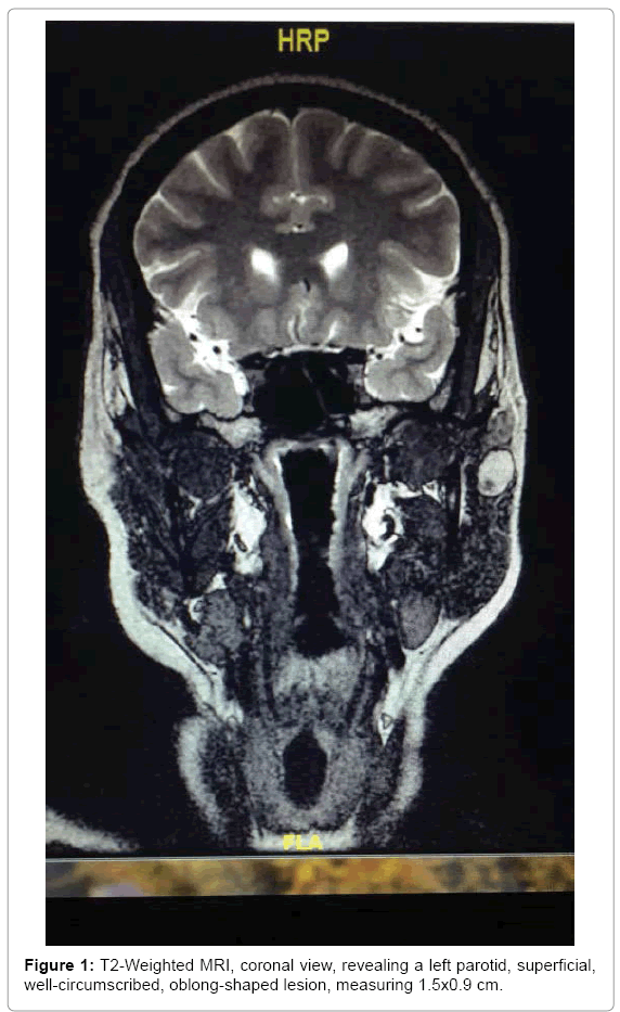
MRI revealed a left parotid lobe with a superficial, wellcircumscribed, oblong-shaped, lesion, measuring 1.5×0.9 cm (Figure 1).
The patient underwent a left superficial parotidectomy, which included an enlarged left superficial parotid lymph node in March 2017.
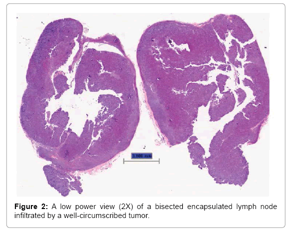
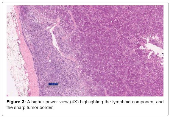
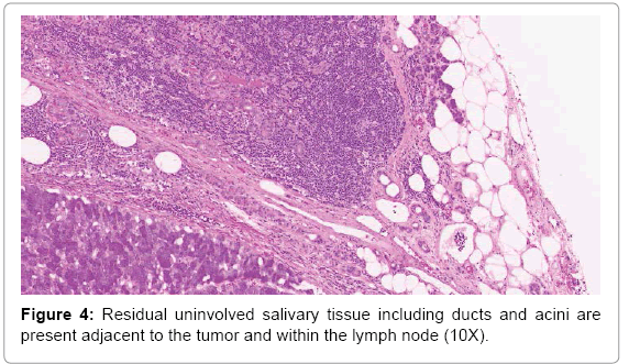
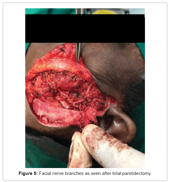
The histopathology specimen, received in 10% buffered formalin, labeled as “Left parotid lymph nodes”, consisted of multiple fragments of brown soft tissue, measuring together as 2 × 1.5 × 0.5 cm. Cut surface of the largest fragment is homogenous grey. Microscopic examination of the Hematoxylin and eosin stained slides reveals a well-circumscribed tumor seen within a lymph node (Figure 2). The tumor forms multiple coalescent nodules separated by thin fibrous septa. The nodules are composed of sheets of tumor cells forming microcysts and show entrapped vascular channels (Figure 3). The tumor cells are polygonal, pleomorphic, with eccentric hyperchromatic nuclei. The cytoplasm was plenty and filled with basophilic granules that are periodic acid Schiff positive, diastase resistant. The features are of a well-differentiated Acinic cell carcinoma. There was no evidence of mitosis, perineural invasion, necrosis or dedifferentiation. As the tumor occupies most of the lymph node, only focal –minimum- extension beyond it as it breaks through the capsule (Figure 4). Within the lymph node there is a rim of uninvolved parotid tissue, which indicates the heterotopic existence. These findings support the fact that the tumor is originating in the heterotopic parotic tissue rather than being an extension from a nearby tumor or tumor metastasis. On April 30th, 2017, total parotidectomy was done with preservation of the facial nerve (Figure 5). The specimen was received fixed in formalin in two containers. The first specimen labeled as “superficial lobe of parotid” consists of two fragments of parotid tissue, measuring 4.0 × 3.5 × 1.5 cm and 3.0 × 2.0 × 1.6 cm. The second specimen labeled as “deep lobe of parotid” consists of two fragments of parotid tissue, measuring 3.5 × 2.5 × 1.5 cm and 2.3 × 0.6 × 0.5 cm. Cut surface of both specimens reveals no evidence of tumor. Both specimens were totally submitted. A thorough microscopic assessment of the resected parotid showed no evidence of malignancy or inflammation. This is to conclude that the acinic cell carcinoma is primarily arising within an ectopic parotid tissue within an intra-parotid lymph node.
Discussion
ACC primarily arising in ectopic parotid gland tissue within the intra-parotid lymph nodes is a rare occurrence with only a few cases reported in the literature, most of which have been reported during the 80’s and 90’s [2-4]. Lidang Jensen and Kiaer reported a poorly differentiated ACC in a hyperplastic intra-parotid lymph node as the primary presentation [2]. They did, however, consider the possibilities of microscopic, clinically undetectable, ACC that metastasized from a primary lesion to the intra-parotid lymph nodes due to the discovery of a single tumor nodulus in the salivary gland tissue outside the lymph node capsule upon multiple tissue sections [2]. Another possibility they have placed was an ectopic origin of salivary tissue inside and outside the intraglandular lymph nodes [2].
Minić also presented a case of ACC of an intra-parotid lymph node. There was no continuity between the tumor and the gland as demonstrated by multiple sections through the tumor and adjacent parotid gland [3]. Minić was able to support the fact that the tumor arose within the lymph node by multiple sections within the node, demonstrating a direct continuity of normal lymphoid tissue with the lesion [3]. However, a postulation was made that the lymphoid tissue found to be in continuity with the lesion might be a result of a local immune response to the tumor [3].
A case report by Perzin and Livlsi described two cases of ACC arising primarily in ectopic salivary gland tissue, located far from normal locations of major and minor salivary glands [4]. In the cases they presented, there was no point of contact between the tumor and parotid gland as revealed by multiple sections through the tumor and gland and it was completely contained within the lymph node. Moreover, there was no evidence of neoplastic tissue in the parotid gland [4].
In order to presume that an ACC, found in the lymph node, is the primary lesion, complete sectioning of the lesion must be done and the lesion must be entirely contained within the lymph node with no evidence of another primary origin [3,4]. In embryologic development, the major salivary glands are formed by the invaginations of the endoderm of the branchial section of the floor of the primitive mouth, which are directly connected with the surrounding lymphatic tissue [5]. These invaginations form solid cords, which then proliferate and canalize to eventually form the excretory ducts [5]. This explains the possibility for lymphoid tissue to appear inside the major salivary glands or ectopic salivary tissue to be found in lymph nodes [5].
In our case the tumor within the intra-parotid lymph node was a primary neoplasm arising from ectopic salivary tissue within the lymph node as the whole parotid gland was free of the tumor as confirmed by histopathology.
Neoplasms primarily arising in intra-parotid lymph nodes should be considered by physicians and pathologists due to the fact that ectopic salivary gland tissue is commonly found in the intra-parotid lymph node and less frequently in the extra glandular cervical nodes [3]. The presence of ectopic tissue in lymph nodes may result in the possibility of malignant transformation [3]. Moreover, a possibility of intra nodal salivary gland tissue, containing immature components that possess the potential to proliferate and differentiate, exists [3]. These lesions are commonly mistaken for metastases from a malignant tumor arising primarily from regional lymph nodes and care should be taken to keep these rare possibilities in mind in order to avoid unnecessary surgical management [3].
Conclusion
ACC primarily arising in ectopic parotid gland tissue within the intra-parotid lymph nodes is a very rare presentation that physicians should be aware of. This type of lesion should be managed aggressively, despite its slow-growing nature, due to its malignant tendencies demonstrated by distant metastasis and recurrence.
References
- Al-Zaher N, Obeid A, Al-Salam S, Al-Kayyali BS (2009) Acinic cell carcinoma of the salivary glands: A literature review. Hematol Oncol Stem Cell Ther 2: 259-264.
- Â Jensen LM, Kiaer H (1992) Acinic cell carcinoma with primary presentation in an intraparotid lymph node. Pathol Res Pract 188: 226-231.
- Minić AJ (1993) Acinic cell carcinoma arising in a parotid lymph node. Int J Oral Maxillofac Surg 22: 289-291.
- Perzin KH, Livolsi VA (1980) Acinic cell carcinoma arising in ectopic salivary gland tissue. Cancer 45: 967-972.
- Barbuscia MA, Caizzone A, Vecchio DA, Mirone S, Putortì A, et al. (2011) Ectopic parotid: Case report. G Chir 32: 429-433.
Citation: Alsayegh RO, Qazi I, Almurshed M, Al Mutairi MM, Alqunaee MA (2018) Acinic Cell Carcinoma Primarily Arising in Ectopic Salivary Gland Tissue within an Intra-parotid Lymph Node: A Case Report. Otolaryngol (Sunnyvale) 8: 346 DOI: 10.4172/2161-119X.1000346
Copyright: © 2018 Alsayegh RO, et al. This is an open-access article distributed under the terms of the Creative Commons Attribution License, which permits unrestricted use, distribution, and reproduction in any medium, provided the original author and source are credited.
Select your language of interest to view the total content in your interested language
Share This Article
Recommended Journals
Open Access Journals
Article Tools
Article Usage
- Total views: 7167
- [From(publication date): 0-2018 - Nov 29, 2025]
- Breakdown by view type
- HTML page views: 6230
- PDF downloads: 937

