An Overview of Dens Invaginatus with Report of 2 Cases
Received: 22-May-2015 / Accepted Date: 08-Jul-2015 / Published Date: 11-Jul-2015 DOI: 10.4172/2161-0681.1000238
Abstract
Dens invaginatus occurs as a result of invagination of the enamel organ. These cases may present difficulties with respect to its diagnosis and treatment because of canal morphology. It frequently leads to caries, pulpal, and periodontal involvement with necrosis and loss of attachment. The knowledge of classification and anatomical variations of teeth with dens invaginatus are of utmost importance for correct treatment. This paper presents two cases of dens invaginatus and its treatment depending on the patient symptoms either by prophylactic sealing or root canal treatment.
Keywords: Dens invaginatus; Enamel organ; Prophylactic sealing; Root canal treatment
319857Introduction
Dens invaginatus is a developmental anomaly resulting from the invaginations of the enamel organ into the dental papilla during the soft tissue stage of development. As the hard tissues are formed, the invaginated enamel organ produces a small tooth within the future pulp chamber. Dens invagination in a human tooth was first described by a dentist named Socrates [1]. The term “dens invaginatus” appears to be most appropriate, as it reflects the invagination of the outer portion (enamel) into the inner portion (dentin) with the formation of a pocket or dead space, which generally occurs before crown calcification [2,3].
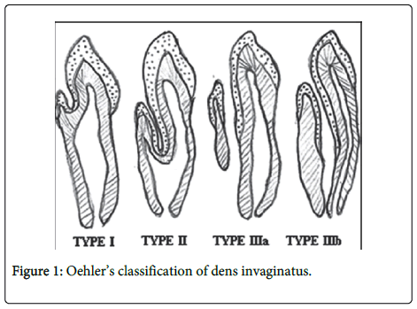
Although the permanent maxillary lateral incisor is the most commonly involved tooth, it may occur in primary teeth as well as in the maxillary and mandibular arches [4]. In most cases, it appears to simply represent an accentuation in the development of a lingual pit [5]. The reported prevalence of adult teeth affected with dens invaginatus is between 0.3% and 10% with the problem observed in 0.25% to 26.1% of individuals examined [6]. The etiology of dens invaginatus is still unclear. Kronfeld suggested that dens invaginatus is caused by a focal failure growth of the internal enamel epithelium [7]. Oehlers suggested that the distortion of the enamel organ occurs during tooth development [8]. Other theories are infection, trauma and genetics as possible contributing factors [9,10]. The system described by Oehler’s appears to be most widely used to classify dens invaginatus on the basis of the radiographic appearance (Figure 1) possibly because of its simple nomenclature (Table 1).
| Type | Description |
|---|---|
| 1 | An enamel-lined cavity confined to the crown and not extending beyond the cementoenamel junction |
| 2 | An enamel-lined cavity extending into the root beyond the cementoenamel junction ending in a blind sac; there may/may not be communication with pulp |
| 3 | An invagination extending beyond the cementoenamel junction perforating laterally (type 3a) or apically (type 3b) at a foramen; usually, there is no communication with the pulp, sometimes it may be completely lined by enamel, and sometimes cementum will be found lining the invagination |
Table 1: Oehler’s classification of dens invaginatus [8,25].
Case Reports
Case report-1
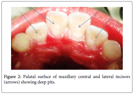
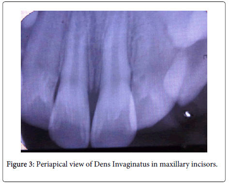
A 10-year-old girl reported with a chief complaint of sensitivity to cold in relation to upper front teeth since 1 week. The patient was in good general health. Extra-oral examination revealed no significant findings. Intra-oral examination revealed deep anatomic pits on palatal surface of maxillary central and lateral incisors (Figure 2). The teeth were non tender on vertical percussion and responded normal to thermal and electric stimuli. The periapical radiograph revealed Dens Invaginatus type II with respect to maxillary lateral incisors and Dens Invaginatus type I with respect to maxillary central incisors (Figure 3).
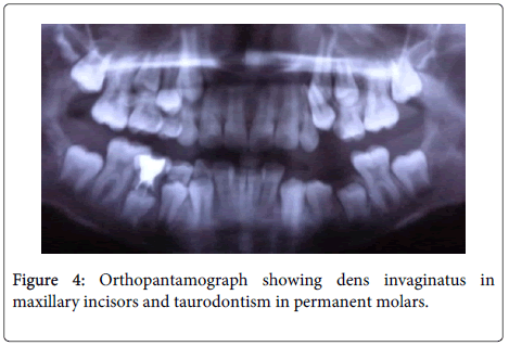
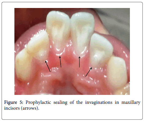
Orthopantomograph examination confirmed the presence of dens invaginatus in maxillary central incisors. On careful observation we noticed presence of taurodontism affecting the permanent both maxillary and mandibular incisors (Figure 4). Prophylactic sealing of all four invaginations was done using flowable composite (Filtek™ Supreme Ultra; 3M ESPE) (Figure 5). The patient remained asymptomatic when she reported 1 week after treatment and continues to be under follow up.
Case report-2
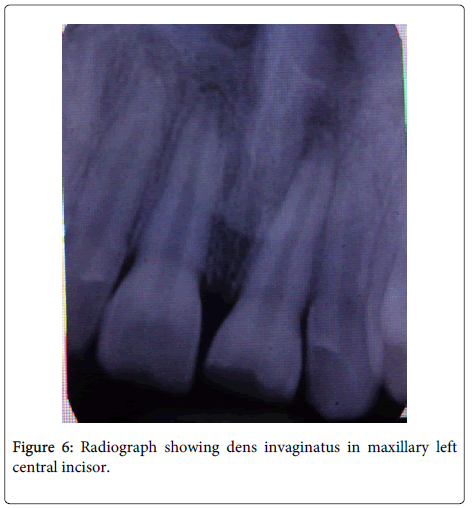
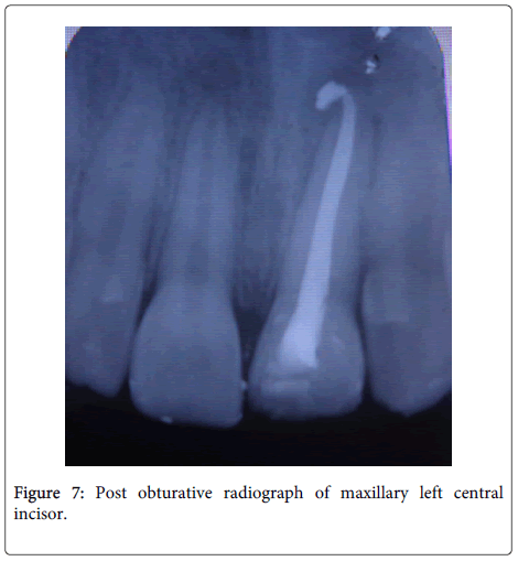
A 12-year-old boy reported with a chief complaint of pain with respect to upper left front tooth since 3 days. The patient’s general health was good. No significant extraoral findings were seen. Intra-oral examination revealed maxillary left central incisor with deep anatomic pit on palatal surface. The tooth was tender on vertical percussion and did not respond to thermal and electric stimuli. The periapical radiograph revealed type I Dens Invaginatus and periapical pathology with respect to maxillary left central incisor (Figure 6). Single-visit endodontics was performed for the involved tooth (Figure 7). The tooth was intact and functional inside the oral cavity when patient came for recall visit after 1 month and is still under follow up.
Discussion
In most cases, a dens invaginatus is noticed by chance on the radiograph. Clinically important hints maybe, unusual crown morphology (‘dilated,’ ‘peg-shaped,’ ‘barrel-shaped’) or a deep foramen coecum but affected teeth may also show no clinical signs of malformation. As pulpal involvement of teeth with coronal invaginations may occur a shortly after tooth eruption, an early diagnosis is essential to instigate preventive treatment [11]. The invagination allows entry of irritants into an area, which is separated from pulpal tissue by only a thin layer of enamel and dentin and presents a predisposition for the development of dental caries. In some cases, the enamel lining is incomplete. Channels may also exist between the invagination and the pulp [12,13]. Therefore, pulp necrosis often occurs rather early, within a few years of eruption, sometimes even before root end closure [14,15] Other reported sequelae of undiagnosed and untreated coronal invaginations include abscess formation, retention of neighbouring teeth, displacement of teeth, cysts and internal resorption [16].
The treatments options include; prophylactic or preventive sealing of the invagination, [17,18] root canal treatment, [19] endodontic apical surgery, [20,21] intentional replantation [22], and extraction [23]. Some authors say there is association between talon cusp and dens invaginatus [18,20]. Nagaveni et al. [18] reported an interesting case showing the occurrence of three unusual anomalies within the same tooth of an Indian patient. These anomalies are talon cusp, dens invaginatus and short root in the lower permanent central incisor which is not reported previously. The literature shows isolated cases or prevalence of taurodontism affecting the permanent molars [24]. In the present report there was concomitant occurrence of dens invaginatus and tarudontism affecting the permanent molars was evident in case 1. In the first case, prophylactic sealing of the invagination was done for all four anterior teeth. In second case, root canal treatment was done for the symptomatic tooth.
Conclusion
The thorough knowledge of classification and anatomic variations of teeth with dens invaginatus is important for its early detection and management.
References
- Schulze C (1970) Development abnormalities of the teeth and the jaws. In. Gorlin O, Goldman H (eds.) Thomas’ Oral Pathology.Mosby, St. Louis, pp. 96-183.
- Pallivathukal RG, Misra A, Nagraj SK, Donald PM (2015) Dens invaginatus in a geminated maxillary lateral incisor. BMJ Case Rep 2015.
- Alani A, Bishop K (2008) Dens invaginatus. Part 1: classification, prevalence and aetiology. IntEndod J 41: 1123-1136.
- Shadmehr E, Kiaani S, Mahdavian P (2015) Nonsurgical endodontic treatment of a maxillary lateral incisor with dens invaginatus type II: A case report. Dent Res J (Isfahan) 12: 187-191.
- Rajendran R (2007) Developmental disturbances of oral and paraoral structures. In: Rajendran R, Shivpathasundram B (eds.) Shafer’s Textbook of Oral Pathology (5thedn.) New Delhi, India
- Thakur S, Thakur NS, Bramta M, Gupta M (2014) Dens invagination: A review of literature and report of two cases. J Nat SciBiol Med 5: 218-221.
- Zubizarreta Macho Ã, Ferreiroa A, Rico-Romano C, Alonso-Ezpeleta LÓ, Mena-Ãlvarez J (2015) Diagnosis and endodontic treatment of type II dens invaginatus by using cone-beam computed tomography and splint guides for cavity access: a case report. J Am Dent Assoc 146: 266-270.
- OEHLERS FA (1957) Dens invaginatus (dilated composite odontome). I. Variations of the invagination process and associated anterior crown forms. Oral Surg Oral Med Oral Pathol 10: 1204-1218 contd.
- Er K, Kustarci A, Ozan U, Tasdemir T (2007) Nonsurgical endodontic treatment of dens invaginatus in a mandibular premolar with large periradicular lesion: a case report. J Endod 33: 322-324.
- Nosrat A, Schneider SC (2015) Endodontic Management of a Maxillary Lateral Incisor with 4 Root Canals and a Dens Invaginatus Tract. J Endod 41: 1167-1171.
- Babaji P (2015) Bilateral supplemental maxillary incisors with both dens invaginatus and dens evaginatus in a non syndromic patient: a rare case report. J ClinDiagn Res 9: ZJ01-02.
- Capar ID, Ertas H, Arslan H, TarimErtas E (2015) A retrospective comparative study of cone-beam computed tomography versus rendered panoramic images in identifying the presence, types, and characteristics of dens invaginatus in a Turkish population. J Endod 41: 473-478.
- Stamfelj I, Kansky AA, Gaspersic D (2007) Unusual variant of type 3 dens invaginatus in a maxillary canine: a rare case report. J Endod 33: 64-68.
- Kasat VO, Singh M, Saluja H, Ladda R (2014) Coexistence of two talon cusps and two dens invaginatus in a single tooth with associated radicular cyst-a case report and review of literature. J ClinExp Dent 6: e430-434.
- Hattab FN (2014) Double talon cusps on supernumerary tooth fused to maxillary central incisor: Review of literature and report of case. J ClinExp Dent 6: e400-407.
- Tiku A, Nadkarni UM, Damle SG (2004) Management of two unusual cases of dens invaginatus and talon cusp associated with other dental anomalies. J Indian SocPedodPrev Dent 22: 128-133.
- Satvati SA, Shooriabi M, Sharifi R, Parirokh M, Sahebnasagh M, et al. (2014) Co-existence of two dens invaginations with one dens evagination in a maxillary lateral incisor: a case report. J Dent (Tehran) 11: 485-489.
- Nagaveni NB, Umashanikara KV, Vidyullatha BG, Sreedevi S, Radhika NB (2011) Permanent mandibular incisor with multiple anomalies - report of a rare clinical case. Braz Dent J 22: 346-350.
- Ceyhanli KT, Celik D, Altintas SH, Tasdemir T, S Sezgin O (2014) Conservative treatment and follow-up of type III dens invaginatus using cone beam computed tomography. J Oral Sci 56: 307-310.
- Nagaveni NB, Umashankara KV (2014) A clinical and radiographic retrospective analysis of talon cusps in ethnic Indian children. J Cranio Max Dis 3:79-84.
- Muppa R, Nallanchakrava HS, Mettu S, Dandu RV, Tadikonda DC (2014) Type III B dens invaginatus: diagnostic and clinical considerations using 128-slice computed tomography. J Indian SocPedodPrev Dent 32: 342-345.
- Teixidó M, Abella F, Duran-Sindreu F, Moscoso S, Roig M (2014) The use of cone-beam computed tomography in the preservation of pulp vitality in a maxillary canine with type 3 dens invaginatus and an associated periradicular lesion. J Endod 40: 1501-1504.
- Brooks JK, Ribera MJ (2014) Successful nonsurgical endodontic outcome of a severely affected permanent maxillary canine with dens invaginatusOehlers type 3. J Endod 40: 1702-1707.
- Nagaveni NB, Radhika NB (2012) Prevalence of taurodontism in primary mandibular first molars of ethnic Indian children. Gen Dent 60: e335-340.
- Oehlers FA (1957) Dens invaginatus (dilated composite odontome). II. Associated posterior crown forms and pathogenesis. Oral Surg Oral Med Oral Pathol 10: 1302-1316.
Citation: Nagaveni NB, Pathak S, Anitha P, Poornima P (2015) An Overview of Dens Invaginatus with Report of 2 Cases. J Clin Exp Pathol 5:238. DOI: 10.4172/2161-0681.1000238
Copyright: © 2015 Nagaveni NB, et al. This is an open-access article distributed under the terms of the Creative Commons Attribution License, which permits unrestricted use, distribution, and reproduction in any medium, provided the original author and source are credited.
Select your language of interest to view the total content in your interested language
Share This Article
Recommended Journals
Open Access Journals
Article Tools
Article Usage
- Total views: 18931
- [From(publication date): 8-2015 - Aug 30, 2025]
- Breakdown by view type
- HTML page views: 14181
- PDF downloads: 4750

