Basaloid Squamous Cell Carcinoma of the Tonsil: A Rare Case
Received: 02-Jul-2015 / Accepted Date: 07-Jul-2015 / Published Date: 14-Jul-2015 DOI: 10.4172/2161-119X.1000199
Abstract
Introduction: Basaloid squamous cell carcinoma is a rare clinical and pathological variant of squamous cell carcinoma. Firstly described in 1986, it is usually found in the upper aerodigestive tract in males over 60 years old.
Case report: We describe the case of a 53-year old male observed in the ENT Department with a left cervical tumefaction with normal ENT examination, despite the evidence of malignant cells in a recently performed ultrasoundguided fine-needle aspiration. Magnetic resonance imaging of the head and neck revealed a potential lesion in the left tonsillar fossa. The patient was submitted to partial pharyngectomy with bilateral neck dissection – histological examination was consistent with a pT1N2aM0 basaloid squamous cell carcinoma, leading to adjuvant chemotherapy and radiotherapy. No signs of active disease have been detected after 12 months of follow up.
Discussion: Basaloid squamous cell carcinoma of the palatine tonsil should be always considered in the differential diagnosis of cervical adenopathy of unknown primary carcinoma.
Keywords: Basaloid squamous cell carcinoma, Tonsil
249742Introduction
Basaloid squamous cell carcinoma (BSSC) is a distinct and rare variant of conventional squamous cell carcinoma (SCC), predominantly localized in the upper aerodigestive tract [1-10]. First described by Wain et al. in 1986, it has a strong predilection for sites such as base of the tongue, hypo pharynx and supraglotic larynx [5,8-10]. BSSC of the palatine tonsil is extremely uncommon with less than 20 cases described in English literature [1,2,5].
Case Report
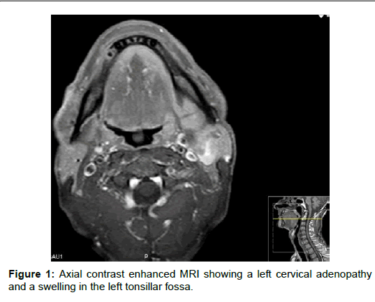
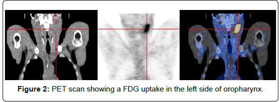
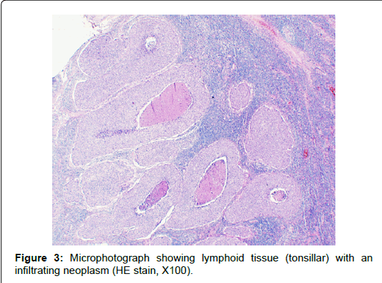
A 53-year-old caucasian man presented in our deparment in December 2013 with a 3-week long history of left neck mass. He had a 45 pack-year history of cigarret smoking, occasional alcohol consumption and no known illness. There was no history of odynophagia, hoarseness, fever or weight loss. On clinical examination there was a 4 cm hard, fixed and non tender mass in left II-III ganglionar group levels. Ultrasoundguided fine-needle aspiration of the neck mass revealed the presence of malignant cells. Magnetic resonance imaging (MRI) of the head and neck (Figure 1) confirmed the presence of metastatic cervical lymph node (size 3,6 × 3,8 cm), and a contrast-enhanced swelling in the left tonsillar fossa. A whole body 18-fluorodeoxyglucose (FDG) positron emission tomography (PET) scan (Figure 2) showed two large foci of intense FDG uptake in left neck region and in the ipsilateral side of oropharynx, respectively. Thoracic tomographic scan, bronchofibroscopy, complete blood count and biochemichal parameters were within normal ranges. The patient underwent partial pharingectomy associated with a left type 3, modified radical neck dissection. The histopathological examination of the pharyngeal specimen showed tonsillar tissue with a infiltrating basaloid squamous cell carcinoma (1,7 × 1,6 cm) with a narrow deep resection margin (0,2 cm) (Figure 3). The immunohistochemical study of malignant cells was positive for cytokeratins AE1/AE3. The neck dissection specimen revealed metastases in 1 of 19 lymph nodes with extracapsular spread. The patient was pathologically staged as T1N2aM0 and received adjuvant chemoradiotherapy. Follow up did not show any evidence of complications, local recurrence or distant metastasis until January 2015.
Discussion
BSCC occurs in both sexes but predominates in males between 60 and 80 years old [1,2,5,8,9], and shows strong association with tobacco and alcohol consumption [1,5,8-10]. The possible relationship of BSCC and viruses was traditionally a matter of debate [5], but recent investigations shown an association between high risk human papillomavirus (HPV) type 16 with oropharynx BSCC [10].
On clinical observation, the tumor frequently presents as an exophytic mass that may have ulcerations, [5] in the case presented, however, the tumor was deep in the tonsil, and completely hidden on physical examination.
BSSC is believed to arise from multipotential primitive cells in the basal layer of the surface epithelium or from the salivary duct lining epithelium [5,7-9]. Its histological diagnosis still remains on hematoxylin and eosin sections by recognizing the typical four diagnostic criteria defined by Wain et al. at 3 decades ago [4,5]: (1) solid group of cells in a lobular configuration, closely opposed to the surface mucosa; (2) small, closely packed cells with scant cytoplasm; (3) hypercromatic nuclei without nucleoli; and (4) small, cystic spaces containing mucin-like material [2,4,5]. By immunohistochemistry, BSCC expresses cytokeratin and epithelial membrane antigens [2,5,8-10], like in the case presented, and which may be of help in the diagnosis at an early stage [2].
The differential diagnosis of FDCS includes adenoid cystic carcinoma, basal cell carcinoma, adenosquamous carcinoma and neuroendocrine carcinoma [1,2,5,7,9,10].
Because most cases of BSSC present with advanced-staged tumors, it was initially believed that the histological features were reflective of its clinical aggressive behavior characterized by extensive nodal involvement and distant metastases [1,4,5,7,8,10]. There are recent studies; however, showing that BSCC may exhibit a dual prognosis based on HPV status, with better prognosis in patients who exhibit high-risk HPV [9,10].
There is no established consensus for treatment of BSCC. While some authors advocate more aggressive treatment with surgical excision and cervical dissection supplemented with adjuvant chemoradiotherapy in all cases [1,5,6,8], others advice a management similar to conventional SCC [10].
Conclusion
BSCC of the tonsil is a rare entity that should be considered in the differential diagnosis of the cervical adenopathy of unknown primary carcinoma.
References
- Chaidas K, Koltsidopoulos P, Kalodimos G, Skoulakis C (2012) Basaloid Squamous cell carcinoma of the tonsil. Hippokratia 16:74-75.
- Shivakumar B, Dash B, Sahu A, Nayak B (2014)Basaloid squamous cell carcinoma: a rare case report with review of literature. J Oral MaxillofacPathol 18: 291-294.
- Raslan W, Barnes L, Krause J, Contis L, Killeen R et al. (1994)Basaloid squamous cell carcinoma of the head and neck: a clinicopathologic and flow cytometric study of a 10 new cases with review of the English literature. Am J Otolaryngol 15: 204-211.
- Wain SL, Kier R, Vollmer RT, Bossen EH (1986)Basaloid squamous cell carcinomas of the tongue, hypopharynx and larynx: report of 10 cases. Hum Pathol 17:1158-1166.
- Shetty D, SubramaniamV, Herale A, Saldanha P, Jain M (2012)Basaloid squamous cell carcinoma of the tonsil: report of a case and review of the literature. ContempOncol (Pozn)16: 447–450.
- Thariat J, Ahamad A, El Naggar AK, Williams MD, Holsinger FC et al.(2008) Â Outcomes after radiotherapy for basaloid squamous cell carcinoma of the head and neck: a case-control study. Cancer112:2698-2709.
- PaulinoA, Singh B, Shah J, Huvos A (2010)Basaloid Squamous Cell Carcinoma of the Head and Neck. Laryngoscope 110: 1479-1482.
- Ereno C, GaafarAgarmendia M, Etxezarraga C, Bilbao F, Lopez J (2008)Basaloid Squamous Cell Carcinoma of the Head and Neck. Head Neck Pathol 2: 83–91.
- Tan S, Chong A, Nazarina A, Prepageran N (2014) Basaloid squamous cell carcinoma of the oropharynx: comparison of two cases and review of the literature. Otolaryngol Pol 68: 268-270.
- Jayasooriva P, Tilakaratne W, Mendis B, Lombardi T (2013) A literature review on oral basaloid squamous cell carcinomas, with special emphasis on etiology. AnnDiagnPathol 17: 547-555.
Citation: Ribeiro L, Sousa M, e Almeida AF, Condé A (2015) Basaloid Squamous Cell Carcinoma of the Tonsil: A Rare Case. Otolaryngology 5:199. DOI: 10.4172/2161-119X.1000199
Copyright: © 2015 Ribeiro L, et al. This is an open-access article distributed under the terms of the Creative Commons Attribution License, which permits unrestricted use, distribution, and reproduction in any medium, provided the original author and source are credited.
Select your language of interest to view the total content in your interested language
Share This Article
Recommended Journals
Open Access Journals
Article Tools
Article Usage
- Total views: 15736
- [From(publication date): 7-2015 - Aug 04, 2025]
- Breakdown by view type
- HTML page views: 11149
- PDF downloads: 4587


