Bullous Lesion of Larynx? An airway Emergency
Received: 10-Aug-2015 / Accepted Date: 20-Aug-2015 / Published Date: 27-Aug-2015 DOI: 10.4172/2161-119X.1000206
Abstract
Bullous pemphigoid (BP) is a chronic autoimmune disease that mainly affects elderly patients. It presents as skin lesions; however, it can affect the eyes and upper aerodigestive system as well. We report a rare case of Bullous pemphigoid that had a laryngeal extension presenting with odynophagia.
Introduction
Bullous pemphigoid is an autoimmune disorder affecting the elderly and commonly involves the skin, mouth, eyes and genitals. Laryngeal involvement in this disorder is albeit rare and may necessitate management of an impending airway emergency. We present a case of laryngeal bullous pemphigoid managed conservatively with steroids and immunoglobulins.
Case Report
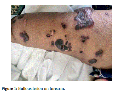
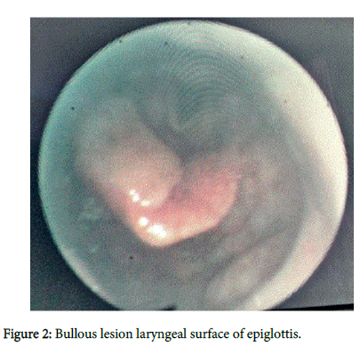
69 years old female patient who was known to have diabetes mellitus type II complicated with diabetic nephropathy, hypertension, dyslipidemia and ischemic heart disease and congestive cardiac failure presented to the cardiology OPD on her regular follow up appointment with a history of vesicular rash on her upper limbs (Figure 1). The vesicles were painful, gradually increasing in size and ends with a burst leaving behind a skin scar. She was admitted for the Manuscript management of congestive cardiac failure and a dermatology opinion was obtained. A provisional diagnosis of Bullous Pemphigoid was made by the dermatologists and a biopsy of the vesicles and autoimmune study was also obtained. Low dose of Prednisolone and betamethasone cream were started. However, 2 days later, the patient developed Odynophagia with a vague discomfort in breathing. ENT opinion was taken and she underwent Fibroptic Laryngoscopy which revealed a fairly large bleb over the laryngeal surface of the epiglottis (Figure 2). Consequent to this finding the Prednisolone dose was increased and nebulized Budesonide was given as well.
Precautions were taken to monitor the patient and a tracheostomy set was prepared in the event of worsening stridor. Within 24 hours, the symptoms of odynophagia improved, yet the skin rashes continued to increase. Repeat Fibroptic Laryngoscopy done two days later showed a reduction in bleb size with no further lesions. However, due to the increase in skin rash, Immunoglobulin was also added to the regime after which the patient started to improve. Skin biopsy and immune studies confirmed the diagnosis of Bullous pemphigoid.
Discussion
Bullous pemphigoid (BP) is a chronic autoimmune disease that mainly affects elderly patients [1]. It has been classified as a Type 2 Hypersensitivity reaction [2]. The bullae are formed by an immune reaction, initiated by the formation of IgG autoantibodies targeting Dystonin also called Bullous Pemphigoid Antigen 1 and/or type XVII collagen also called Bullous Pemphigoid Antigen [3]. The disease is usually characterized by periods of relapses and remissions. It involves the skin as 1-3 cm blisters in all cases, while in one third of the cases there is involvement of the oral mucosa. In 50 percent of cases with oral cavity involvement there can be involvement of the larynx, hypo pharynx or esophagus. The diagnosis is based on the clinical presentation, biopsy of the bullous lesions and immunopathological studies [4]. The lesions tend to dominate on the lower trunk, axilla, groin or flexor surface of extremities. The differential diagnosis of BP would include auto immune diseases like Epidermolysis bulla aquisita, bullous lesions in SLE, dermatitis herpetiformis and pemhigoid gestationis IgA dermatosis.Immunopathological staining may be useful in differentiating these disorders. Non immunological disorders mimicking bullous pemphigoid may include bullous erythema multiforme, drug reactions and porphyria. Despite all the investigations a clear distinction may not be made between these conditions clinically. Immunosuppressive medication in the form of systemic steroids form the mainstay of therapy for this lesions. The involvement of the larynx may in some cases compromise the airway and may necessitate a tracheostomy in some cases. In some cases Helium oxygen mixtures have been used an adjunctive therapy in case of critical airway compromise [5]. Steroids are used in the dose of 0.75 to 1 mg/kg per day in case of ocular, laryngeal or recurrent disease while topical steroids may be used in case of milder disease. Immunosupressive therapy in the form of azathioprine, cyclophosphamide and mycophenolate mofetil have also been used in case of extensive disease. The use of Intravenous immunoglobulin is usually restricted to 24 percent of patients with bullous pemphigoid who do not respond to conventional therapy [6]. It is important to start Immunoglobulin therapy for patients who are at risk of developing fatal side effects from conventional immunosuppressive therapy. The IV immunoglobulin needs to be gradually withdrawn after a clinical control has been achieved. In case of extensive laryngeal disease with airway compromise, it would be prudent to perform an elective tracheostomy to secure the airway [7]. The management of the airway involves a close monitoring for new laryngeal lesions through flexible fibre optic laryngoscopy and to secure the airway early in case of additional laryngeal lesions and worsening stridor. The long term outlook in laryngeal bullous pemphigus seems to be good in contrast to cicatrical type of pemphigus where scarring may lead to laryngeal stenosis leaving permanent sequelae [8]. In our case, the timely use of IV immunoglobulin produced a noticeable improvement in the patient’s condition. This case also highlights the requirement of close monitoring of airway in patients with flexible fibre optic scopy in patients with bullous pemphigoid to rule out laryngeal involvement which may compromise the airway.
Conclusion
Bullous lesions of the larynx can present with airway compromise and early otolaryngology referrals would be prudent to monitor the airway and secure the airway if required. There is a role for conservative management with steroids and immunoglobulins provided early airway compromise can be followed by using repeated fibreoptic scopies to watch out for the appearance of new bullous lesions in the larynx.
References
- Stanley JR (1993) Cell adhesion molecules as targets of autoantibodies in pemphigus and pemphigoid, bullous diseases due to defective epidermal cell adhesion. Adv Immunol 53: 291-325.
- Kasperkiewicz M,Zillikens D (2007) The pathophysiology of bullous pemphigoid. Clin Rev Allergy Immunol 33: 67-77.
- Stanley JR, Hawley-Nelson P, YuspaSH, ShevachEM, Katz SI (1981) Characterization of bullous pemphigoid antigen: a unique basement membrane protein of stratified squamous epithelia. Cell 24: 897-903.
- Yancey KB, Egan CA (2000) Pemphigoid: clinical, histologic, immunopathologic, and therapeutic considerations. JAMA 284: 350-356.
- Iqbal M, Ahmed R, KashefSH, Bahame PS (2006) Laryngeal involvement in a patient with bullous pemphigoid. Ann Saudi Med 26: 152-154.
- Wetter DA, Davis MD, Yiannias JA, Gibson LE, Dahl MV, et al. (2005) Effectiveness of intravenous immunoglobulin therapy for skin disease other than toxic epidermal necrolysis: a retrospective review of Mayo Clinic experience. Mayo ClinProc 80: 41-47.
- Sato K,Hanazawa H, Sato Y, Watanabe J (2005) Initial presentation and fatal complications of linear IgA bullous dermatosis in the larynx and pharynx. J LaryngolOtol 119: 314-318.
- Wilhelm T (1992) [Cicatricial mucous membrane pemphigoid with laryngeal involvement. Case report and review of the literature]. HNO 40: 495-499.
Citation: Belushi F, Chaly VA, Abri RA (2015) Bullous Lesion of Larynx? An airway Emergency. Otolaryngol (Sunnyvale) 5:206. DOI: 10.4172/2161-119X.1000206
Copyright: © 2015 Belushi F, et al. This is an open-access article distributed under the terms of the Creative Commons Attribution License, which permits unrestricted use, distribution, and reproduction in any medium, provided the original author and source are credited.
Select your language of interest to view the total content in your interested language
Share This Article
Recommended Journals
Open Access Journals
Article Tools
Article Usage
- Total views: 16211
- [From(publication date): 9-2015 - Aug 19, 2025]
- Breakdown by view type
- HTML page views: 11471
- PDF downloads: 4740


