Epistaxis Related to Internal Carotid Artery Cavernous Sinus Aneurysm
Received: 25-Jan-2015 / Accepted Date: 15-Feb-2015 / Published Date: 23-Feb-2015 DOI: 10.4172/2161-119X.1000188
Abstract
Background: Nasal bleeding is one of the most common clinical presentation in the Otolaryngology Department. Epistaxis related to internal carotid artery aneurysm is a rare cause of epistaxis. In this case we report a case of recurrent epistaxis for more than 7 months in a previously healthy Chinese man.
Case report: Patient complained recurrent nasal bleeding for seven months without any inducing factors, with no history of trauma to the nose, congestive heart failure, diabetes mellitus, hypertension and other diseases. Head and neck computed tomography revealed left maxillary sinus, sphenoid sinus and the right frontal sinus inflammation and nasal septum deviation to the right. Head and neck angiography revealed left internal carotid artery cavernous sinus aneurysm.
Conclusion: Epistaxis related to internal carotid artery aneurysm is quite rare but it is important to consider aneurysms in the etiology of epistaxis. Which may be fatal if the cause of nasal bleeding cannot be identified. We managed our patiently conservatively using nasal packing, hemostatic, anti-inflammatory drugs. The aneurysm was managed by using stent.
Keywords: Epistaxis, Sphenoid sinus, Internal carotid artery,Cavernous sinus, Aneurysm
249306Introduction
Epistaxis is simply known as nose bleeding, presented as hemorrhage from the nasal cavity which is noticed when blood flows out through nostrils. Nasal bleeding may occur due to nasal cavity and sinus disease, nasopharyngeal lesions, cavernous sinus lesions, rupture of artery and false aneurysm, some systemic diseases can also cause nasal bleeding. We describe an uncommon cause of epistaxis in this report and discuss the imaging findings, treatment course of our patient.
Case Report
A previously healthy 71-year old Chinese man was admitted in our department for recurrent epistaxis. He complained of recurrent left nasal bleeding for seven months without any inducing factors, amount of blood loss about 400 ml, the bleeding was fresh blood. After pressing the nasal cavity by fingers bleeding stopped. One month before the admission patient again suffered from left nasal bleeding, fresh blood, continuous bleeding, amount of blood loss was about 600 ml, accompanied by dizziness, nausea, thirst, sweating. Patient sought medical consultation went to local hospital diagnosed as Epistaxis. Patient underwent endoscopic guided nasal packing. When the nasal packing was removed after 7 days, epistaxis still continued, mainly from left nostril. For the better diagnosis and treatment patient came to our hospital diagnosed as EPISTAXIS and admitted to our department. Since the onset of illness patient has no history of coma and loss of consciousness, no fever, no head and face pain and no visual changes, no nasal itching, sneezing, purulent nasal discharge and other symptoms. No history of nasal bone trauma and fracture. The general mental state, sleep and appetite are normal, no significant changes in body weight. No other surgical and medical history revealed by patient. Physical examination revealed no external nose deformity slightly nasal mucosa edema, bilateral nasal turbinates, concha olfactory groove, nasopharyngeal structures were seemed to be normal, no any continuous bleeding spot and old bleeding lesion was seen. Blood test showed slight anemia (103. 00 g/l) and elevated white cell count (14. 40*10~9/L). The Patient tested negative for all bleeding disorder examinations
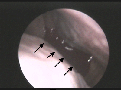
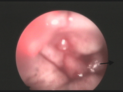
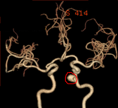
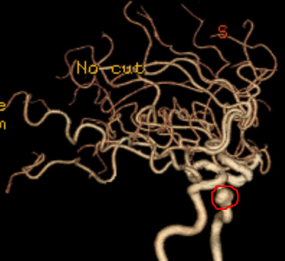
Urgent Head and neck Computed Tomography (CT) revealed Left maxillary sinus, sphenoid sinus and the right frontal sinus inflammation and nasal septum deviation towards right. The patient was treated with hemostatic, anti-inflammatory drugs and other symptomatic treatment. On the 3rd and 4th day of admission patient again suffered from nasal bleeding, the amount of bleeding was about 5-6 ml, after applying pressure for about 1 minute bleeding stopped itself. At that time anterior and posterior rhinoscope reveal no active bleeding lession, no any further medical intervention was taken and continued previous medication. On 7th day of admission patient suffered from massive epistaxix, under the guidance of nasopharyngeal endoscope nasal cavity and nasopharynx was explored, saw bleeding from left side olfactory grove (Figures 1 and 2), anterior and posterior nasal packing was done and was able to control bleeding. 3 days after nasal packing patient removed the packing himself, but patient still conitnue to have slight nasal bleeding. Head and neck angiography was done in order to find the condition of blood vessels in that region. Angiography revealed internal carotid artery cavernous sinus segment aneurysm (Figures 3 and 4). Patient was transferred to the departent of neurosurgery and underwent Digital substraction angiography and stent surgery. The patient had no further episodes of nasal bleeding and other complications..
Discussion
Patient had been suffering from recurrent epistaxix for 7 months . Inspite of medical and surgical intervention was not been able to control bleeding . Nasal bleeding is a common medical problem, the cause of nasal bleeding may be local cause (trauma, inflammation, nasal septum disease, tumor, foreign bodies, etc. ) or systemic cause(cardiovascular disease, blood diseases, acute febrile diseases and severe nutritional disorders and vitamin deficiency, drug poisoning, endocrine disorders, vascular dilatation disease, liver and kidney chronic disease. In 90% of case bleeding occur from little’s area [1]. Children have mild anterior nasal bleeding while elderly have profuse posterior nasal bleeding. Bleeding site may be (1) above the level of middle turbinate from anterior and posterior ethmoidal branches of ophthalmic artery (branch of internal carotid artery). (2) Below the level of middle turbinate from sphenopalatine branch of maxillary artery (branch of external carotid artery). (3) Some time bleeding site may be hidden in middle and inferior turbinates [2]. Approximately 60% of population suffer nasal bleeding, about 6% require formal medical intervention, of those episode of epistaxis 10% can be serious and life threatening [3]. Males are affected more than female, but after the age of 50 both sexes are affected equally. Hypertension is the most common cause in the elderly population [2]. As increase in blood pressure the duration and the amount of nasal bleeding also increase. The elderly populations are more prone to nasal bleeding because their nasal mucosa tens to become dry and thin and the mucosal blood vessels are also not able to contract easily to control bleeding. In 90% of population nasal bleeding can be easily controlled, but under certain circumstances and in the presence of various undertaking conditions life itself may be at risk. Some time because of an inadequate history, the use of inappropriate or wrongly positioned packing, poor supervision after hemostasis, and the mistaken use of chemical cautery may leads to fatal complications and management will be stressful [4].
Cavernous carotid aneurysm is divided into mainly two types; traumatic and non-traumatic. Cavernous segment carotid artery aneurysm is a rare occurrence, represents less than 2% of all intracranial aneurysm [5]. Because of its close anatomical relationship with the sphenoid sinus as well as the nasal fossae in some cases may cause epistaxis. Non-traumatic carotid artery aneurysms are a rare cause of epistaxis [6]. The first case of epistaxis caused by intracranial aneurysm was published at a lecture at the Royal College of Surgeons of England by Dr. Beadles in 1907 [6]. Mass effect is the most common presentation for non-traumatic cavernous internal carotid artery aneurysms, with only 3% presenting with hemorrhage [7]. In those case which present with hemorrhage, severe, delayed, life-threatening, epistaxis often occurs after latent period, which sometimes make it very difficult to identify the relation between underlying cause and epistaxis [8]. Therefore early suspicion and detection is necessary for timely management. Goldenberg-Cohen et al. reported in 40 cavernous sinus aneurysms (27 women, 4 men), Mean age at diagnosis was 60. 4 years (range 25 to 86; median 64). The most common symptoms were diplopia (61%), headache (53%), and facial or orbital pain (32%). Fifteen patients (48%) were diagnosed after they developed cranial nerve pareses, four (13%) after they developed Carotid–Cavernous Sinus Fistulas (CCFs), and 12 (39%) by neuroimaging studies done for unrelated symptoms [9].In another study conducted by Higashida, Halbach, Dowd, et al. in 87 patients, the most common presentation was mass effect (79%) rupture of aneurysm causing carotid –cavernous fistula (9%), trauma resulting to Cavernous pseudoaneurysm (8%) and hemorrhage (3%). Diplopia is secondary due to the compression of cranial nerves by aneurysm [10]. Variety of neurological deficits, related to vision, including diplopia from single or multiple oculomotor nerve pareses, decreased visual acuity from compressive or ischemic optic neuropathy, corneal and facial anesthesia or hyperesthesia from involvement of the trigeminal nerve, and facial pain may be related to cavernous carotid aneurysm. Like other intracranial aneurysms, these aneurysms can rupture, but this is a rare event and usually does not produce a subarachnoid or intracerebral haemorrhage. Instead, rupture of a Cavernous Carotid Aneurysm (CCA) usually causes a carotid–cavernous sinus fistula or, rarely, epistaxis [7].
The common cause of epistaxis; nasal perforation, nasal septum deviation, rhinitis, sinusitis, and upper respiratory tract infection, can be managed by nasal packing, or use of hemostatic agent or by chemical cautery or by use of a folley’s catheter. That epistaxis which cannot be controlled by good nasal packing can be treated under endoscopic evaluation where nasal cavity is explored to indentify bleeding point and the bleeding vessels can be ligated. This commonly seen epistaxis has less risk of recurrence. Recurrent epistaxis other than cavernous carotid aneurysm may be due to congestive heart failure, diabetes mellitus, hypertension, and a history of anemia. Epistaxis related to cavernous carotid aneurysm is difficult to manage by nasal packing, even posterior packing is not sufficient to control bleeding, under certain circumstances and in the presence of various undertaking conditions life itself may be life threatening, so special attention and measures should be undertaken to manage such case of recurrent epistaxis. The clinical presentation of recurrent epistaxis which occurs in absence of any history of trauma and surgery may lead to miss diagnosis or delay the definitive diagnosis. The diagnosis of clinically suspected aneurysm causing nasal bleeding should be confirmed using ultrasonogrophy of neck with colored Doppler flow, CT imaging, CT angiography or Magnetic resonance inages, Angiography provides the most reliable and definitive diagnostic information and offers the opportunity of performing endovascular treatment [11]. High index of suspicion is required for the diagnosis of carotid cavernous aneurysm causing epistaxis and internal carotid artery angiography should be performed in those cases and when considering embolization [12]. Endovascular embolizations, clipping of aneurysm neck or ligation of internal carotid artery are few treatment options.
Conclusion
Although epistaxis is the most common clinical presentation in the department of Otolaryngology and the management of epistaxix is not very difficult, but some time management may be stressful for those epistaxis of unknown origin and recurrent episodes and may be fatal in case of massive bleeding. Aneurysm of the internal carotid artery is rarely mentioned as a cause of epistaxis. This condition is quite rare but it is important to consider aneurysms in the etiology of epistaxis because of their high mortality rate and since they require management quite different from that of epistaxis of other origins. We may consider the internal carotid artery aneurysm as the cause of epistaxis. Similar cases of epistaxis have been reported. Those are due to traumatic rupture of aneurysm or radiotherapy induced aneurysm. We described the rare cause of epistaxix which is related to non-traumatic cavernous carotid aneurysm in a healthy patient with no history of congestive heart failure, diabetes mellitus, hypertension and other diseases and its management. As for otolaryngologist medical intervention and nasal packing are the management option. The stent placement is quick, safe, rapid and long term treatment of carotid artery aneurysm.
References
- Jiakong W, Zhou L, Tang A (2010) Otolaryngology-Head and Neck Surgery,Beijing,People’s Medical Publishing House 303-306.
- Bansal M (2013) Disease of External Nose and Epistaxis.Disease Of Ear ,Nose and Throat,Jaypee Brothers Medical Publishers(P) Ltd. 289-297
- Thomas A, Tami, James A, Marrell, Ballengera’s (2009) Otorhinolaryngology Head and Neck Surgery,Shelton, People’s Medical Publishing House 551-555.
- Schreiner L (1976) Complications and errors in the management of nose bleeding (author's transl). LaryngolRhinolOtol (Stuttg) 55: 257-263.
- Handa J, Handa H (1976) Severe epistaxis caused by traumatic aneurysm of cavernous carotid artery. SurgNeurol 5: 241-243.
- Beadles CF (1907) Aneurysm of larger cerebral arteries.Brain30:285-336.
- Chaboki H, Patel AB, Freifeld S, Urken ML, Som PM (2004) Cavernous carotid aneurysm presenting with epistaxis. Head Neck 26: 741-746.
- Bavinzski G, Killer M, Knosp E, Ferraz-Leite H, Gruber A, et al. (1997) False aneurysms of the intracavernous carotid artery--report of 7 cases. ActaNeurochir (Wien) 139: 37-43.
- Goldenberg-CohenN, CurryC, MillerNR, TamargoRJ, Murphy KPJ (2004) Long term visual and neurological prognosis in patients with treated and untreated cavernous sinus aneurysms.JNeurolNeurosurg Psychiatry 75:863-867
- Higashida RT, Halbach VV, Dowd C, Barnwell SL, Dormandy B, et al. (1990) Endovascular detachable balloon embolization therapy of cavernous carotid artery aneurysms: results in 87 cases. J Neurosurg 72: 857-863.
- da Silva PS, Waisberg DR (2011) Internal carotid artery pseudoaneurysm with life-threatening epistaxis as a complication of deep neck space infection. PediatrEmerg Care 27: 422-424.
- Andres Maldonado-Naranjo,Varun R,Kshettry, Gabor Toth, et al. (2013) Non-traumatic superior hypophyseal aneurysm associated with pseudoaneurysm presenting with massive epistaxis. Clinical Neurology and Neurosurgery 115:2251-2253.
Citation: Gyanwali B, Wu H, Zhu M, Tang A (2015) Epistaxis Related to Internal Carotid Artery Cavernous Sinus Aneurysm. Otolaryngology 5:188. DOI: 10.4172/2161-119X.1000188
Copyright: © 2015 Gyanwali B, et al. This is an open-access article distributed under the terms of the Creative Commons Attribution License, which permits unrestricted use, distribution, and reproduction in any medium, provided the original author and source are credited.
Select your language of interest to view the total content in your interested language
Share This Article
Recommended Journals
Open Access Journals
Article Tools
Article Usage
- Total views: 20888
- [From(publication date): 5-2015 - Sep 02, 2025]
- Breakdown by view type
- HTML page views: 16095
- PDF downloads: 4793


