Middle Cranial Fossa Defect in Atypical Presentation: Spontaneous CSF Rhinorrhea with Left Meningo-Encephalocele of Temporal Bone in Idiopathic Intracranial Hypertension: A Case Report
Received: 10-Mar-2019 / Accepted Date: 21-Mar-2019 / Published Date: 28-Mar-2019 DOI: 10.4172/2161-119X.1000366
Abstract
The development of Spontaneous cerebrospinal fluid (CSF) leakage has been related to various factors. It may occur in patients with normal Intracranial pressure (ICP) and in only a minority with elevated ICP. Meningo-encephaloceles (MEC) of the temporal bone are mostly as result of otologic surgery or head trauma. The spontaneous type of MEC is further very rare. We describe here a rare case of CSF rhinorrhea as a presenting symptom of Idiopathic intracranial hypertension (IIH). The site of leakage found to be at left Middle cranial fossa (MCF) “left tygmen tympani” which was associated with left temporal MEC and CSF leakage into middle ear space. This rare complication of IIH represents a difficult condition to deal with, in regard to its diagnosis and management and carries a serious sequalae if it is mismanaged. Diagnostic process and management outlines were addressed. Management of IIH plus surgical repair of the skull base defect with middle ear reconstruction were applied here. Surgically, we intervene through transmastoid approach with exploratory left modified radical mastoidectomy; which showed two big holes in tegmen tympani measured about 8 × 4 mm and 5 × 4 mm with herniated MEC. Cauterizations of the herniated cele then closure of the defects were applied. Followed by middle ear reconstruction. The bony defects closed in multi-layered fashion. We used temporalis fascia, tragal cartilage, perichondtrium, bone dust, bone wax (hydroxyapatite bone cement) and fibrin glue. The posterior wall of external auditory canal was reconstructed by helical cartilage.
Keywords: Middle cranial fossa defect; Tegmen tympani; Spontaneous CSF leak; Encephalocele; Idiopathic intracranial HTN; Surgical approaches
Introduction
The development of Spontaneous cerebrospinal fluid (CSF) leakage has been related to various factors. It may occur in patients with normal Intracranial pressure (ICP) and in only a minority with elevated ICP [1]. Furthermore, spontaneous herniation of Meningo-encephaloceles (MEC) through Middle cranial fossa (MCF) defect is very rare. Idiopathic intracranial hypertension (ITH) is well known cause of increased ICP. Other includes intracranial tumors and hydrocephalus.
Encephalocels are categorized into occipital, fronto-ethmoidal, temporal encephaloceles. Skull base encephaloceles account for 5% only [2], with temporal bone encephaloceles are being the most common skull base encephalocele.
On other hand, skull base defect is categorized into primary (including congenital and spontaneous) or secondary (include traumatic, iatrogenic, or due to tumor). They occur predominantly in the floor of the middle cranial fossa at the level of tegmen [3]. The spontaneous temporal bone defects present in 2 major distinct categories based on the age of onset. Congenital temporal bone abnormalities in children include defect in the hyrtl fissure, wide fallopian canal, mondini dysplasia & patent cochlear acqueduct [4]. Spontaneous temporal bone defects in adults are less common and generally present without previous history of petrous pyramid disease [5]. However, the majority of the defects are secondary and related to head trauma [6]. Usually the defect is single; as single tegmen defect of temporal bone was reported in 15-34% of the autopsies [7]. And the brain tissue usually becomes necrotic after herniation [8,9].
We describe here a rare case of CSF rhinorrhea as a presenting symptom of Idiopathic intracranial hypertension (IIH); in which the CSF passed from the middle ear cleft, through the Eustachian tube into the nasal cavity. In addition, MEC was found herniated through left tegmen tympani.
Case Report
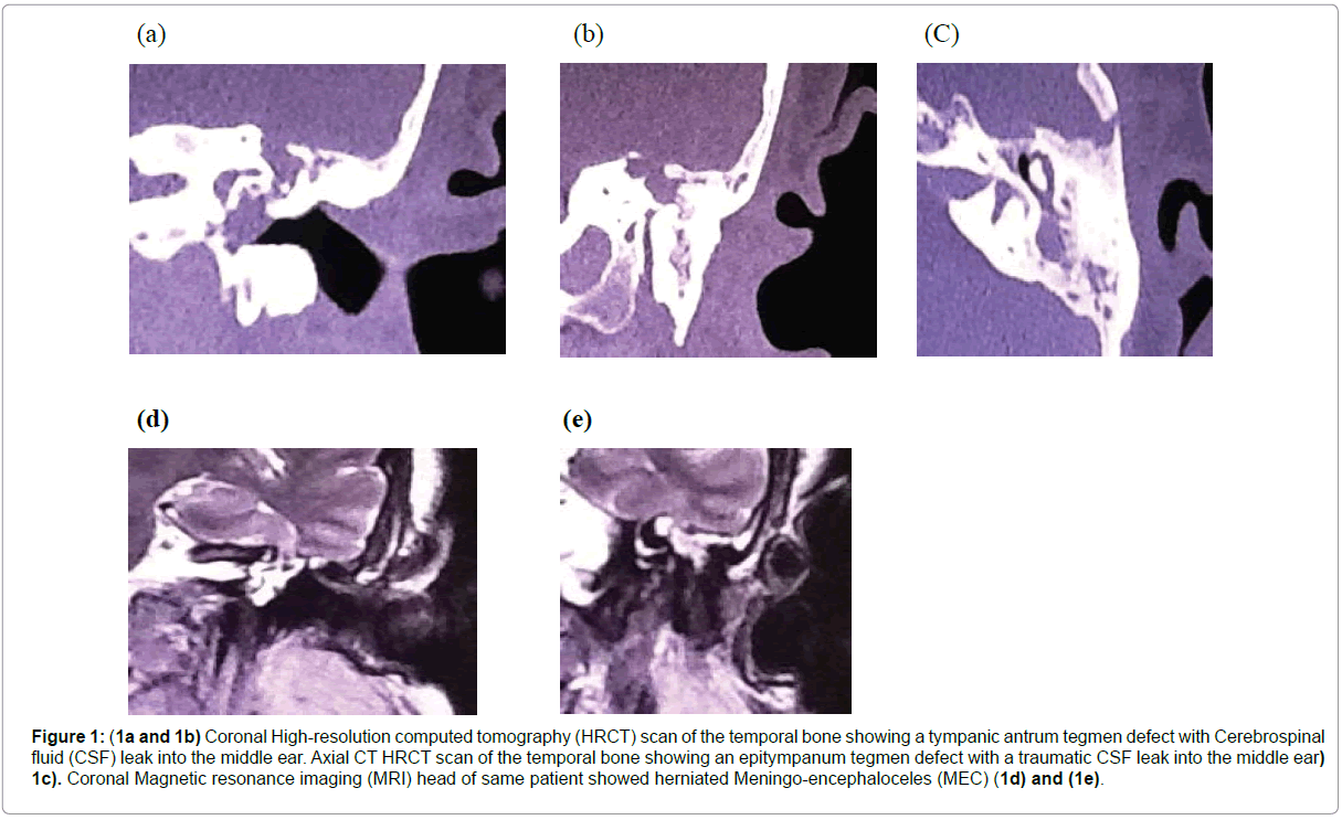
A 59-year-old Saudi Arabian obese woman; presented with on/ off left side clear watery rhinorrhea for 1-year duration. Her complain was associated with on/off headache and left aural fullness. Her Body mass index (BMI) is 30. She was referred to our institution for further evaluation and management as case of query CSF leak as her watery rhinorrhea persist despite anti-allergic medications. Her examinations were unremarkable, apart from positive beta-2 transferrin results from fluid collected from her nostril. Audiometric evaluation showed left side type B (flat curve) tympanometry with Air Bone Gap (ABG) of 20 dbl. Computed tomography (CT) of the Paranasal sinus (PNS) was unremarkable. CT PNS cisternography failed to localize the site of the leakage. MRI head cisternography revealed MEC descending through left MCF defect. CT temporal bone showed empty sella+opacified left middle ear cavity+bony defect at middle ear roof. Because of persistent headaches, she was sent to a neuro-ophthalmologist, who found mild papilledema on examination. The patient underwent a microscopic middle ear exploration then reconstruction via trans-mastoid approach. During exploratory left modified canal wall down mastoidectomy, two bony holes at tegmen tympani with herniated MEC were found. Cauterizations of the herniated cele then closure of the defects were applied. Followed by middle ear reconstruction. The bony defects at tegmen closed by undelay technique and in multi-layered fashion. We used temporalis fascia, tragal cartilage, perichondtrium, bone dust, bone wax (hydroxyapatite bone cement) and fibrin glue. The posterior wall of external auditory canal was reconstructed by helical cartilage. Lumbar puncture (LP) (subdural drain) was inserted in same setting. LP’s opening pressure could not be measured although normal CSF composition was seen. The drain was kept for three days and CSF fluid was frequently aspirated. Post operatively; she was started on acetazolamide; in addition to routine postoperative measures for elevated ICP. The patient was diagnosed with IIH with spontaneous MEC of left temporal bone. On post operative follow up, the headache and CHL improved. The CSF rhinorrhea resolved. She expressed mild dizziness in immediate postoperative period, which was disappeared within 48 hours. She was discharged from our hospital 1-week post surgery. No other complications were noticed. Acetazolamide was given to her by neurology team to treat IIH. She was followed for 1-year post surgery by otolaryngology, neuro-opthalmology and neurology. Her condition showed stable course (Figure 1).
Figure 1: (1a and 1b) Coronal High-resolution computed tomography (HRCT) scan of the temporal bone showing a tympanic antrum tegmen defect with Cerebrospinal fluid (CSF) leak into the middle ear. Axial CT HRCT scan of the temporal bone showing an epitympanum tegmen defect with a traumatic CSF leak into the middle ear) 1c). Coronal Magnetic resonance imaging (MRI) head of same patient showed herniated Meningo-encephaloceles (MEC) (1d) and (1e).
Discussion
The existence of spontaneous Meningo-encephaloceles (MEC) in the middle cranial fossa due to dehiscence of the tegmen tympani is rare and their etiology is unknown [10,11]. There are two main theories concerning the etiology of middle fossa bony defect, which considered as the basics for pathophysiology of spontaneous CSF leakage. The first is the congenital defect theory, which posits that tiny defects within the tegmen caused by aberrant embryologic development enlarge over time secondary to constant CSF pressure. This enlargement leads to eventual dural herniation and subsequent bone and dural thinning with resultant CSF otorrhea [12]. The second is the arachnoid granulation theory postulates that arachnoid granulations that do not find a venous termination during embryonic development come to lie in a blind end against the inner bony surface of the skull [13]. These granulations enlarge with age and may eventually erode bone after decades by the action of intracranial pressure and pulsatility, causing a thinning of the dura mater and ultimately causing MEC [13,14]. The resulting communication within the central nervous system can lead to a multitude of infectious complications, with significant morbidity, potentially disastrous long-term neurologic deficits, and even death [15].
The diagnosis of spontaneous temporal bone MECs may be quite challenging. The clinical presentation of MECs include conductive hearing loss, intermittent/continuous CSF otorrhoea or rhinorrhoea, serous otitis media, headache, meningitis, temporal lobe seizures [16], expressive aphasia & facial nerve weakness [17]. Up to 25% of adult-onset CSF leaks present with meningitis [8]. Testing of beta-2- transferrin in samples of otorrhea/ rhinorrhea fluid is highly sensitive and specific for identifying CSF [18]. Another more recently utilized marker, b-trace protein, has higher specificity than beta-2-transferrin and potentially allows for earlier result interpretation due to more rapid laboratory processing [19].
Imaging studies are essential for determining the site of the CSF leak in the temporal bone [20]. High-resolution computed tomography (HRCT) will usually identify the bony defect of the temporal bone but will not demonstrate the site of the dural tear. In this situation, CT cisternography can play an important role and can demonstrate the leak [21]. However, even with CT cisternography, determination of the location of a CSF leakage is not possible in all cases [22].
MRI is noninvasive, offers excellent anatomic detail, and has no radiation risk. It can distinguish middle ear/mastoid encephalocele from chronic ear disease, as the CSF signal will be bright on T2- weighted images, and an encephalocele will appear contiguous with the brain [23].
Empty sella probably represents a sign of elevated intracranial pressure that leads to idiopathic, spontaneous CSF leaks. Spontaneous CSF leaks are strongly associated with the radiographic finding of an empty sella and are more common in obese females, similar to benign intracranial hypertension [5].
In one study, it has been concluded that benign intracranial hypertension is prevalent in a significant number of patients presenting with spontaneous encephalocele and CSF otorrhea at a rate much higher than general population. They suggest that all patients with spontaneous encephalocele/CSF leak warrant evaluation for benign intracranial hypertension [24]. Zhijun et al studied the association between primary spontaneous cerebrospinal fluid rhinorrhea and elevated ICP. They concluded that elevated ICP has been associated with all patients which were included in the study. And symptoms of IIH were present in most of them. In addition, the majority were women and obese [25].
Selection of surgical approach and the materials for repair were based on several factors including the location of the temporal bone defect, presence of a recurrent leak, and surgeon preference. The surgical approach should enable adequate exposure of the area for accurate localization of the temporal bone defect and facilitate middle ear reconstruction. The most commonly used methods are trans-mastoid, middle fossa, or combined approaches. The trans-mastoid approach has the advantage of better localization of the dural defect in the temporal bone while avoiding brain retraction and potential neurological complications. Potential disadvantages of the trans-mastoid approach include decreased exposure of the defect, lower initial success rate and durability of the repair, and increased risk of hearing loss [26].
The herniated MEC had been considered to be devitalized; and not functional [27] and extradural excision is recommended; except for large celes where it would be better to reintroduce it [11]. The excision of the cele should be done by coagulating the neck. Direct suture is the best option for the repair of the dura mater if feasible. It is also possible to employ dural plasties or autologous tissue. Autologous materials include abdominal fat, free or pedicled temporalis muscle/fascia, conchal cartilage/perichondrium, and calvarial bone/pericranium. Synthetic materials include fibrin glue, hydroxyapatite bone cement, vicryl mesh, and a cellular dermis (Alloderm, LifeCell, Branchburg, NJ) [28].
In the present case, we intervene through trans-mastoid approach with exploratory left modified radical mastoidectomy. We found two big holes in tegmen tympani measured about 8 × 4 mm and 5 × 4 mm with herniated MEC. Cauterizations of the herniated cele then closure of the defects were applied. Followed by middle ear reconstruction. The bony defects closed in multi-layered fashion. We used temporalis fascia, tragal cartilage, perichondtrium, bone dust, bone wax (hydroxyapatite bone cement) and fibrin glue. The posterior wall of external auditory canal was reconstructed by helical cartilage.
In study done by Pelosi et al. trans-mastoid approach is also the preferred approach as the initial route of access to CSF leaks of the temporal bone in most cases. As it provides a sufficient exposure to precisely define the defect and repair it. In the study, they suggest that the intracranial approach is needed in cases of large, multiple, or medial defects and for recurrent CSF leaks as revision procedures. They often utilize a combined transmastoid/transcranial approach to bolster the security of the repair, and this technique has a low demonstrated risk of failure [29].
The evidence for external lumbar drainage usage is still lacking; but it may be useful in order to decrease CSF pressure [27,30], and should be maintained for 3-5 days postoperatively.
Conclusion
We report a rare case of spontaneous CSF rhinorrhea as presentation for middle cranial fossa defect with MEC. We need to be aware that CSF leak could be a rare complication of IIH. Early recognition is essential to avoid devastating sequelae. Evaluation includes both laboratory tests and imaging. Optimal therapy includes lowering of ICP and surgical repair of bony defect.
References
- Allen KP, Perez CL, Kutz JW, Gerecci D, Roland PS, et al. (2014) Elevated intracranial pressure in patients with spontaneous cerebrospinal fluid otorrhea. Laryngoscope 124: 251-254.
- Ba MC, Kabre A, Badiane SB, Ndoye N, Ly Ba A, et al. (2003) Fronto- ethmoidal encephaloceles in Dakar. Report of 9 cases. Dakar Med 48: 131-133.
- Lundy LB, Graham MD, Kartush JM, LaRouere MJ (1996) Temporal bone encephalocele and cerebrospinal fluid leaks. Am J Otol 17: 461-469.
- Quiney RE, Mitchell DB, Djazeri B, Evans JN (1989) Recurrent meningitis in children due to inner ear abnormalities. J Laryngol Otol 103: 473-480.
- Schlosser RJ, Bolger WE (2013) Significance of empty sella in cererbrospinalfliud leaks. Otolaryngol Head Neck Surg 128: 32-38.
- Connor SE (2010) Imaging of skull-base cephalocoeles and cerebrospinal fluid leaks. Clin Radiol 65: 832-841.
- Brown NE, Grundfast KM, Jabre A, Megerian CA, O’Malley BW Jr, et al. (2004) Diagnosis and management of spontaneous cerebral fluid–middle ear effusion and otorrhoea. Laryngoscope 114: 800-805.
- Kamerer DB, Caparosa RJ (1982) Temporal bone encephalocele-diagnosis and treatment. Laryngoscope 92: 878-882.
- Glasscock ME, Dichins JR, Jackson CG, Wiet RJ, Feenstra L (1979) Surgical management of brain tissue herniation into the middle ear and mastoid. Laryngoscope 89: 1743-1754.
- Papanikolaou V, Bibas A, Ferekidis E, Anagnostopoulou S, Xenellis J (2007) Idiopathic temporal bone encephalocele. Skull Base17: 311-316.
- Ahron C, Thulin CA (1965) Lethal intracranial complications following inflation in the external auditory canal in treatment of serous otitis media and due to defects in the petrous bone. Acta Otolaryngol 60: 407-421.
- Pappas DG Jr, Hoffman RA, Cohen NL, Pappas DG Sr (1992) Spontaneous temporal bone cerebrospinal fluid leak. Am J Otol 13: 534-539.
- Wilkins RH, Radtke RA, Burger PC (1993) Spontaneous temporal encephalocele: Case report. J Neurosurg 78: 492-498.
- Nahas Z, Tatlipinar A, Limb CJ, Francis HW (2008) Spontaneous meningoencephalocele of the temporal bone: clinical spectrum and presentation. Arch Otolaryngol Head Neck Surg 134: 509-518.
- Hyson M , Andermann F, Olivier A, Melanson D (1984) Occult encephaloceles and temporal bone epilepsy: developmental and acquired lesions in the middle fossa. Neurology 34: 363-366.
- Uri N, Shupak A, Greenberg E, Kelner J (1991) Congenital middle ear encephalocele initially seen with facial paresis. Head Neck 13: 62-67.
- Skedros DG, Cass SP, Hirsch BE, Kelly RH (1993) Beta-2 transferrin assay in clinical management of cerebral spinal fluid and perilymphatic fluid leaks. J Otolaryngol 22: 341-344.
- Raine C (2005) Diagnosis and management of otologic cerebrospinal fluid leak. Otolaryngol Clin North Am 38: 583-595.
- Rao AK, Merenda DM, Wetmore SJ (2005) Diagnosis and management of spontaneous cerebrospinal fluid otorrhea. Otol Neurotol 26: 1171-1175.
- Eberhardt KE, Hollenbach HP, Deimling M, Tomandl BF, Huk WJ (1997) MR cisternography: a new method for the diagnosis of CSF fistulae. Eur Radiol 7: 1485-1491.
- Dietrich U, Feldges A, Sievers K. Kocks W (1993) Localization of frontobasal traumatic cerebrospinal fluid fistulas. Comparison of radiologic and surgical findings. Zentralbl Neurochir 54: 24-31.
- Tyagi I, Syal R, Goyal A (2005) Cerebrospinal fluid otorhinorrhoea due to inner-ear malformations: clinical presentation and new perspectives in management. J Laryngol Otol 119: 714-718.
- Brainard L, Chen DA, Aziz KM, Hillman TA (2012) Association of benign intracranial hypertension and spontaneous encephalocele with cerebrospinal fluid leak. Otol Neurotol 33: 1621-1624.
- Zhijun Y, Bo W, Chungcheng W, Pinan L (2011) Primary spontaneous cerebrospinal fluid rhinorrhea: a symptom of idiopathic intracranial hypertension? J Neurosurg 115: 165-170.
- Gacek RR (1990) Arachnoid granulation cerebrospinal fluid otorrhea. Ann Otol Rhinol Laryngol 99: 854-862.
- Wind JJ, Caputy AJ, Roberti F (2008) Spontaneous encephaloceles of the temporal lobe. Neurosurg Focus 25: 11.
- Savva A, Taylor MJ, Beatty CW (2003) Management of cerebrospinal fluid leaks involving the temporal bone: report on 92 patients. Laryngoscope 113: 50-56.
- Stanley P, Joshua BB, Eric ES (2010) Cerebrospinal Fluid Leaks of Temporal Bone Origin: Selection of Surgical Approach. Skull Base 20: 253-259.
- Pappas DG Jr, Hoffman RA, Holliday RA, Hammerschlag PE, Pappas DG Sr, et al. (1995) Evaluation and management of spontaneous temporal bone cerebrospinal fluid leaks. Skull Base Surg 5: 1-7.
Citation: Alshaikh M, Mandura R (2019) Middle Cranial Fossa Defect in Atypical Presentation: Spontaneous CSF Rhinorrhea with Left Meningo-Encephalocele of Temporal Bone in Idiopathic Intracranial Hypertension: A Case Report Otolaryngol (Sunnyvale) 9: 365. DOI: 10.4172/2161-119X.1000366
Copyright: © 2019 Alshaikh M, et al. This is an open-access article distributed under the terms of the Creative Commons Attribution License, which permits unrestricted use, distribution, and reproduction in any medium, provided the original author and source are credited.
Select your language of interest to view the total content in your interested language
Share This Article
Recommended Journals
Open Access Journals
Article Tools
Article Usage
- Total views: 3720
- [From(publication date): 0-2019 - Nov 06, 2025]
- Breakdown by view type
- HTML page views: 2846
- PDF downloads: 874

