Case Report Open Access
Periapical Healing of a Mandibular Molar with Middle Mesial Canal: A Case Report
Stacey M Woo*Woo Dental Corporation, Anaheim, California, USA
- *Corresponding Author:
- Stacey M Woo
Woo Dental Corporation, Anaheim
California, USA, 92808
Tel: 530-428-5443
Fax: 949-305-5201
E-mail: staceywoodds@gmail.com
Received Date: February 20, 2017; Accepted Date: February 28, 2017; Published Date: March 07, 2017
Citation: Woo SM (2017) Periapical Healing of a Mandibular Molar with Middle Mesial Canal: A Case Report. J Interdiscipl Med Dent Sci 5: 209. doi:10.4172/2376-032X.1000209
Copyright: © 2017 Woo SM. This is an open-access article distributed under the terms of the Creative Commons Attribution License, which permits unrestricted use, distribution, and reproduction in any medium, provided the original author and source are credited.
Visit for more related articles at JBR Journal of Interdisciplinary Medicine and Dental Science
Abstract
Introduction: Complex root canal anatomies challenge the limits of our skills, techniques, and abilities to clean the root canal system and achieve a successful endodontic outcome. Background: The following case report depicts a first mandibular molar indicated for root canal treatment after diagnosis of pulpal necrosis and asymptomatic apical periodontitis due to caries. Pre-operative radiographic analysis revealed two distinct periapical lesions and a Periapical Index (PAI) Score of 3. Methods: The tooth was accessed for root canal treatment and instrumented to a final apical size of #20. Additional cleaning and disinfection were performed utilizing the GentleWave® System. After the GentleWave® Procedure, the tooth was obturated with gutta-percha and an epoxy resin based sealer by warm vertical condensation and thermoplasticized gutta-percha backfill. Post-operative radiographs revealed a middle mesial canal not previously visualized during instrumentation or prior to performing the GentleWave Procedure. The newly located, uninstrumented, middle mesial canal was filled with sealer. Results: Recall was performed over a 9-month period. Both clinical and radiographic assessments showed complete healing, no clinical signs or symptoms, and a PAI score of 1 at the 9-month recall. This case illustrates healing after root canal treatment utilizing minimal instrumentation and the GentleWave Procedure, suggesting that GentleWave Procedure can clean and disinfect complex root canal anatomy.
Keywords
Uninstrumented canals; Middle mesial canal; GentleWave; Apical periodontitis; Periapical healing
Introduction
Many factors affect the successful outcome of root canal treatment (RCT) and promote the healing of periradicular lesions [1-7]. Root canal systems with complex anatomies present particular challenges for access, instrumentation, and irrigation, especially roots with configurations such as lateral canals, isthmi, fins, apical delta, Cshaped systems, furcation canals, and multiple apical foramina [8-12]. Clinicians often aim to strike a balance between effective cleaning, shaping, and preservation of tooth structure to avoid weakening the root structure [13].
A new technology, the GentleWave® Procedure, has recently been developed that uses Multisonic UItracleaning™. Multisonic Ultracleaning creates cavitation implosions that generate multisonic waves, which propagate throughout the root canal system and enhance root canal cleaning and disinfection by advanced fluid dynamics, acoustics, and tissue dissolution chemistry [14-16]. Studies have shown that the GentleWave Procedure efficiently removes tissue debris from the root canal system in vitro [17]. Clinical studies in patients have shown a high rate of healing of periapical lesions and favorable outcomes at 6 and 12 months [18,19]. This case report describes a necrotic mandibular first molar in which a middle mesial canal in the apical third and isthmi in the mesial and distal roots were revealed after the GentleWave procedure, despite minimal instrumentation.
Case Presentation
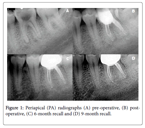
A 25-year-old male with a non-contributory medical history presented to the clinic with a chief complaint of occasional mild sharp pain when biting and chewing. Clinical examination of the left mandibular posterior found tooth #19 with gross caries extending into the pulp. Tooth #19 was not sensitive to percussion or palpation and did not respond to vitality testing with Endo-Ice. Radiographic analysis concluded a pre-operative Periapical Index (PAI) Score 3 (Figure 1A) [20]. Based on clinical and radiographic findings, the diagnosis was Pulpal Necrosis and Asymptomatic Apical Periodontitis (AAP). Root canal treatment was recommended in an attempt to extend the life of the tooth. The prognosis was fair. The patient consented to treatment.
Local anesthesia was administered using 4% articaine (72 mg) with 1:100,000 epinephrine and 2% lidocaine (72 mg) with 1:100,000 epinephrine via inferior alveolar nerve block and long buccal nerve block. The tooth was isolated with a rubber dam. The carious lesion was excavated and verified with caries indicator dye. Absent tooth structure was built up with micro-hybrid composite. The tooth was accessed with round carbide and EndoZ burs in a water-cooled hand piece. Four orifices were identified in the pulpal floor. Patency was gained with a size #10 K-file, and working lengths (WL) were measured using an electronic apex locator and confirmed radiographically. A glide path was created with K-files up to size #20. Canals were instrumented up to ProTaper® F1 (Dentsply, Tulsa Dental Specialties, Tulsa, OK) powered by an electric, motor-driven, contraangle handpiece under copious 0.5% sodium hypochlorite (NaOCl) irrigation. Following minimal instrumentation, debridement and disinfection were completed utilizing the GentleWave® Procedure (Sonendo®, Laguna Hills, CA), in which delivery of sodium hypochlorite, ethylenediaminetetraacetic acid (EDTA) and Multisonic UItracleaning technology were employed. The root canal system was rinsed with 100% ethanol, dried with a microsuction tip and absorbent paper points. Obturation was completed with gutta-percha and AH Plus® Sealer (Dentsply, Tulsa Dental Specialties, Tulsa, OK) by warm vertical condensation with a System B heat source and thermoplasticized gutta-percha backfill with an Obtura Max III System. The access cavity was sealed with a microhybrid composite build-up, and the patient was advised to return to the referring general dentist for crown placement. The root canal treatment was completed in a single visit. Recall visits were scheduled for 6 and 9 months to monitor healing and assess clinical and radiographic outcomes.
All treatment was completed under high power magnification with a dental operating microscope. Cone beam computed tomography (CBCT) scans were completed utilizing the Carestream 8100 3D (Carestream Health, Inc. Rochester, NY) with a focused field of view of the region of interest.
Results
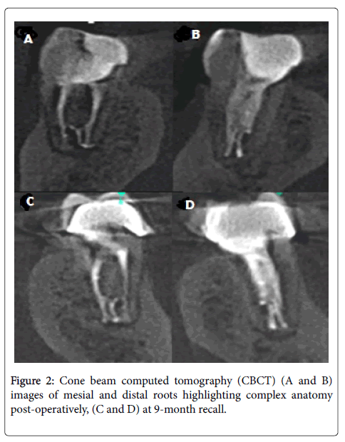
Pre-operatively, the tooth was diagnosed with Pulpal Necrosis and Asymptomatic Apical Periodontitis (AAP) and had a Periapical Index (PAI) Score of 3 (Figure 1A). Post-operative radiographs (Figure 1B) show complex anatomies in the root canal system that were not previously realized, including a middle mesial canal and isthmi in the mesial and distal canals filled with obturation materials. These complex anatomies were further confirmed on a post-operative CBCT scan as seen in Figures 2A and 2B. Figures 1A-1D show radiographs of tooth #19 at pre-operative, post-operative and 6- and 9-month recall visits, respectively.
At the 6-month recall, the patient was asymptomatic and the tooth had been restored with a full coverage crown. The tooth was functional. Radiographic analysis showed healing of the periapical lesions (Figure 1C). Complete resolution of apical periodontitis was noted at the 9-month recall and no clinical signs or symptoms were present (Figure 1D). The 9-month post-operative PAI score was 1. CBCT analysis further confirmed periapical healing (Figures 2A and 2B). The patient was prescribed no medications and was advised to return to the general dentist for continued comprehensive dental care.
Discussion
One of the many goals of root canal treatment is to remove as much of this debris as possible, as close to the apex as possible. Studies have examined the relationship between the apical size of instrumentation and cleaning in the apical third.
After endodontic instrumentation, anatomical variations typically contain tissue remnants, bacteria, and dentin shavings that inhibit the ability of irrigation fluids to reach areas of the root canal system [21,22]. Khademi et al. found that the minimum instrumentation size needed for penetration of irrigants to the apical third is #30 [22]. However, endodontic irrigants have limited access to the apical 3 mm with standard root canal treatment [23]. Studies found that a canal instrumented to a size #35 allows greater irrigation in the apical third [24]. Peters, et al. found that the tested endodontic rotary instrumentation techniques leave 35% or more of the canals surface area unchanged [25]. With increasing file size, there is also an increasing reduction in bacteria [26]. Ricucci et al. found that lateral canals that appear filled after standard root canal treatment are usually a mix of sealer, smear layer, and bacteria, not necessarily cleaned and filled [27].
Studies have looked at ultrasonically-activated acoustic streaming as a technique to augment the ability of irrigants to reach beyond the instrumented canal walls [21,28-31]. However, studies show that while acoustic streaming significantly improves the cleanliness of canals and isthmi over traditional side-vented needle irrigation, an ultrasonicallyactivated instrument can only remove debris up to 3 mm in front of the file tip and debris still persists near the apex, even after instrumenting up to a ProTaper size F4 [29,32,33].
In this case report, minimal instrumentation to a size #20 was utilized, which under standard root canal treatment would be insufficient to permit irrigants to access the entire root canal system, would leave debris within the root canal system, and would block the ability of obturation materials to reach these areas.
Obturation of complex anatomies suggests that despite minimal instrumentation, cleaning occurred in areas of the root canal system not touched by endodontic files. In this case, complex anatomies were revealed in post-operative imaging, including a middle mesial canal in the apical third, mesial isthmus, and distal isthmus (Figures 1B, 2A and 2B), suggesting that the GentleWave Procedure enhanced cleaning in the apical third and in areas not previously realized. Together, this likely contributed to the patient’s favorable endodontic outcome.
The success rate of root canal treatment depends on many factors [4-7]. Root canal systems with complex canal morphologies such as lateral canals, isthmi, fins, C-shaped canals, and varying canal configurations present special challenges in cleaning, shaping, and obturation [11,12,34-38]. For example, 29.4% of maxillary molars have lateral canals, 20.2% of distal roots in molars exhibit isthmi, and middle mesial canals, located between the mesiobuccal and mesiolingual canals of a mandibular molars, occur in 1-20% of the population [39-43].
Conclusion
In the present case report, the middle mesial canal originates in the apical region and would not be accessible for instrumentation or irrigation with standard endodontic techniques. To instrument and irrigate a middle mesial canal in the apical third would require further dentin removal to access, putting the tooth at higher risk for file separation, strip perforations, and ledges and compromising the integrity of the tooth structure. However, through minimal instrumentation and Multisonic UItracleaning of the root canal system using the GentleWave Procedure, the middle mesial canal was cleaned and dentin was preserved.
In root canal treatment, cleaning is often a balance between dentin preservation and instrumentation to allow irrigants to reach the apical portions of the root canal system. This case report shows that with the GentleWave Procedure, it may be possible to clean and disinfect the apical portions of the root canal system while preserving more tooth structure.
References
- Sjogren U, Hagglund B, Sundqvist G, Wing K (1990) Factors affecting the long-term results of endodontic treatment. J Endod 16: 498-504.
- Ng YL, Mann V, Rahbaran S, Lewsey J, Gulabivala K (2007) Outcome of primary root canal treatment: Systematic review of the literature-Part 2. Effects of study characteristics on probability of success. IntEndod J 40: 921-939.
- Nair PN (2004) Pathogenesis of apical periodontitis and the causes of endodontic failures. Crit Rev Oral Biol Med 15: 348-381.
- Friedman S, Abitbol S, Lawrence HP (2003) Treatment outcome in endodontics: The toronto study,phase 1: Initial treatment. J Endod 29: 787-793.
- Farzaneh M, Abitbol S, Lawrence HP, Friedman S (2004) Treatment outcome in endodontics-the toronto study, Phase II: Initial Treatment. J Endod 30: 302-309.
- Marquis VL, Dao T, Farzaneh M, Abitbol S, Friedman S (2006) Treatment outcome in endodontics: The toronto study, Phase III: Initial Treatment 32: 299-306.
- De Chevigny C, Dao TT, Basrani BR, Marquis V, Farzaneh M, et al. (2008) Treatment outcome in endodontics: The toronto study-Phase 4: Initial treatment. J Endod 34: 258-263.
- Weine FS, Healey HJ, Gerstein H, Evanson L (2012) Canal configuration in the mesiobuccal root of the maxillary first molar and its endodontic significance 1969. J Endod 38: 1305-1308.
- Gutmann JL (1978) Prevalence, location, and patency of accessory canals in the furcation region of permanent molars. J Periodontol 49: 21-26.
- Estrela C, Rabelo LE, De Souza JB, Alencar AH, Estrela CR, et al. (2015) Frequency of root canal isthmi in human permanent teeth determined by cone-beam computed tomography. J Endod 41: 1535-1539.
- Christie WH, Peikoff MD, Fogel HM (1991) Maxillary molars with two palatal roots: a retrospective clinical study. J Endod 17: 80-84.
- Von Arx T, Steiner RG, Tay FR (2011) Apical surgery: endoscopic findings at the resection level of 168 consecutively treated roots. IntEndod 44: 290-302.
- Lertchirakarn V, Palamara JE, Messer HH (2003) Patterns of vertical root fracture: factors affecting stress distribution in the root canal. J Endod 29: 523-528.
- Wang Z, Shen Y, Haapasalo M (2014) Dental materials with antibiofilm properties. Dent Mater 30: e1-16.
- Charara K, Friedman S, Sherman S, Kishen A, Malkhassian G, et al. (2016) Assessment of apical extrusion during root canal irrigation with the novel gentlewave system in a simulated apical environment. J Endod 42: 135-139.
- Vandrangi P (2016) Evaluating penetration depth of treatment fluids into dentinal tubules using the gentlewave system. Dentistry 6: 3-7.
- Molina B, Glickman G, Vandrangi P, Khakpour M (2015) Evaluation of root canal debridement of human molars using the gentlewave system. J Endod 41: 1701-1705.
- Sigurdsson A, Garland RW, Le KT, Woo SM (2016) 12-Month healing rates after endodontic therapy using the novel gentlewave system: A prospective multicenter clinical study. J Endod 42:1040-1048.
- Sigurdsson A, Le KT, Woo SM, Shahriar RA, McLachlan K, et al. (2016) Six-Month healing success rates after endodontic treatment using the novel gentlewave system: The pure prospective multi-center clinical study. J ClinExp Dent 8: e290-298.
- Orstavik D, Kerekes K, Eriksen HM (1986) The periapical index: a scoring system for radiographic assessment of apical periodontitis. Endod Dent Traumatol 2:20-34.
- Gu LS, Kim JR, Ling J, Choi KK, Pashley DH, et al. (2009) Review of contemporary irrigant agitation techniques and devices. J Endod 35: 791-804.
- Khademi A, Yazdizadeh M, Feizianfard M (2006) Determination of the minimum instrumentation size for penetration of irrigants to the apical third of root canal systems. J Endod 32:417-420.
- Senia ES, Marshall FJ, Rosen S (1971) The solvent action of sodium hypochlorite on pulp tissue of extracted teeth. Oral Surg Oral Med Oral Pathol 31:96-103.
- Salzgeber RM, Brilliant JD (1977) An Invivoevaluation of the penetration of an irrigating solution in root canals. J Endod 3:394-398.
- Peters OA, Schönenberger K, Laib A (2001) Effects of four ni-ti preparation techniques on root canal geometry assessed by micro computed tomography. IntEndod J 34:221-230.
- Dalton BC, Orstavik D, Phillips C, Pettiette M, Trope M (1998) Bacterial reduction with nickel-titanium rotary instrumentation. J Endod 24:763-767.
- Ricucci D, Siqueria JF (2010) Fate of the tissue in lateral canals and apical ramifications in response to pathologic conditions and treatment procedures. J Endod 36: 1-15.
- Ahmad M, Pitt Ford TR, Crum LA (1987) Ultrasonic debridement of root canals: Acoustic streaming and its possible role. J Endod 13:490-499.
- Kahn FH, Rosenberg PA, Gliksberg J (1995) An Invitroevaluation of the irrigating characteristics of ultrasonic and subsonic handpieces and irrigating needles and probes. J Endod 21:277-280.
- Burleson A, Nusstein J, Reader A, Beck M (2007) The Invivoevaluation of hand rotary ultrasound instrument in necrotic human mandibular molars. J Endod 33:782-787.
- Cunningham WT, Martin H, Forrest WR (1982) Evaluation of Root canal débridement by the endosonic ultrasonic synergistic system. Oral Surg Oral Med Oral Pathol 53:401-404.
- Malki M, Verhaagen B, Jiang LM, Nehme W, Naaman A, et al. (2012) Irrigantflow beyond the insertion depth of an ultrasonically oscillating file in straight and curved root canals: Visualization and cleaning efficacy. J Endod 38:657-661.
- Adcock JM, Sidow SJ, Looney SW, Kiu Y, McNally K, et al. (2011) Histologic evaluation of canal and isthmus debridement efficacies of two different irrigant delivery techniques in a closed system. J Endod 37:544-548.
- Cleghorn BM, Christie WH, Dong CC (2006) Root and root canal morphology of the human permanent maxillary first molar: A literature review. J Endo 32:813-821. DOI:10.1016/j.joen.2006.04.014
- Vertucci FJ (2005) Root canal morphology and its relationship to endodontic procedures. Endo Topics 10:3-29.
- Cooke HG, Cox FL (1979) C-shaped canal configurations in mandibular molars. J Am Dent Assoc 99:836-839.
- Kulild JC, Peters DD (1990) Incidence and configuration of canal systems in the mesiobuccal root of maxillary first and second molars. J Endod 16:311-317.
- Martins JN, Mata A, Marques D, Anderson C, Carames J (2016) Prevalence and characteristics of the maxillary c-shaped molar. J Endod 42:383-389.
- Herbranson E (2014) The anatomy of the root canal system as a challenge to effective disinfection, in disinfection of root canal systems: The treatment of apical periodontitis (ed N. Cohenca), John Wiley & Sons, Inc, Hoboken, NJ, USA.
- Baugh D, Wallace J (2004) Middle mesial canal of the mandibular first molar: A case report and literature review. J Endod 30: 185-186.
- Goel NK, Gill KS, Taneja JR (1991) Study of root canal configuration in mandibular first permanent molars. J Indian SocPedodPrev Dent 8: 12-14.
- Azim AA, Deutsch AS, Solomon CS (2015) Prevalence of middle mesial canals in mandibular molars after guided troughing under high magnification: An Invivo Investigation. J Endod 41:164-168.
- Nosrat A, Deschenes RJ, Tordik PA, Hicks ML, Fouad AF (2014) Middle mesial canals in mandibular molars: incidence and related factors. J Endod 41:28-32.
Relevant Topics
- Cementogenesis
- Coronal Fractures
- Dental Debonding
- Dental Fear
- Dental Implant
- Dental Malocclusion
- Dental Pulp Capping
- Dental Radiography
- Dental Science
- Dental Surgery
- Dental Trauma
- Dentistry
- Emergency Dental Care
- Forensic Dentistry
- Laser Dentistry
- Leukoplakia
- Occlusion
- Oral Cancer
- Oral Precancer
- Osseointegration
- Pulpotomy
- Tooth Replantation
Recommended Journals
Article Tools
Article Usage
- Total views: 4025
- [From(publication date):
April-2017 - Sep 03, 2025] - Breakdown by view type
- HTML page views : 3074
- PDF downloads : 951


