Prediction of Fluid Characteristics in Pediatric Otitis Media with Effusion from the Tympanometry
Received: 24-Sep-2017 / Accepted Date: 20-Oct-2017 / Published Date: 27-Oct-2017 DOI: 10.4172/2161-119X.1000325
Abstract
Objective: To correlate the characteristics of type B tympanogram with the type of middle ear fluid in children suffering from persistent OME.
Methodology: A prospective study was conducted on children with persistent OME for more than 3 months who failed to respond to medical treatment with type B tympanogram in one or both ears and were candidate for myringotomy. The middle ear fluid was aspirated and the viscosity of the fluid was evaluated objectively by measuring the diameter of the diffusion circle on a filter paper in mm.
Results: Thirty eight children (70 ears) were enrolled in this study. The shape of type B tympanogram was either flat (67.14%) or peaked (32.86 %).Three fluid types were noticed in OME namely serous, mucoid and glue. The shape was highly correlated with the type of secretions. Compliance and gradient of type B tympanogram were negatively correlated with the viscosity of the secretions.
Conclusion: Tympanometry is a beneficial clinical tool in prediction, diagnosis and management of OME. Characteristics of type B tympanogram were correlated with the viscosity of middle ear fluid. Detailed analysis of type B tympanogram can assist the clinicians in determining the indication of comprehensive treatment.
Keywords: OME; Tympanometry; Gradient; Compliance
Introduction
Otitis media with effusion (OME) is one of the most common disorders of childhood [1]. Bluestone and Klein defined OME as an inflammation of the middle ear with a collection of fluid in the middle ear space [2]. The amount and nature of fluid found in OME is highly variable. The fluid may partly or completely fill the tympanomastoid compartment and can be serous, mucoid or glue like [3,4].
Diagnosis of OME is based on three factors: patient history, otoscopic examination of the TM and audiological evaluation that includes tympanometry and pure tone audiometry [5]. Tympanometry is the best objective evidence of a middle ear effusion [6,7]. It measures changes in the acoustic admittance of the ear as a function of the pressure in the sealed external ear canal. The curve of a tympanogram is defined by 3 major characteristics: 1) the maximum acoustic admittance of the ear, which is the value of the admittance at the peak of the tympanogram (may be compensated for the ear canal volume); 2) the pressure at the peak of the tympanogram, which reflects the pressure in the middle ear; and 3) the gradient, which represents the flatness of the curve [5,8].
Jerger [8] classified tympanograms as types A, B and C. Types A and C are characterized by the presence of a peak, which is close to 0 decapascals (dapa) in type A and negative (equal to or less than -100 dapa) in type C. In type B tympanogram, there is no distinct peak and the admittance does not significantly change during pressure variations in the external ear canal. Type B tympanogram may be either straight or with a pseudopeak (round B). In clinical practice, type B is generally related to fluid in the middle ear [5,9].
Objective
To correlate the characteristics of type B tympanogram with the type of middle ear fluid in children suffering from persistent OME.
Materials and Methods
A prospective study was done in the ENT Department at Sohag University Hospital, Egypt, over one year duration between September 2015 and September 2016.
Subjects
Children (under the age of 12 years, either males or females) with persistent OME for more than 3 months who failed to respond to medical treatment, with type B tympanogram in one or both ears and are candidate for myringotomy were randomly included in our study. Those children complaint mainly from, diminution of hearing, nasal obstruction or snoring. The exclusion criteria include children with extensive tympanosclerosis, scarred TM, adhesive otitis media or those who revealed purulent aspirate.
Methods
The study was done after approval from Sohag Faculty of Medicine Ethics Committee. Informed consents were obtained from the parents of all children who agreed to participate in this study. All children were subjected to a careful history taking and full ENT examination including otoscopic examination, pneumatic otoscopic examination (Welch Allyn otoscope).
The tympanometer (Mico 41) was performed using a 226 Hz probe frequency with positive to negative pressure sweeps of 600 daPa/s. Only type B tympanograms were interpreted as pathological. Those with type B tympanogram and failed to respond to medical treatment for 3 months were included in the study. At the day of surgery tympanometry was repeated and those with type B were subjected to myringotomy under general endo-tracheal anesthesia and the middle ear fluid was aspirated into a 3 ml syringe mounted on the aspiration cannula and the viscosity of the fluid was evaluated objectively to add to the subjective surgeon's opinion. 0.2 ml of the aspirated fluid was applied to the center of a filter paper and left flat for 5 min inside a glass container to allow for diffusion of fluid contents of the aspirate. Then, the diameter of the diffusion circle was measured in mm. Similarly, 0.2 ml of water and of aspirated nasal mucus in normal volunteers was applied to filter papers to measure the diameter of diffusion circles of both as a control. The mean water diffusion diameter is taken as a control reference of serous MEE and the nasal aspirate diffusion diameter is taken as a control reference of mucoid MEE. Correlation between the tympanometric results and type of ME secretions was done using the correlation coefficient test via the SPSS program. P-values<0.05 were considered significant.
Results
Over a one year period, 38 children (70 ears) were included in this study with an age range from 1.25 to 12 years with a mean of 4.61 years. They were 22 males (57.89%) and 16 females (42.11%).
In our study, there were 28 (73.68%) cases having adenoid with encroachment on the nasopharyngeal opening of the ET, five patients of those with adenoids presented only with snoring/nasal obstruction with no otologic symptoms despite having OME. Seven (18.42%) cases had previous history suggestive of AOM.
Characters of tympanometry
Shape: Shape of type B tympanogram in this study was either flat in 47 ears (67.14%) or peaked tympanogram in 23 ears (32.86%).
Pressure: Mean value was -17
Compliance: Mean value was 0.14
Gradient: Mean value was 312
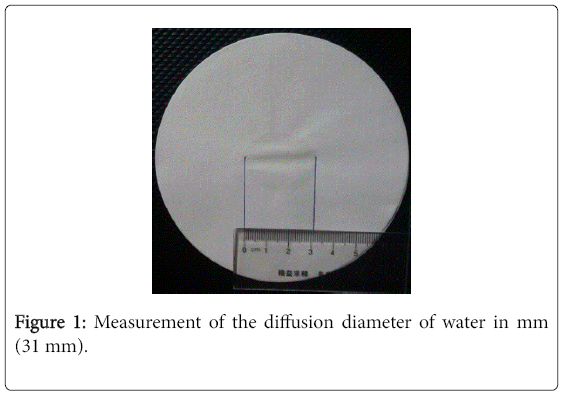
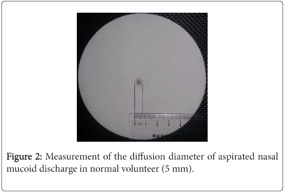
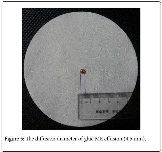
Characters of the fluid: The mean diffusion diameter of water was 30 mm (Figure 1) and the mean diffusion diameter of nasal aspirate was 5 mm (Figure 2). The diffusion diameter ranged from 4-31 mm. We classified the type of secretions according to the diffusion diameter into:
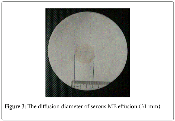
Serous effusion: With a diffusion diameter of 25-31 mm (Figure 3)
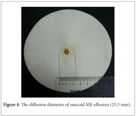
Mucoid effusion: With a diffusion diameter of >5-<25 mm (Figure 4)
Glue effusion: With a diffusion diameter of ≤ 5 mm (Figure 5)
Table 1 demonstrates the number and percentage of each type of effusion in the 70 ears.
| Type of fluid | Number | Percentage |
|---|---|---|
| Serous | 16 | 22.86 % |
| Mucoid | 26 | 37.14 % |
| Glue | 20 | 28.57 % |
| No secretion | 8 | 11.43 % |
Table 1: The number and percentage of the type of secretions in the 70 ears.
Correlation study
The correlation between the tympanometric results and types of effusion was demonstrated in Table 2. There was a high statistically significant positive correlation between the type of secretions and the shape of tympanometry. On the other hand, there was a high statistically significant negative correlation between the type of secretions and the character of tympanometry (gradient and compliance).
| Type of fluid | Shape | Compliance | Gradient | |
|---|---|---|---|---|
| Shape | ||||
| R | 0.91 | |||
| P value | <0.0001* | |||
| Compliance | ||||
| R | -0.87 | 0.998 | ||
| P value | <0.0001* | <0.0001* | ||
| Gradient | ||||
| R | 0.84 | 0.94 | -0.96 | |
| P value | <0.0001* | <0.0001* | <0.0001* |
Table 2: The correlation between the tympanometric results and type of secretions.
Discussion
The question examined in the present study is whether tympanometry, can clearly differentiate cases with thin fluid from those with thick fluid. If feasible, this differentiation might assist the otologist in determining whether medications or surgery would be more efficient treatment as regards the minimization of delay in the resolution of the effusion and subsequent hearing loss.
The main function of the ET is middle ear ventilation to equalize the middle ear pressure with atmospheric pressure [10]. Mechanical obstruction of the ET because of adenoid hypertrophy may be an important factor in OME [11,12]. Kinderman et al. found that obstruction of the ET orifice by adenoid tissue was associated with tympanograms suggestive of abnormal pressure in the middle ear [13]. In our study, there were 28 (73.68%) cases having adenoid with encroachment on the nasopharyngeal opening of the ET. 7 (18.42%) cases had previous history suggestive of AOM. AOM is more often present in age below 2 years as reported by many authors, 20% of those transforms into OME [14,15].
Variable fluid types of middle ear effusion and variable tympanometric results of type B tympanograms were documented in our study. Difference in the fluid type in different patients and in some cases in different ears in the same patient is without convincing evidence in the literature. In our study, we found tympanometry to be an accurate tool in assessing patients for the presence of fluid in a preoperative setting. Shape of type B tympanogram was either flat in the larger percentage of cases (67.14%) or peaked in the remaining ones. In a correlation study between the shape of type B tympanogram and the type of secretion there was a highly statistically significant positive correlation (Table 2) which means that the more flatness of type B tympanogram the more the viscosity of the secretions. This is in agreement with Maw et al. [16] who reported that when an effusion is present with a peaked tympanogram, it is more likely that the effusion will be serous, whereas with a flat tympanogram the majority of effusions are mucoid. It has been reported that the three parameters of tympanograms, pressure, compliance and gradient correlated with effusion within the middle ear [9]. In the present study, there was a highly statistically significant negative correlation between the type of secretions and the character of tympanometry (gradient and compliance); which means that when compliance and gradient decreased, the secretion will be more viscous. This could be explained by that the value at maximum compliance (peak) on tympanogram presents the pressure within the middle ear. Compliance is associated with mobility of the TM. Since effusion within the middle ear decreases mobility of the TM, low compliance and gradient tympanograms usually imply effusion within the middle ear [17]. Therefore, it has been found that type B tympanograms in Jerger’s classification [8] were associated with effusion.
Conclusion
Tympanometry is a beneficial clinical tool in prediction, diagnosis and management of OME. Characteristics of type B tympanogram were correlated with the viscosity of middle ear fluid. Detailed analysis of type B tympanogram can assist the clinicians in determining the indication of comprehensive treatment.
References
- Bondy J, Berman S, Glazner J, Lezotte D (2000) Direct expenditure related to otitis media diagnosis: Extrapolation from pediatric medicated cohort. Pediatrics 105: E72.
- Bluestone C, Klein J (2001) Otitis media in infants and children. WB Saunders, Philadelphia.
- Gates GA (1987) Surgical management of otitis media with effusion. Adv Otolaryngol Head Neck Surg 1: 127-142.
- Cunningham M, Eavey R (1993) Otitis media with effusion. In: Nadol JB Jr, Schuknecht HF (eds.) Surgery of the ear and temporal bone. New York (NY): Raven Press, pp: 205-222.
- Bluestone CD, Beery QC, Paradise JL (1973) Audiometry and tympanometry in relation to middle ear effusion in children. Laryngoscope 83: 594-604.
- Gates GA, Avery C, Cooper JC, Hearne EM, Holt GR (1986) Predictive value of tympanometry in middle ear effusion. Ann Otol Rhinol Laryngol 95: 46-50.
- Sassen ML, van Aarem A, Grote JJ (1994) Validity of tympanometry in the diagnosis of middle ear effusion. Clin Otolaryngol Allied Sci 19: 185-189.
- Jerger J (1970) Clinical experience with impedance audiometry. Arch Otolaryngol 92: 311-324.
- Brooks DN (1968) An objective method of detecting fluid in the middle ear. Int J Audiol 7: 280-286.
- Chauhan B, Chauhan K (2013) Comparative study of eustachian tube functions in normal and diseased ears with tympanometry and video nasopharyngoscopy. Indian J Otolaryngol Head Neck Surg 65: 468-476.
- Maw AR, Parker A (2009) Surgery of the tonsils and adenoids in relation to secretory otitis media in children. Acta Otolaryngol Suppl 454: 202-207.
- Di Francesco R, Paulucci B, Nery C, Bento RF (2008) Craniofacial morphology and otitis media with effusion in children. Int J Pediatr Otorhinolaryngol 72: 1151-1158.
- Kindermann CA, Roithmann R, Lubianca Neto JF (2008) Obstruction of the eustachian tube orifice and pressure changes in the middle ear: Are they correlated? Ann Otol Rhinol Laryngol 117: 425-429.
- Marchisio P, Principi N, Passali D, Salpietro DC, Boschi G, et al. (1998) Epidemiology and treatment of otitis media with effusion in children in the first year of primary school. Acta Otolaryngol 118: 557-562.
- Park CW, Han JH, Jeong JH, Cho SH, Kang MJ, et al. (2004) Detection rates of bacteria in chronic otitis media with effusion in children. J Korean Med Sci 19: 735-738.
- Maw AR, Bawden R, O'Keefe L, Gurr P (1993) Does the type of middle ear aspirate have any prognostic significance in otitis media with effusion in children? Clin Otolaryngol Allied Sci 18: 396-399.
- Watters GW, Jones JE, Freeland AP (1997) The predictive value of tympanometry in the diagnosis of middle ear effusion. Clin Otolaryngol Allied Sci 22: 343-345.
Citation: Ali AHA, Sayed RH, Mawgoud SMA, Ghayes D (2017) Prediction of Fluid Characteristics in Pediatric Otitis Media with Effusion from the Tympanometry. Otolaryngol (Sunnyvale) 7:325. DOI: 10.4172/2161-119X.1000325
Copyright: © 2017 Ali AHA, et al. This is an open-access article distributed under the terms of the Creative Commons Attribution License, which permits unrestricted use, distribution and reproduction in any medium, provided the original author and source are credited.
Select your language of interest to view the total content in your interested language
Share This Article
Recommended Journals
Open Access Journals
Article Tools
Article Usage
- Total views: 6010
- [From(publication date): 0-2017 - Dec 15, 2025]
- Breakdown by view type
- HTML page views: 5000
- PDF downloads: 1010

