Proboscis Lateralis: A Unique Case with Choanal Atresia and Bilateral Ophthalmopathy
Received: 18-Aug-2014 / Accepted Date: 10-Dec-2014 / Published Date: 20-Dec-2014 DOI: 10.4172/2161-119X.1000183
Abstract
Proboscis Lateralis (PL) is a rudimentary nasal appendage which is located off-center from the vertical midline of the face. It is a very rare craniofacial malformation frequently associated with abnormalities of the eyes and/or ocular adnexa. We report a unique case with bilateral ophthalmopathy and choanal atresia.
Case Report
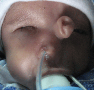
A 36-day-old male, and 2.9 kg infant who was born at 38 weeks’ gestation by uncomplicated spontaneous vaginal delivery with Apgar scores of 7 and 9 at 1 and 5 minutes was transferred to the Otolaryngology-Head and Neck Surgery of our academic, tertiary-care center for evaluation of a soft, approximately 2.5 cm long and 1.25 cmdiameter tube like process originating from the region of the left medial canthus. There was a clear, mild mucoid discharge from an orifice at its distal end. A diagnosis of proboscis lateralis was made on the basis of the characteristic appearance of the anomaly (Figure 1). Associated anomalies included left nasal hypoplasia, left ocular hypoplasia, bilateral choanal atresia, and contralateral ophthalmic agenesis and blindness. Prenatal history was negative for consanguinity, exposure to alcohol, ionizing radiation, prescription medications, or toxic drugs. The patient’s parents denied any family history of blindness, craniofacial abnormalities, mental retardation, or other congenital defects. He was a first child of the family. Findings from examination of the eyes were remarkable for complete agenesis of the right eyes and microphthalmia on the left with ocular hypoplasia and no functional eye. The anterior chamber was normal.
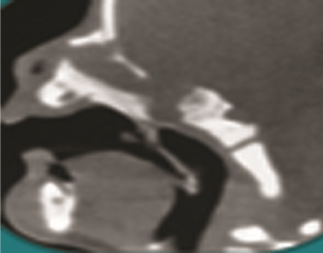
Diagnostic imaging consisting of Computer Tomography (CT) and Magnetic Resonance Imaging (MRI) was obtained to evaluate the full extent of the congenital anomaly and to rule out communication to the cranium before doing any surgical procedure. Spiral CT Scan was performed in the direct axial and coronal planes with 2.5-mm collimation. Sagital and tridimentional were reconstructed .This evaluation demonstrated normal cerebral parenchyma, ventricular architecture, and mid-line anatomy. Hypoplasia of the left nasal passage (undeveloped ipsilateral middle turbinates, inferior turbinates, and ethmoid air cells) with bilateral choanal atresia was present (Figure 2). We performed MRI of the brain with high-resolution images through the orbits with and without gadolinium, which demonstrated enhancement of the proboscis central tract without evidence of intracranial communication (Figure 2).
Discussion
For the first time Forster reported proboscis lateralis in 1861 by his monograph Congenital Malformations of the Human Body [1]. It is a rare congenital anomaly in which a tubular, nose-like structure is seen to arise from the medial canthal area [2]. PL is embryologically related to the median facial cleft that finally may result in incomplete formation of the nose [3]. This appendage is generally 2 to 3 cm in length and 1 cm in diameter and has a central tract lined with respiratory epithelium that typically expresses clear mucus from the blind dimple at its distal end and may be continuous with the paranasal sinuses proximally. Generally, it arises from the area of the medial canthus. As expected for respiratory epithelium, the central tract of the proboscis enhanced with gadolinium on MRI. However, enhancement after gadolinium may be mistaken for dural enhancement and viewed as a sign of intracranial involvement [4,5].
There is not any racial predilection in proboscis lateralis. Review of 9 cases by Boo-Chai’s supports the notion of a male preponderance with a 3:1 male female ratio [6]. Embryologic development of the face and nose is a complex process. The nasal placodes appear to function as the primary organizers for the developing nose. The nasal placodes invaginate into the frontonasal process, separating this structure into the medial and lateral nasal processes. The precise embryologic mechanism responsible for the development of PL has not been defined [6]. Although PL has been described in the absence of other congenital anomalies, it is characteristically accompanied by ipsilateral heminasal hypoplasia or aplasia and rarely by choanal atresia [7]. Ocular and/ or ocular adnexal (anophthalmia, microphthalmia, microcornea, lenticular opacities, cyclopean eye, and colobomas of the choroid, retina, iris, and eyelids) findings as well as cleft lip and/or palate are the most common anomalies seen in conjunction with PL and have been used as the basis for a classification system [8,9]. Group 1 consists of isolated PL without other associated anomalies (9%). Group 2 consists of PL with associated ipsilateral nasal defect (23%). Group 3 consists of PL with associated ipsilateral nasal and ocular and/or ocular adnexal defect (47%). Group 4 includes the features of group 3 with the addition of cleft lip and/or palate (21%). However Sakamoto, et al. redefined classification and added two new groups including group 5 as hypertelorism with encephalocele and group 6 as hypotelorism [10].
Although most patients with proboscis lateralis do not have serious central nervous system abnormalities, proboscis lateralis may coexist with central nervous system anomalies and early neuro-imaging is indicated to rule out intracranial abnormalities [11].
Initial reports regarding the treatment of PL recommended simple surgical excision of the proboscis. More recently, surgical management of PL has been approached with reconstruction in mind. Because there is some variability in facial anomalies and the degree of nasal hypoplasia seen with proboscis lateralis, management must be individualized [4,5]. Due to the variety of maxillofacial and ocular disease seen with proboscis lateralis, optimal care of the patient warrants a multidisciplinary approach that may involve an otolaryngologist or oromaxillofacial surgeon, plastic surgeon, and ophthalmologist.
Conclusion
PL is a rare congenital anomaly with a characteristic appearance. Computed tomography and MRI are complementary in determining the extent of the bony and soft tissue components of the anomaly. Proboscis lateralis might be associated with developmental anomalies of the ipsilateral nasal and ocular structures. Complete surgical excision at the base of the proboscis is desirable as a primary procedure if there is adequate ipsilateral nasal development or as a delayed excision if the proboscis is used in nasal reconstruction.
References
- Forster A (1861) Die Missbildung des Menschen Syttematisch Dargestellt. Jena, Germany: F Mauke
- Martin S, Hogan E, Sorenson EP, Cohen-Gadol AA, Tubbs RS, et al. (2013) Proboscis lateralis. Childs Nerv Syst 29: 885-891.
- Chauhan DS, Guruprasad Y (2010) Proboscis lateralis. J Maxillofac Oral Surg 9: 162-165.
- Thorne MC, Ruiz RE, Carvalho J, Lesperance MM (2007) Proboscis lateralis: case report and review. Arch Otolaryngol Head Neck Surg 133: 1051-1053.
- Guerrero JM, Cogen MS, Kelly DR, Wiatrak BJ (2001) Proboscis lateralis. Arch Ophthalmol 119: 1071-1074.
- Khoo BC (1985) The proboscis lateralis--a 14-year follow-up. Plast Reconstr Surg 75: 569-577.
- Chen MT, Yeong EK (1992) Supernumerary nostrils. Br J Plast Surg 45: 557-558.
- Khoo BC (1985) The proboscis lateralis--a 14-year follow-up. Plast Reconstr Surg 75: 569-577.
- Magadum SB, Khairnar P, Hirugade S, Kassa V (2012) Proboscis lateralis of nose-a case report. Indian J Surg 74: 181-183
- Sakamoto Y, Miyamoto J, Nakajima H, Kishi K (2012) New classification scheme of proboscis lateralis based on a review of 50 cases. Cleft Palate Craniofac J 49: 201-207.
- Meeker Lh, Aebli R (1947) Cyclopean eye and lateral proboscis with normal one-half face: report of a case. Arch Ophthal 38: 159-173.
Citation: Mohsen N, Mehdi B (2015) Proboscis Lateralis: A Unique Case with Choanal Atresia and Bilateral Ophthalmopathy. Otolaryngology 5:183. DOI: 10.4172/2161-119X.1000183
Copyright: © 2015 Mohsen N, et al. This is an open-access article distributed under the terms of the Creative Commons Attribution License, which permits unrestricted use, distribution, and reproduction in any medium, provided the original author and source are credited.
Select your language of interest to view the total content in your interested language
Share This Article
Recommended Journals
Open Access Journals
Article Tools
Article Usage
- Total views: 15495
- [From(publication date): 3-2015 - Sep 01, 2025]
- Breakdown by view type
- HTML page views: 10868
- PDF downloads: 4627


