Research Article Open Access
Radiographic Density and Contrast of Images of Individual Films from Double Film Packets
Plauto Christopher Aranha Watanabe1*, Angela Jordão Camargo2 and Luiz Carlos Pardini1
1Department of Stomatology, Public Oral Health and Forensic Dentistry, Ribeirão Preto Dental School, University of São Paulo, Ribeirão Preto, São Paulo, Brazil
2Department of Stomatology, School of Dentistry, University of São Paulo, São Paulo, São Paulo, Brazil
- *Corresponding Author:
- Plauto Christopher Aranha Watanabe
Department of Stomatology, Public Oral Health and Forensic Dentistry
Ribeirão Preto Dental School-University of São Paulo, Cafe Avenue
Zip Code: 14040-904, Ribeirão Preto, SP, Brazil
Tel: +55 16 3315-3993
E-mail: watanabe@forp.usp.br
Received date: August 02, 2016; Accepted date: September 24, 2016; Published date: September 29, 2016
Citation: Watanabe PCA, Camargo AJ, Pardini LC (2016) Radiographic Density and Contrast of Images of Individual Films from Double Film Packets. Cosmetol & Oro Facial Surg 2:110.
Copyright: © 2016 Watanabe PCA, et al. This is an open-access article distributed under the terms of the Creative Commons Attribution License, which permits unrestricted use, distribution, and reproduction in any medium, provided the original author and source are credited.
Visit for more related articles at Cosmetology & Oro Facial Surgery
Abstract
The aim of this study was determined whether individual films from unexpired and expired double packets present the same image quality in terms of radiographic density and contrast. Methods: An aluminum step wedge was radiographed using 20 Agfa periapical double film packets. 10 of them unexpired and 10 used 5 years after the expiration date, maintained under refrigeration at 10°C. Results: Statistical analysis showed a significant difference in radiographic density (p<0.01) and contrast (p<0.05) between individual films from unexpired double packets, whereas the individual films of the expired packets did not differ. Compared the individual films without using the values of direct film exposure (zero) the difference in contrast between individual films was no longer present, but optic density was still significantly different (p<0.05) for the unexpired group. Conclusion: Within the limitations of this study, the results suggest there is a difference in quality of the radiographic image between the individual films of double packets when submitted to laboratory analysis.
Keywords
Double film packet; Radiographic density; Radiographic contrast
Introduction
Brazil is the country with the largest number of world dental professionals in absolute numbers: there are 219,575 registered professionals [1]. The Global Atlas of Dentistry, "published in 2009 by Dental Federation, some estimates more than one million dentists worldwide. Thus, the radiographic films, analogical, still are the base of the main tool to assist diagnosis in dentistry [2].
Several factors may affect the diagnostic ability of the radiographic image such as kVp, time of exposure, mA, filtration, collimation, grids, and types of the position indicating device. In general, operating protocols for a given radiographic film are based on the nominal kVp of the equipment and X-ray units with the same nominal kVp may present quite different spectra [3].
Another important factor is the radiographic film, due to its properties, can significantly alter the quality of the radiographic image, affecting interpretation and planning of the case, and/or this would require new exposure of the patient to obtain new image. Oftentimes, small differences in image quality can be a significant difference in the caries diagnosis, i.e., carious interproximal lesions. Dental caries, also known as tooth decay or cavities, is the most common disorder affecting the teeth and the radiographic exam is a important diagnostic tool Legacy of the evolution of ever faster film has been a concomitant lower patient exposure [4]. These changes in the film speed accompanied, film base was also undergoing improvements in the image characteristics as density and contrast. Also, restaurative materials, as some types of cement commonly used for the cementation of implant-supported prostheses have poor radiodensity and may not be detectable following radiographic examination [5].
The double radiographic films allow the professional to store a radiograph and leave the other with patient, thus solving the dilemma of the property rights of a radiograph and without expose the patient to a radiation dose. In addition, the dentist can get different images, making changes in radiographic processing, to verify specific structures, mainly by changing the radiographic density [6]. The relative positions of these films in double film packets can also affect quality of the radiographic image although are juxtaposed one in front of another. Films contained in double packets present different mean densities when exposed to X-rays [7,8].
Expired films maintained under refrigeration according to manufacturer instructions presented the highest optic density as well as the lowest radiographic contrast [9]. Expired double film packets can still be used clinically for several years although a loss of speed and a gain in contrast should be taken into consideration [10]. The image quality of individual films in double packets had variation, the front films (those further from the source) had significantly superior image quality compared to back films [11]. No studies are available about comparison of radiographic density and contrast (physical measurements) between individual films contained in double film packets. It is not our intention in this study to evaluate other radiographic quality parameters, such as image resolution, because the films are identical and juxtaposed on the packaging of films. The aim of present study was to determine whether the individual films contained in expired and unexpired duplicating film packets present the same image quality in terms of radiographic density and contrast when analyzed in the laboratory, and to compare these factors between expired and unexpired groups.
Materials and Methods
20 Agfa periapical double film packets (31 × 41 mm, ANSI 1#2) of group D was used, 10 of them unexpired and 10 used 5 years after the expiration date after storage in an environment maintained at 10°C. The films were removed from storage and left at room temperature 20 min before exposure. Immediately before each exposure, the values of the exposure factors of the X-ray machine were determined with the NERO device (Non Evaluated Radiation Output) and the following mean values were obtained: effective kVp 71.82, exposure time 1.61 sec and radiation dose 158.7 mR. The X-ray machine used was a Weber type 11R apparatus nominally calibrated for 70 kVp, 10 mA, 1.6 sec and a focusfilm distance of 40 cm. As recommended by Manson-Hing and Bloxon [12], was used an aluminum step wedge with 2 mm steps varying in thickness from 0 to 16 mm. The geometric factors of exposure were the same as used by Watanabe et al. [9].
After exposure, the 20 double films were submitted to radiographic processing using fresh Kodak reagents and the temperature/time method. Both films from the double film packets were developed simultaneously in a solution at a temperature of 25°C and using two and a half minutes of processing in a dark room which was also devoid of a safety light. The films were carefully separated and hung on different holders to avoid confusion between the first and the second film. The remaining procedures for radiographic processing followed manufacturer recommendations.
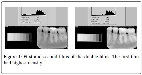
The films were then dried in an incubator with circulating hot air. The radiographs, which had been identified with lead letters before radiographic exposure, were mounted on cards for photodensitometric reading with a Digital densitometer II, model 0.7-424 (Victoreen, Inc.) with a diaphragm aperture of 1 mm. 360 values obtained were recorded on appropriate cards and represented the experimental sample. These original data referred to four groups, i.e., 1st unexpired film, 2nd unexpired film, 1st expired film, and 2nd expired film. Data were analyzed statistically for intragroup comparison unexpired and expired films [6,9,13] i.e., the data were submitted to hyperbolic transformation and a straight line was obtained for the evaluation of radiographic density and contrast values (Figure 1).
In Figure 1 it is possible to see with some effort that the left image (first in the involucre) have more density than the right image.
Results
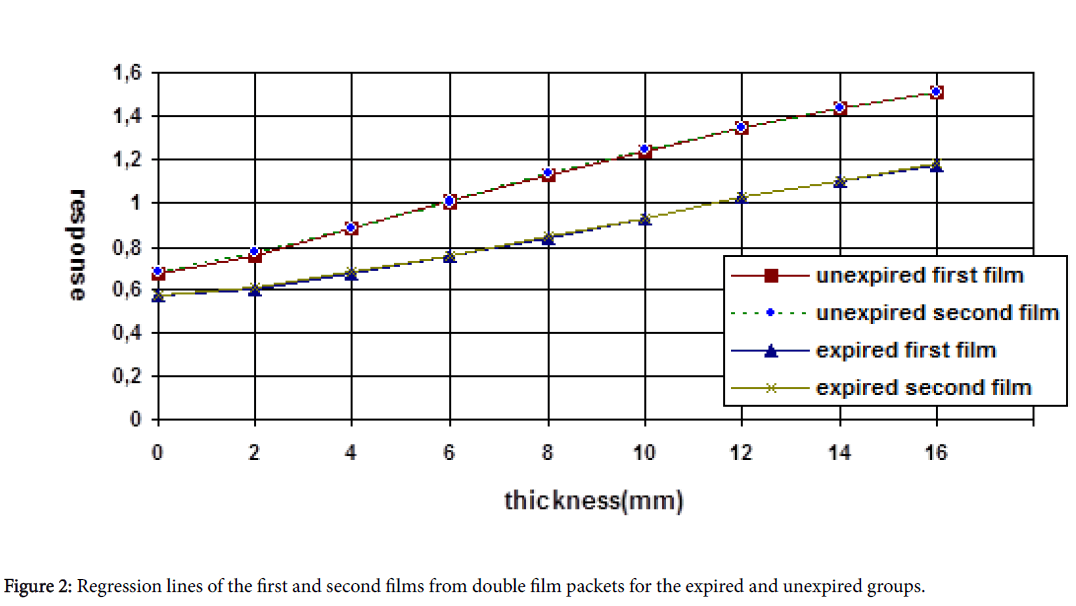
The densitometric measurements of the aluminum step wedge are described in Table 1 for both groups. These measurements are plotted on the graph of Figure 2. In the regression line format. The densitometric readings of the stepwedge images are independent data. Moreover, these data were cumulative since the thickness of each step increased according to a constant additive factor in relation to the previous step. On this basis, the nature of these data required a more adequate statistical analysis for the correct interpretation of the responses [9].
| Mean optic density | |||||||||
|---|---|---|---|---|---|---|---|---|---|
| Thickness of the aluminum step wedge (mm) | |||||||||
| Groups | 0 | 2 | 4 | 6 | 8 | 10 | 12 | 14 | 16 |
| Unexpired film | |||||||||
| first film | 0.68 ± 0.02 | 0.76 ± 0.01 | 0.89 ± 0.01 | 1.01 ± 0.02 | 1.12 ± 0.01 | 1.23 ± 0.02 | 1.34 ± 0.02 | 1.44 ± 0.01 | 1.51 ± 0.02 |
| second film | 0.69 ± 0.01 | 0.78 ± 0.02 | 0.89 ± 0.01 | 1.01 ± 0.01 | 1.13 ± 0.01 | 1.24 ± 0.01 | 1.34 ± 0.01 | 1.44 ± 0.01 | 1.51 ± 0.01 |
| Expired film | |||||||||
| first film | 0.58 ± 0.02 | 0.60 ± 0.01 | 0.68 ± 0.01 | 0.76 ± 0.02 | 0.84 ± 0.02 | 0.93 ± 0.02 | 1.02 ± 0.02 | 1.10 ± 0.03 | 1.17 ± 0.03 |
| second film | 0.58 ± 0.02 | 0.61 ± 0.01 | 0.69 ± 0.02 | 0.76 ± 0.02 | 0.85 ± 0.02 | 0.93 ± 0.02 | 1.02 ± 0.03 | 1.10 ± 0.03 | 1.18 ± 0.03 |
Table 1: Mean (+SD) values of optic density for the first and second radiographic films contained in double film packets for the 10 replications after hyperbolic transformation of the data.
We used the same method initially outline in a previous papers (Watanabe et al., 1994) and it was subsequently improved to permit the simultaneous setting of the characteristic optical density and contrast of a roentgenogram, having in view of the unique mathematical transformation (Table 2). In this way, to analize the experimental data we used the hyperbolic equations that gave excellent “r” values, indicating concordance between the experimental dates and the derived equations.
| "a" Values - Radiographic Density | "b" Values - Radiographic Contrast* | ||||||
|---|---|---|---|---|---|---|---|
| unexpired film* | expired film++ | unexpired film** | expired film++ | ||||
| first film | second film | first film | second film | first film | second film | first film | second film |
| 0.67102 ± 0.0152 | 0.68817 ± 0.0085 | 0.53999 ± 0.0131 | 0.54598 ± 0.0147 | 0.05452 ± 0.0015 | 0.05341 ± 0.0004 | 0.03921 ± 0.0018 | 0.0386 ± 0.0027 |
* p<0.01 and **p<0.05 by the Mann-Whitney U test; ++The Mann-Whitney U test showed statistically identical values.
Table 2: Mean (+SD) values of radiographic density ("a") and contrast ("b") for the experimental groups
So, the hyperbolic transformation of the original data transforms the original hyperbolic curve into a straight line (equation y=a+bx), which permits a much easier interpretation of the radiographic quality of the images [9]. This value can be seen in the Table 3.
| "a" Values - Radiographic Density | "b" Values - Radiographic Contrast* | ||||||
|---|---|---|---|---|---|---|---|
| unexpired film* | expired film++ | unexpired film** | expired film++ | ||||
| first film | second film | first film | second film | first film | second film | first film | second film |
| 0.67704 ± 0.00938 | 0.68693 ± 0.00951 | 0.51593 ± 0.01282 | 0.51743 ± 0.01447 | 0.05406 ± 0.00099 | 0.05345 ± 0.00050 | 0.04137 ± 0.00202 | 0.04145 ± 0.00222 |
*p<0.05 by the Mann-Whitney U test; **A parametric test showed statistically identical values; ++The Mann-Whitney U test showed statistically identical values.
Table 3: Mean (+SD) values of radiographic density ("a") and contrast ("b") for the 10 replications for the 4 experimental groups without considering zero (0) values, i.e., direct film exposure.
Discussion
The expired films were more radiolucent and had much lower contrast values compared to unexpired films. This was probably due to the organic deterioration of the emulsion of the films [9]. Thunthy and Weinberg [10] detected increased radiographic contrast and decreased speed in expired double films packets, but even so indicated their clinical utilization. This difference compared to our findings is due to the fact that individual films in double films packets have different sensitometric patterns.
The four groups of experimental films did not present normality when submitted to the normality test, so that a nonparametric statistical treatment was deemed necessary. The Mann-Whitney U test was then chosen for intragroup comparison. The difference between expired and unexpired films can be clearly seen in Figure 1 and is confirmed by the numerical values presented in Table 2. Also, a significant difference was detected between the first and second film of unexpired duplicating films for radiographic density (p<0.01) and contrast (p<0.05) (Table 2).
The two films from expired double films packets were statistically identical, probably due to the organic deterioration of their emulsion [9,10]. The present results disagree from those reported by Razmus et al. [14] with respect to the quality of duplicating films. It should be pointed out that these investigators only performed clinical analyses using observers. We detected statistically significant differences in radiographic quality image between the 1st and 2nd film of double packets, but only under laboratory conditions. Thus, in a future study we shall determine whether these differences are also detected by the visual acuity of observers, as done by Razmus et al. [14].
Our findings agree with those reported by Sewerin [7], especially when we eliminated the values of direct film exposure (zero). This investigator also detected a significant difference in radiographic density between the 1st and 2nd films in double films packets. This is explained by the fact that when the film is exposed to X-rays, the film located in the anterior part of the packet (1st) acts as a barrier against the X-rays, thus reducing the intensity of the latter when they reach the film located posteriorly (2nd), even when we eliminate the values of direct film exposure (zero), as recommended by Oishi and Parfitt [15]. These direct exposure values are extremely important for radiographic interpretation. For example, when we analyze an interproximal radiograph for the early diagnosis of incipient carious lesions, we perform a visual analysis of the region contrasting dental enamel with the space that surrounds the point of contact of enamel surfaces. This space is an area of direct exposure to X-rays since X-rays cross soft tissue (interdental papilla) only inferiorly. On this basis, we decided to interpret our results using the direct exposure (zero) values of the contrast scale, thus confirming the presence of significant differences in radiographic density (p<0.01) and contrast (p<0.05) between the individual films in unexpired double films packets.
Also Jarvis et al. [11] agree with our findings but author’s found not significant difference in laboratory analysis. On the other hand, the clinical phase reflected the observer’s opinions that image quality varied between front and back image films. The author has judged that the major contributor to differences in detail and definition between front and back films is parallax unsharpness that is an evident geometric aspect.
Araki et al. [8] studied the effects of lead lamina in dental X-ray film Packets on radiographic image quality and two features emerge from these results: (I) the front film has a better image quality than the back film; (II) one of the effects of lead foils is to improve image quality by shielding films from back-scattered radiation. We didn’t study the interference of the lead lamina but we agree with authors. Most textbooks, when describing radiographic films in terms of their packaging, state that double films packets permit the professional to obtain two identical radiographs of the same case because of the packaging of two identical films in the same packet and point out the advantages of these films such as obtaining duplicates for the files with a single patient exposure, the possibility of obtaining images of different densities during radiographic processing which may greatly facilitate the radiographic diagnosis according to the clinical requirements of the professionals. The results of the present study should be taken into consideration when using this type of films. We should point out the necessity to evaluate these results clinically and to consider other exposure factors for analysis.
The results found by Malezan et al. [3], with reference to possible differences que X-ray units with the nominal same kVp may present quite different spectra that it could modify the quality of the image and the dose to the patient will be significantly altered by the spectrum's shape. Expired radiographic films, even kept in refrigerated conditions, have lower quality of the radiographic image in relation to nonoverdue films and thus should not be used, according to our experimental conditions.
Conclusion
• Both unexpired radiographic films from the double packets presented little laboratory differences, but significant differences in radiographic density (p<0.01) and contrast (p<0.05) to each other, when exposed to X-rays under the conditions of the study.
• Both expired radiographic films from the double packets did not present significant differences in radiographic density or contrast to each other, when exposed to X-rays under the conditions of the study.
• The unexpired individual films from the double packets did not differ from each other in terms of radiographic contrast when exposed to X-rays under the experimental conditions of the study and when the direct exposure values were excluded.
• The expired individual films from the double packets showed larger density and less contrast than unexpired films when exposed to X-rays under the present experimental conditions.
• The method proposed for the qualitative and quantitative interpretation of the experimental data obtained with an aluminum step wedge permitted an objective analysis of double films packets both in terms of optic density and contrast.
Acknowledgments
This study was supported by a grant provided by Coordination of Improvement of Higher Education Personnel (CAPES).
References
- Morita MC, Haddad AE, Araujo ME (2010) Perfil Atual e Tendências do Cirurgião-Dentista Brasileiro. Ed Dental Press Int Maringá, 96.
- Beaglehole R, Benzian H, Crail J, Mackay J (2009) The Oral Health Atlas. Paris: FDI World Dental Federation.
- Malezan A, Poletti ME, Tomal A, Watanabe PCA, Albino LD (2015) Spectral reconstruction of dental X-ray tubes using laplace inverse transform of the attenuation curve. Radiat Phys Chem Oxf Engl 116: 278-281.
- Peter T (2014) Image Receptors: An update. International Journal of Innovation and Applied Studies 7: 205-212.
- Wadhwani C, Hess T, Faber T, Piñeyro A, Chen CS (2010) A descriptive study of the radiographic density of implant restorative cements. J Prosthet Dent 103: 295-302.
- Watanabe PCA, Issa JPM, Pardini LC, Monteiro SAC, Benites ABCE (2007) A singular method to compare dental radiographic films used to study maxillofacial structures. Int J Morphol 25: 573-578.
- Sewerin I (1986) Reporting radiographic methods in dental epidemiologic and experimental studies. Community dent oral epidemiol 14: 90-93.
- Araki k, Kand S, Toyofuku F (1993) A study of the effects of lead lamina in dental X-ray film Packets on radiographic image quality. Dentomaxillofac Radiol 22: 179-182.
- Watanabe PCA, Tamburus JR, Maia-Campos G (1989) Influence of storage temperature on the density and radiographic contrast overdue and non-overdue movies. Rev Odontol Univ 8: 137-143.
- Thunthy KH, Weinberg R (1990) Sensitometric comparison of unexpired duplicating films used in dentistry. Oral Surg Oral Med Oral Pathol 69: 374-377.
- Jarvis WD, Pifer RG, Griffin JA, Skidmore AE (1990) Evaluation of image quality in individual films of double film packets. Oral Surg Oral Med Oral Pathol 69: 764-767.
- Manson-Hing LR, Bloxon RM (1985) A stepwedge quality assurance test for machine and processor in dental radiography. J Am dent Assoc 110: 910-913.
- Maia-Campos G, Tamburus JR (1991) A method to evaluated and compare roentgenograms. Braz Dent J 2: 95-102.
- Razmus T, Williamson GF, Bricker SL (1989) Clinical avaliation of individual films from double film packets. J Indiana Dental Assoc 68: 17-21.
- Oishi TT, Parfitt GJ (1976) Effects of varying peak kilovoltage and filtration on diagnostic dental radiographs. J Can Dent Assoc 42: 449-452.
Relevant Topics
- Blepharoplasty
- Bone Anchored Hearing Aids
- Chemical peel
- Cleft Surgery
- Congenital Craniofacial Malformations
- Cosmetic Facial Surgery
- Craniofacial Surgery
- Dental Orofacial Surgery
- Dentoalveolar Surgery
- Head and Neck Reconstruction
- Injectable Cosmetic Treatments
- Lip Reconstruction
- Mandibular Nerve Surgery
- Maxfax Surgery
- Maxillofacial Surgery
- Neck Liposuction
- Oral and Maxillofacial Surgery
- Oral Surgery Surgeon
- Orofacial Surgery Braces
- Pediatric Maxillofacial Surgery
- Rhytidectomy
- Sleep Apnea Orofacial Surgery
- Temporomandibular Joint Disorders
- Upper Jaw Surgery
Recommended Journals
Article Tools
Article Usage
- Total views: 16023
- [From(publication date):
December-2016 - Aug 19, 2025] - Breakdown by view type
- HTML page views : 15044
- PDF downloads : 979


