Research Article Open Access
Relationship of Facial Skin Complexion with Gingiva and Tooth Shade on Smile Attractiveness
Bushra Ghani1*, Rizwan Jouhar2 and Naseer Ahmed31BDS, FCPS-II Dental Resident, Department of Operative Dentistry, Altamash Institute of Dental Medicine, Karachi, Pakistan
2BDS, FCPS, Assistant Professor, Department of Operative Dentistry, Altamash Institute of Dental Medicine, Karachi, Pakistan
3BDS, FCPS, Assistant Professor, Department of Prosthodontics, Altamash Institute of Dental Medicine, Karachi, Pakistan
- *Corresponding Author:
- Dr. Bushra Ghani. BDS
FCPS-II Dental Resident
Department of Operative Dentistry
Altamash Institute of Dental Medicine
2-R Sunset Boulevard
Defence Housing Authority, Karachi-75500, Pakistan
Tel: (+92) 0311-325 9596
E-mail: bushra.shekhani.aidm.edu@gmail.com
Received Date: August 31, 2016; Accepted Date: October 01, 2016; Published Date: October 07, 2016
Citation: Ghani B, Jouhar R, Ahmed N (2016) Relationship of Facial Skin Complexion with Gingiva and Tooth Shade on Smile Attractiveness. J Interdiscipl Med Dent Sci 4: 205. doi:10.4172/2376-032X.1000205
Copyright: © 2016 Ghani B, et al. This is an open-access article distributed under the terms of the Creative Commons Attribution License, which permits unrestricted use, distribution, and reproduction in any medium, provided the original author and source are credited.
Visit for more related articles at JBR Journal of Interdisciplinary Medicine and Dental Science
Abstract
Background: A Smile is the most visible record of the dentist care. There are three important factors that play an important role when planning and designing any fixed or removable aesthetic and functional restoration. They include; Facial skin complexion, gingival tissue pigmentation and Tooth shade respectively. Objective: The objective of this study was to determine a relationship of facial skin complexion with the gingiva and tooth shade on smile attractiveness. Methods: This cross-sectional, analytical study was conducted at the Operative Dentistry Department of Altamash Institute of Dental Medicine, Karachi, Pakistan. It included 250 patients from 18-68 years of age of either gender over a period of 1 year from 15th October 2014 to 15th October 2015 respectively. The Facial skin complexion was determined by the Ideal Balance Quick Stick Makeup shade guide manufactured by (Revlon Foundation Makeup Shade Guide, USA) and was divided into three skin tone groups (Fair, Medium and Dark) respectively. The intensity and location of Gingival tissue pigmentation was determined by Dummett-Oral Pigmentation Index (DOPI). Lastly, the shade of middle one-third of labial surface of maxillary teeth was recorded visually with the help of a Vitapan 3D- Master shade Guide (VITA Zahnfabrik, Bad Sackingen, Germany) under a standardized natural day lightening procedure. Results: The data was analyzed by using the SPSS version 20.0 (Statistical Package for Social Sciences for Windows, SPSS Inc. Chicago). Out of 250 participants; 162 were male and 88 were female participants. The most common shade recorded was 2M1 (14.4%) followed by 3M2 (13.6%), 2L1.5 (10.8%) and 2R1.5 (9.6%). Among the males, the most common shade recorded was 3M2 (18.5%) and among the females the most common shade recorded was 2M1 (21.5%). The Value 2 shade was commonly seen in (45.2%), followed by Value 3; (31.2%), Value 1; (13.2%) and Value 4 (10.1%) of patients; However no shade was recorded for Value 5 shade group. The Medium skin complexion was more commonly seen in 60.4% of male participants and a Fair skin complexion was seen in 60.2% of female participants respectively. Conclusion: The prevalence of gingival tissue pigmentation was more found on the attached gingiva and interdental papilla (class II) and more severe in dark-skinned people and lesser in fair-skinned people. People having a fair skin complexion had a lower tooth shade value with the teeth appearing more darker in color whereas, people having a darker skin complexion had a higher tooth shade value with the teeth appearing more lighter in color. The respective male gender had a lower tooth shade value whereas females had a higher tooth shade value. The aging patients had a darker shade of teeth because of a lower tooth shade value respectively.
Keywords
Facial skin complexion; Gingival tissue pigmentation; Tooth shade; Shade guide
Clinical Relevance
This research study highlights the fact that the Restorative dental practitioners and Prosthodontists need to properly understand and be aware of the relationship of facial skin complexion with the gingival tissue pigmentation and tooth shade, when designing and fabricating any fixed or removable restoration in order to achieve excellence and success in aesthetic and functional parameters of contemporary and modern dentistry.
Introduction
Smile is a facial expression formed by flexing the muscles near both ends of the mouth [1-3]. It is considered as the most important of facial expression and is essential in expressing friendliness, agreement and appreciation [4,5]. The word “AESTHETICS” means beauty or appreciation of beauty; is regularly used in contemporary dentistry to describe restorations and artificial teeth replacements [3,6,7]. In today’s beauty-conscious society, the demand for aesthetic dentistry has considerably been increased in last few years and moreover people are often judged, and therefore judge themselves, by the smile. An attractive smile results in achieving an excellent aesthetics, gain of self-esteem, confidence and an excellent dental and mental health [4]. Proper analysis and understanding of psychology, health, function and aesthetics (PHFA) are the most essential components of smile design that particularly dominate in accomplishing patient’s needs and expectations respectively [2,8].
There are certain important factors that predominantly affect a beautiful and a life-like smile. They include; Facial skin complexion, Gingival tissue pigmentation and Tooth shade respectively. The color of the facial skin serves as a basic guide to the gingival display and Tooth shade [2,5,9]. The facial skin tone (complexion) is essential when planning and designing any fixed or removable restorations of teeth and considering shades of natural teeth [10]. Restorative dental practitioners and Prosthodontists have always faced the challenge of harmonizing facial skin complexion with the tooth shade when fabricating any fixed or removable restoration. It has been suggested that the hue (basic color) of the artificial teeth should harmonize with the patient’s facial skin complexion and moreover, has emphasized on the importance of facial skin complexion (color) as a basic guide in selecting the color of the artificial teeth in Caucasian population.
Many research studies have drawn a valuable attention towards the importance of distributive pattern of tooth shade color in order to achieve an adequate knowledge regarding the aesthetic dentistry. Brien et al. reported that a statistically significant difference exists between gingival to incisal region of the teeth and they are clinically significant when fabricating any aesthetic restoration respectively. A study by Richardson has reported a significant contrast between the facial skin complexion and tooth shade; showing that the individuals with a darker skin complexion tend to have a lighter tooth shade.
In a recent study conducted at New Jercy, it was found that people having a medium to darker facial skin complexion were more likely to have teeth with a higher tooth shade value (lighter shades) where as individuals with a fair or lighter skin complexion tend to have teeth with a lower tooth shade value (darker shades) regardless of their age or gender [4]. Natural teeth are known to possess different shades in their surfaces [2,8,11]. Teeth shade vary with skin color, age and gender [3,10,12]. However, it has been found that the color of natural teeth is influenced by many factors including the advancing age, light, color blindness (Physiological factors), [8,13,14] extrinsic factors including; smoking, betal quid, beverages, alcohol, xerostomia, diet, restorations [3,5] and intrinsic factors including congenital defects of enamel or dentin such as amelogenesis and dentinogenesis imperfecta, hypoplasia, hypocalcification or hypomaturation or environmental factors such as tetracycline staining, traumatic injuries to the teeth, dental caries; affecting the shades of the teeth in vivo respectively [3,5,15,16].
Aging has a significant impact on the tooth shade selection as with the advancing age; [12,17,18] the teeth become darker with a lower tooth shade value (decrease in lightness and increase in yellowness) and less translucent, more opaque since with the advancing age, [19,20] there is more deposition of secondary dentine resulting in a smaller pulp chamber therefore lighter shade of teeth recommended for younger people and darker shades for geriatric patients [10,12,18]. Moreover gender also has a significant impact on the tooth shade selection as many studies have shown that women have lighter and less yellow teeth than men where as men tend to have darker teeth [3,12,14]. An attractive, beautiful life-like smile is perceived by the appearance of gingiva; [9,21] a fibrous mucosa that is surrounding the teeth and covering the coronal portion of alveolar process [11,22]. Dummett [15,23] in 1966 queried that the healthy gingival color varies from pale pink and coral color in Causacians to brownish and blue blackish areas in Asian or African population [11,23,24]. The most frequently pigmented and readily seen pigmented intraoral tissue is the gingiva. The gingival pigmentation is caused by the cells called melanocytes [12,15,25,26]. The melanocytes secrete a non-hemoglobin derived pigment called Melanin [21,25,27] that is primarily responsible for the gingival tissue pigmentation [23,25]. In the past, various research studies [15,23] have advocated the presence of a relationship of facial skin complexion with the gingival tissue pigmentation [27]. In 1975, Ibuski, reported that the color of the gingiva varied with the position of the papillary, marginal and attached gingiva [9].
Many research studies [21,24,28] have emphasized on the significance of a strong relationship of facial skin complexion with the severity of gingival tissue pigmentation, concluding that people having a fair skin complexion tend to have a mild gingival tissue pigmentation where as people having a darker skin complexion tend to have a moderate to severe gingival tissue pigmentation respectively [14,15,29].
The word; “MAKEUP” here refers to the Revlon Foundation Makeup Shade Guide, USA (The Ideal Balance Quick Stick Makeup). The word; “INTENSITY OF GINGIVAL TISSUE PIGMENTATION” here refers to the qualitative evaluation of gingival tissue pigmentation by the Dummett Gupta Oral Pigmentation Index (DOPI). The word; “SHADE” here in this study refers to the shade of middle one-third of the labial surface of maxillary central incisor’s clinical crown. Lastly, the word; “SHADE GUIDE” here refers to the Vitapan 3D- Master Shade Guide (VITA Zahnfabrik, Bad Sackingen, Germany) respectively.
The rationale of this study is to determine a relationship of facial skin complexion with the gingival tissue pigmentation and tooth shade when designing and fabricating any fixed or removable restoration. Moreover, no local or international study has identified this particular relationship as a whole collectively. To our knowledge, our study is the first one that is determining this specific relationship on the basis of age and gender of the participants in Pakistan with a good response rate. This particular research study will definitely prove to be a valuable aid for the Restorative dental practitioners and Prosthodontists, when planning and fabricating any aesthetic or functional restorations for individuals belonging to different age groups, thereby accomplishing patient’s needs and expectations and achieving excellence in aesthetic and functional parameters of modern dentistry.
Materials and Method
This was a cross-sectional, analytical study with a non-probability convenient sampling technique, conducted in the Department of Operative Dentistry at Altamash Institute of Dental Medicine, Karachi (Pakistan), for a duration of 1 year from 15th October 2014 to 15th October 2015 respectively. Our Sample size consisted of 250 patients, who visited the Dental Opd for a routine dental care.
Ethical Considerations and Inclusion and Exclusion Criteria:
Prior to the treatment, an official approval protocol was sought from the Ethical Review Committee of the respective institute before recruitment of patients to the study. Participants were informed about the nature and purpose of the study and a written informed mutual consent was taken from them. The patients were selected according to the inclusion and exclusion criteria after a detailed history taking procedure and a thorough clinical extra and intra oral examination. Confounding variables as well as bias were controlled by strictly following the exclusion criteria. For inclusion criteria, patients of both genders were selected who presented with completely erupted maxillary central incisors and within the age range starting from 18 years up to 68 years, having a good periodontal health and a good oral hygiene, healthy patients with uniformly pigmented and non-mottled gingiva. The exclusion criteria included; patients having any systemic disorders; Diabetes Mellitus, Peutz Jeghers syndrome, Addisons disease, Albright syndrome, Melonoma, Periodontitis or any gingival pathology inducing color changes. Patient’s with permanent maxillary central incisors (right or left) exhibiting any of the following findings were excluded from this study: Dental caries, any type of restorations, endodontic therapy, extrinsic or intrinsic staining (e.g. betal quid chewing, smoking, tetracycline staining, etc.) dental abrasion, attrision, erosion, craze or fracture lines, developmental anomalies like dental fluorosis, orthodontic bands or brackets. Patients who presented with xerostomia, having history of radiation therapy and tooth bleaching procedures. Female patients were excluded from the study who were not willing to remove their lipstick, lip gloss and makeup prior to shade evaluation procedure.
Data Collection Procedure
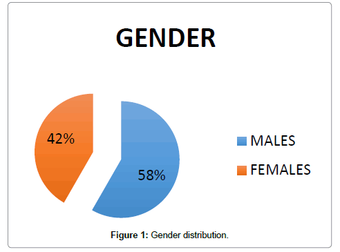
Two hundred and fifty (250) patients visiting the Department of Operative Dentistry for a routine dental care whose permanent maxillary central incisors met the selection criteria mentioned above, were included in this study. In this respective study, 58% were male and 42% were female participants (Figure 1). They were divided into 5 groups of 50 each, depending on their chronological age. The groups included; Group I, 18-27 years, Group II, 29-38 years, Group III, 39-48 years, Group IV, 49 -58 years and lastly Group V, 59-68 years respectively.
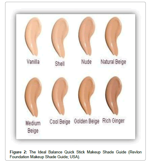
The facial skin complexion/tone was matched by using The Ideal Balance Quick Stick Makeup Shade manufactured by (Revlon Foundation Makeup Shade Guide, USA) (Figure 2). The facial skin complexion was divided into three categories; Fair, Medium and Dark. The distribution of various shades of makeup was arranged into respective facial skin complexion groups. The Vanilla, shell and nude shade group of the Revlon compact correspond to the fair skin group, the natural beige, medium beige and cool beige shades of the compact correspond to the medium skin group and the golden beige, rich ginger shades of the compact correspond to the darker shade group. The shades beyond the deeper shades of the compact were categorized in the darker facial skin complexion group. The skin tone determinations were acquired from the back of the hands and back of the ear skin so that the area was free from the makeup or any residues respectively.
The intensity of physiological gingival tissue pigmentation evaluation in this study was determined by the Dummett-Gupta Oral Pigmentation Index (DOPI) index. The DOPI index represents the assignment of a composite numerical value to the total melanin pigmentation clinically seen on a thorough intra oral examination of gingival tissues. It is represented as follow:
0 = Pink tissue (No gingival pigmentation)
1 = Mild, light brown tissue (Mild gingival pigmentation)
2 = Medium brown or mixed pink or brown tissue (Moderate gingival pigmentation)
3 = Dark brown or blue/black tissue (heavy gingival pigmentation)
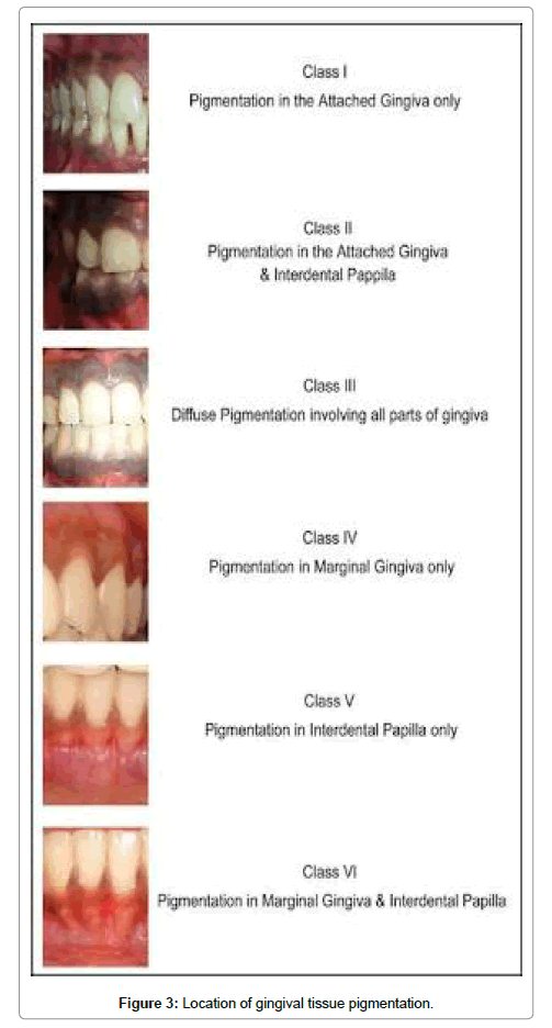
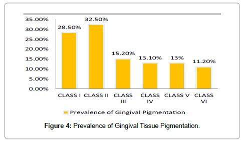
A higher number represents more severity and intensity of gingival tissue pigmentation. A normal color vision and color aptitude of the investigator was tested using the Ishihara color blindness test [30], before the commencement of the gingival tissue examination. The investigator was also seen to be adapted to day light because higher intensity of light available from the day light sources may produce a considerable change of color. The location of gingival tissue pigmentation was evaluated (Figure 3) and the distributive pattern and prevalence of gingival tissue pigmentation was examined throughout the anatomical areas of gingiva (Figure 4). The intraoral photographs were obtained with a digital camera (Canon EOS 5D Mark III Japan) with specific standardized settings for grey, white and black and a centimeter scale with standard lighting and backdrop conditions. The Photographs were reproduced in a computer display and the location of the gingival tissue pigmentation was assessed in the anterior, posterior teeth and in the entire anatomical areas of the gingiva respectively.
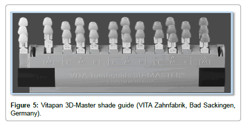
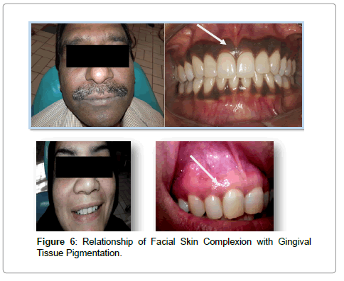
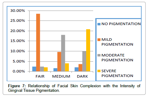
The shade of middle one third of labial surface of permanent right or left maxillary central incisor was taken by using the Vitapan 3D-Master Shade Guide (VITA Zahnfabrik, Bad Sackingen,Germany) (Figure 5). The individual shade numbers as mentioned on the shade guide were noted (For e.g. 3M1, 3M2). The shade readings were taken after passing the Ishihara Color- Blindness test [30]. The investigator was properly educated about the shade guide matching procedure. Care was taken to take the shade readings at the start of the appointment to avoid dehydration. Shades were particularly taken in the morning time for around 2-3 minutes and at least 5 patients were seen by the investigator in a day. A grayish to bluish napkin was draped around the patient to cover any brightened or dark colored clothes. They were asked to remove spectacles or glasses. Female patients were advised to remove the lipstick or lip gloss. The patients were placed in an upright position and their entire gingival tissue was wiped with a 2-2 gauze or cotton to dry the entire area prior to shade selection. They were viewed at eye level of the investigator so that the most color sensitive part of retina was used. Dental unit light was turned off and shades were selected using the artificial light source (Philips Cool Day Light Energy Saver Lamp; 23 W; 6500 K; 50-60 Hz; Philips Co; China) keeping at a distance of 2-3 feet that stimulated natural day light. The relationship of facial skin complexion with the location and intensity of gingival tissue pigmention was evaluated and observed respectively (Figure 6 and 7).
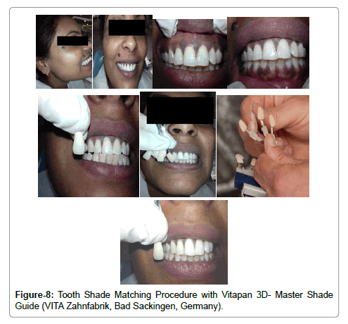
The shade readings were taken from an arm’s length distance to the patient’s mouth. The shade tabs were moistened, and were positioned towards the maxillary central incisor and the investigator concentrated only on the middle one-third of the facial surface to determine the correct shade. First of all the lightness level (Value) was determined (1,2,3,4,5) starting from the darkest group. Next Chroma (Saturation) was selected from the same value group in the middle (M) column on the basis of the determined lightness level and samples were spread out like a fan. One of the three shade samples were selected (L,M,R) and lastly the Hue (Basic Color) was determined by determining whether the natural tooth was more reddish (R-column) or yellowish (L-column) as compare to the shade sample selected (Figure 8) respectively.
Data Analysis
The data was analyzed using the Statistical Package for Social Sciences (SPSS) for Windows, (Version 20.0, SPSS Inc. Chicago). Baseline information on demographics was analyzed using descriptive statistics. Descriptive statistics were calculated for both qualitative and quantitative variables. Independent Sample T-test at 95% confidence level was used to compare the mean of each variable between males and females. Correlation of shades of the teeth with respect to age, gender and facial skin complexion was determined using the by Pearson’s Chisquare test separately for males, females, younger and older age groups. A P-value of < 0.05 was taken as statistically significant.
Results
Our research study comprised of 250 patients. Out of which 162 were males and 88 were females participants respectively (Table 1). Based on a thorough clinical intraoral examination, the severity and location of gingival pigmentation; Six categories of pigmentation were defined. It was seen that gingival tissue pigmentation was more found on the attached gingiva and interdental papilla (Class II) 53.0% in males and 51.1% in females. It was least found on the marginal gingiva and interdental papilla (Class VI) 6% in males and 5.6% in females (Table 2). The relationship of facial skin complexion with the intensity of gingival pigmentation was observed with respect to Dummett-Gupta Oral Pigmentation index (DOPI). It was observed that people having fair or lighter skin complexion had mild gingival pigmentation (28.4%) where as people having darker skin complexion had severe gingival pigmentation (20.8%) (Table 3). However, as such no significant correlation was seen with respect to gender or age. Frequency and Percentages were calculated for the qualitative variables.
| Gender | |||
|---|---|---|---|
| Age group | Male | Female | Total |
| Group I | 38 | 14 | 50 |
| Group II | 32 | 20 | 50 |
| Group III | 38 | 18 | 50 |
| Group IV | 32 | 16 | 50 |
| Group V | 22 | 20 | 50 |
| Total | 162 | 88 | 250 |
Table 1: Patient distribution according to age group and gender.
| Class I | Class II | Class III | Class IV | Class V | Class VI | Total | |
|---|---|---|---|---|---|---|---|
| Male | 32 | 86 | 18 | 12 | 8 | 6 | 162 |
| 19.70% | 53.00% | 11.10% | 7.40% | 4.30% | 4.30% | ||
| Female | 13 | 45 | 10 | 8 | 7 | 5 | 88 |
| 14.70% | 51.10% | 11.30% | 9.00% | 7.90% | 5.60% | ||
| Total | 45 | 51 | 28 | 20 | 15 | 11 | 250 |
| Prevalence | 18% | 52.40% | 11.20% | 8% | 6% | 4.40% | 100% |
Table 2: Prevalence of location of gingival tissue pigmentation.
| Dummetts Oral Pigmentation Index | |||||||
|---|---|---|---|---|---|---|---|
| Frequency | No Pigmentation | Mild Pigmentation | Moderate Pigmentation | Severe Pigmentation | Total | ||
| Fair | 6 | 71 | 6 | 5 | 88 | ||
| 2.40% | 28.40% | 2.40% | 2% | 35.20% | |||
| Medium | 4 | 24 | 45 | 10 | 83 | ||
| 1.60% | 9.60% | 18.00% | 4% | 33.20% | |||
| Dark | 5 | 9 | 13 | 52 | 79 | ||
| 2% | 3.60% | 5.20% | 20.80% | ||||
| Total | 15 | 104 | 64 | 67 | 250 | ||
| 6% | 41.60% | 25.60% | 26.80% | 100% | |||
Table 3: Relationship of facial skin complexion with the intensity of gingival tissue pigmentation.
Patients were equally divided into 5 groups depending on the chronological age. Overall 20 shades were recorded from the Vitapan 3D Master Shade Guide out of 26 shades respectively. 20 shades recorded were present among the male patients and 15 among the female patients. The 6 shades that were not recorded included 4L1.5, 4R1.5, 4R2.5, 5M1, 5M2 and 5M3. The most commons shade recorded was 2M1 (14.4%) followed by 3M2 (13.6%), 2L1.5 (10.8%) and 2R1.5 (9.6%). Among the males most common shade recorded was 3M2 (18.5%), 1M1 (11.7%), 2M1 (10.4%), 2M2 (9.8%) and 2R1.5 (9.2%) and among the females the most common shades recorded were 2M1 (21.5%), 3M1 (18.1%) and 2L1.5 (15.9%) respectively as shown in table 04. Among the participants, “Value 2” shade group of the shade guide was more commonly seen in 113 patients (45.2%), Value 3 (31.2%), Value 1 (13.2%) and Value 4 (10.1%) (Table 4), However no shade was recorded for Value 5 shade group. Value 2 shade group was much more common and out of 250 participants, 62 (31%) were males and 51 (20.4%) were females respectively. Tooth Shade Value 2 was more commonly seen in Group I (66%) and Group II (50%) and Tooth Shade Value 3 was seen in Group III (50%), IV (40%) and V (34%) (Table 5).
| Tooth ShadeValue | Male | Female | Total | Percentage % |
|---|---|---|---|---|
| 1 | 28 (11.2%) | 5 (2%) | 33 | 13.20% |
| 2 | 62 (31%) | 51 (20.4%) | 113 | 45.20% |
| 3 | 50 (25%) | 28 (11.2%) | 78 | 31.20% |
| 4 | 22 (8.8) | 4 (1.6%) | 26 | 10.40% |
| Total | 162 | 88 | 250 | 100% |
Table 4: Patients distribution according to gender and tooth shade value.
| Age Group | |||||||
|---|---|---|---|---|---|---|---|
| Tooth Shade Value | Group I (18-28 years) | Group II (29-38 years) | Group III (39-48 years) | Group IV (49-58 years) | Group V (59-68 years) | Total | Percentage % |
| 1 | 11 | 13 | 5 | 4 | 2 | 35 | 14% |
| 2 | 33 | 25 | 18 | 10 | 15 | 101 | 40.40% |
| 3 | 3 | 10 | 25 | 20 | 17 | 75 | 30% |
| 4 | 3 | 2 | 2 | 16 | 16 | 39 | 15.60% |
| Total | 50 | 50 | 50 | 50 | 50 | 250 | 100% |
Table 5: Patients distribution according to age group and tooth shade value.
With respect to gender, Medium Facial Skin complexion was commonly seen in 123 cases (49.2%). The Medium skin complexion was more commonly found in males 60.4% and a lighter or fair skin complexion was found in females (60.2 %) (Table 6). With respect to Value (Brightness), Value 2 was more commonly seen in fair skin complexion (45%) and (47.1%) in medium skin complexion (Table 7). The independent Sample T-test was applied at 95% confidence level to enhance the study objective. A strong relationship of facial skin complexion with gingival tissue pigmentation and tooth shade with respect to age and gender was significantly seen with regard to this specific research study. This study was highly significant (P-value<0.001) when either the facial skin complexion was correlated with the gender, gingival tissue pigmentation, tooth shade or age of the patient.
| Gender | ||||||
|---|---|---|---|---|---|---|
| Facial Skin Complexion | Male | % | Female | % | Total | % |
| Fair (light) | 49 | 30.2 | 53 | 60.2 | 102 | 40.8 |
| Medium | 98 | 60.4 | 25 | 28.4 | 123 | 49.2 |
| Dark | 15 | 9.2 | 10 | 11.3 | 25 | 10 |
| Total | 162 | 100 | 88 | 100 | 250 | 100 |
Table 6: Patients distribution according to facial skin complexion and gender.
| Facial Skin Complexion | ||||
|---|---|---|---|---|
| Tooth Shade Value | Fair | Medium | Dark | Total |
| 1 | 10 (9.8%) | 14 (11.3%) | 5 (20%) | 29 (11.6) |
| 2 | 46 (45.0%) | 58 (47.1%) | 15(60%) | 119 (47.6) |
| 3 | 38 (37.2%) | 36 (29.2%) | 3 (12%) | 77 (30.8%) |
| 4 | 8 (7.8%) | 15 (12.1%) | 2 (8%) | 25 (20%) |
| Total | 102 | 123 | 25 | 250 |
Table 7: Patients distribution according to facial skin complexion and tooth shade value.
Discussion
To our knowledge, this study is the very first research study that has tried to establish a significant relationship of facial skin complexion with the location and intensity of gingival tissue pigmentation and tooth shade with respect to age and gender. The skin color can be an important indicator of oral mucosal and gingival pigmentation [2,24,25,27]. The hyperpigmentation of the gingiva could be a genetic trait in some populations [22,29]. The incidence of melanin gingival pigmentation is reported to be 0-89% in regard to genetic and secondary factors [12,23,28,31] like smoking habits [26,31]. In the present study, it was noted that majority of participants had melanin pigmentation on the attached gingiva and interdental papilla (class II) respectively. However, this finding was in contrast with a Jewish population [32] study where they found attached gingiva, to be the only most common pigmented anatomic division and in a South African population, where the melanin pigmentation was most frequently found in the interdental papilla [13]. This produces a strong valuable fact that there is racial variation in the pigmentation of the gingiva [26].
This study established the fact that people having a darker skin complexion had heavy gingival melanin pigmentation whereas people having a fair skin complexion had mild gingival melanin pigmentation [15]. These findings are similar to a previous research study carried on in an Indian population [25], where the prevalence of gingival melanin pigmentation increased as the complexion changed to darker shades [27,33]. Apart, from gingival tissue pigmentation, the facial skin complexion shares a valuable relation with tooth shade; when aesthetic parameters of contemporary dentistry are concerned. Currently there are two methods widely used to determine the tooth shade in vivo; One, commercially available in the form of a visual shade [5,17,34] guide and secondly a hi-tech instrument, Spectrophotometer [5]. However visual shade guide remains the most commonly used method [13]. A correct color for a successful aesthetic restoration is a mandatory requirement [26,32,35]. The middle one third region of the tooth was taken as a representative of color shade selection as there is a marked degradation of color changes from the cervical to incisal region of the tooth due to scattered modification of light and enamel translucency [2,8,36]. The age of an individual has shown a marked influence on the tooth shade [3,13,18]. Numerous studies have been reported to acknowledge the respective finding. A clinical study by Jahangiri et al. [4] reported that a considerable relationship exists between the tooth color and age. Teeth tend to become darker with increase age with a lower tooth shade value [4]. Another clinical study reported conducted in Baghdad by Hassan [18] reported that individuals having colors of grey and redgrey increased with the advancing age. Furthermore, Goodkind et al. [36] stated that in late 30s, the teeth tend to start becoming much yellow, with an increase in saturation (Chroma) centrally, decrease in the brightness level of the tooth shade and the cervical portion of the tooth remains the same due to considerable presence of thin enamel. Essan et al. [19] also found that aging has a strong impact on the color shade selection, planning and fabricating any removable prosthesis for completely edentulous geriatric patients. In Japanese population, Hasegawa et al, [8] stated that the value of the teeth decreased in the middle on third portion of the teeth with the advancing age. A recent study by Hartmann et al. [10] has mentioned that with the advancing age, the teeth tend to become darker due to the gradual darkening of the dentin with the age. These results of the respective studies have provided a valuable support to our study that tooth shade is affected by the increase age of the individuals.
This study also draws a valuable attention that the tooth shade can also be influenced by the gender. This finding is supported by a research study conducted by Guo et al. [37] and Essan et al. [19] reporting that gender is strongly associated with the tooth shade, the men are more likely to have a lower tooth shade value with a darker color of teeth and Women tend to have a higher tooth shade value with a lighter color of teeth of the same age group respectively. However one study by Zhu et al. [7] concluded that men and women of the same age group had the same tooth color as that particular research only recorded the tooth color and not the particular CIE lab values and the spectrophotometer only recorded the positive correlation whereas the negative correlation was not specified.
The tooth shade is also influenced by the facial skin complexion (Tone). It was concluded that individuals with darker skin complexion tend to have a higher tooth shade value with a lighter shade of teeth where as individuals having a fair skin complexion tend to have a lower tooth shade value with their teeth appearing more darker. This finding is well supported by Jahangiri et al. [4] who concluded that people having a medium to darker skin complexion have lighter teeth with a higher tooth shade value where as people having a fair skin complexion have darker teeth with a lower tooth shade value. In our research study, Vitapan 3D-Master Shade Guide, a valuable aid has been used to cover the entire range of complete tooth shade matching system and our present study is more valuable since it covers all aspects of tooth shade matching procedures unlike the previous studies by Dummett et al. [15] and Essan et al. [19] who found no satisfactory correlation of facial skin complexion with tooth shade.
A life-like beautiful smile is the most valuable asset for the patient visiting the restorative dental practitioner to achieve an excellent aesthetic restoration [37]. When designing, planning and fabricating any aesthetic or functional restorations or any dental treatment, care should be taken to keep certain important considerations in mind that can surely achieve the desired objective; including Facial skin complexion, age and gender respectively.
Moreover; there is a lack of research and literature regarding this particular aspect; It is suggested that further research studies should be accomplished with different shade guide systems that are commercially available to achieve more reliable shades and to manufacture some innovative shade guides that can represent more local tooth shades and play a valuable role in contributing more and more in future aesthetic zones of contemporary dentistry, thereby accomplishing the goal of achieving patient’s satisfaction and fulfilling patient’s needs and expectations in future.
Limitations of Our Research Study:
The limitations of our research study were that there was an unequal distribution of male and female participants that were selected primarily by a non-probability convenience sampling technique respectively. Moreover, different shade guide systems should be commercially available and manufactured so that more reliable shades can be achieved.
It is recommended that such research studies should be more often conducted with a much larger sample size so that more accurate results can be achieved in future, that can definitely be a highly valuable aid in enhancing the contemporary zones of modern dentistry.
Conclusion
The following conclusions are made from the present respective study conducted, keeping the limitations of our research study in mind :
People having a darker skin complexion have moderate to severe gingival tissue pigmentation and people having a fair/lighter skin complexion have mild gingival tissue pigmentation. The gingival tissue pigmentation is more commonly seen on the attached gingiva and interdental papilla (class-II) and it is less commonly seen on the marginal gingiva.
People having a fair or lighter skin complexion tend to have a darker shade of teeth, having a lower tooth shade value whereas People having a medium or darker skin complexion tend to have a lighter shade of teeth, having a higher tooth shade value. It was reported that with the advancing age, the teeth appear more darker in color as the tooth shade value decreases. The males tend to have a darker tooth color with a lower tooth shade value as compare to the females, having a lighter color of teeth with a higher tooth shade value of the same age group. However, the tooth shade shares a highly strong relationship with respect to gender, age and facial complexion of the patient respectively.
Conflict of Interest
The authors declare that there are no conflicts of interests related to this research study and the authors have no financial affiliations associated with this study.
Author’s Contribution
1Research article study manuscript write up, 1Data collection, 1Methodology, 1Statistical analysis and 1Final Review of the manuscript. However Dr. Bushra Ghani conducted the entire research study under the guidance of Dr. Rizwan Jouhar and Dr. Naseer Ahmed and this research study was accomplished successfully with with their wonderful supervision, appraisal and a strong moral support respectively.
References
- Koirala S (2010) Maximum Esthetics with Minimal intervention. Contemporary Clinical Dentistry 1:59-61.
- Sabherwal RS, Gonzalez J, NainiFB (2009) Assessing the influence of skin color and tooth shade value on perceived smile attractiveness. J Am Dent Assoc 140: 696-705.
- Hassel AJ, Nitchke I, Dreyhaupt J, Wegener I, Rammelsberg P, et al. (2008) Predicting tooth color from facial features and gender: results from a white elderly cohort. J Prosthet Dent 99:101-106.
- Jahangiri L, Reinhardt SB, Mehra RV, Matheson PB (2002) Relationship between tooth shade value and skin color: an observational study. J Prosthet Dent 87:149-152.
- Analoui M, Papkosta E, Cochran M, Matis B (2004) Designing visually optimal shade guides. J Prosthet Dent 92: 371-376.
- Schwabacher WB, Goodkind RJ, Lua MJR (1994) Interdependence of the hue, value and chroma in the middle side of anterior human teeth. J Prosthod 3:188-192.
- Zhu H, Lei Y, Liao N (2001) Color measurements of 1,944 anterior teeth of people in southwest of China-discreption. ZhonghuakouQiang Yi QueZaZhi 36: 285-288.
- Hasegawa A, HYPERLINK "http://linkinghub.elsevier.com/retrieve/pii/S0022391300564749"Ikeda I, Kawaguchi S (2000)Color and translucency of in vivo natural central incisors. J HYPERLINK "http://linkinghub.elsevier.com/retrieve/pii/S0022391300564749"Prosthet Dent 83:418-423.
- Ibusuki M (1975) The color of gingiva studied by visual color matching Part II. Kind, location and personal difference in color of the gingiva. Bulletin Tokyo Medical and Dental University 22:281.
- Hartmann R, Muller F (2004) Clinical studies on the appearance of natural anterior teeth in young and old adults. Gerodontol 21:10-16.
- Dummett CO, Barens G (1967) Pigmentation of oral tissues: a review of literature. Journal of Periodontology 39:369-378.
- Vandhana KL, Savitha B (2005) Thickness of gingiva in association with age, gender and dental arch location. J ClinPeriodontol22: 828-830.
- Gozalo-Diaz D, Johnston WM, Wee AG (2008) Estimating the color of maxillary central incisors based on age and gender. J Prosthet Dent 100: 93-98.
- Hassel AJ, Nitschke I, Dreyhaupt J, Wegener I, Rammelsberg P (2008) Predicting tooth color from facial features and gender:results from a white elderly cohort. J Prosthet Dent 99:101-106.
- Dummett CO, Sakumura JS, Barens G (1980) The relationship of facial skin complexion to oral mucosa pigmentation and tooth HYPERLINK "http://www.thejpd.org/article/0022-3913(80)90207-3/abstract"color. J Prosthet Dent 43:392-396.
- Watts A, Addy M (2001) Tooth discoloration and staining: a review of the literature. Br Dent J 190:309-316.
- California: HYPERLINK "http://cerec-pdf.s3.amazonaws.com/3DMaster%5b1%5d.pdf"Vident Inc. (2005) VidentInc Vita System 3D-Master: the tooth shade system that makes shade matching simple.
- Hassan AK (2000) Effect of age on color of dentition of Baghdad patients. East Mediterr Health J 6:511-513.
- Essan TA, Olusile AO, AkeredoluPA (2006) Factors influencing tooth shade selection for completely edentulous patients. J Contemp Dent Prac 7:80-87.
- Frush JP, Fisher RD (1957) The age factor in dentogenics. J Prosthet Dent 7:5-13.
- Dummett CO (1946) Physiological pigmentation of the oral and cutaneous tissue in the Negro. J Dent Res 25:0.
- Manson JD, Eley BM (2000) Outline of Periodontics. (4thedn), Butterworth Heinemann: Oxford.
- Dummett CO, Gupta OP (1966) The DOPI assessment in gingival pigmentation. Journal of Dental Research 45: 122.
- Krom C, Waas M, Oosterveld P, Koopmans A, Garrett N (2005) The oral pigmentation chart: A clinical adjunct for oral pigmentation in removable prosthesis. International Journal of Prosthodontics 18: 66-70.
- Raut RB, Baretto MA, Mehta FS, Sanjana MK, Shourie KL (1954) Gingival pigmentation: Its incidence amongst the Indian adults. Journal of the Ameican Dental Association 26: 9-10.
- Oluwole D, Elizabeth D (2010) Gingival tissue color related with facial skin and acrylic resin denture base color in a Nigerian population. Afr J Biomed Res 13: 107-111.
- Prasad D, Sunil S, Mishra R, Sheshadri (2005) Treatment of ginigval pigmentation: A case series. Indian J Dent Res 16:171-176.
- Wright SM (1974) Prosthetic reproduction of gingival pigmentation. Br Dent J 136:367-372.
- Aina TO, Wright SM, Pullenwarner E (1978) The reproduction of skin color and texture in facial prosthesis for Negro patients. Journal of Prosthetics and Dentistry 39:74-99.
- (2007) Ishihara color test. Wikipedia, the free encyclopedia [online].
- Dummett CO, Barens G (1971)Oromucosal pigmentation. An updated review. Journal of Periodontology 42: 726-736.
- Gorsky M, Buchner A, Fundoianu-Dayan D, Aviv I (1984) Physiologic pigmentation of the gingival in Israeli Jews of different ethnic origin. Oral Surgery Oral Medicine Oral Pathology 58:506-509.
- Kaur H, Jain S, Sharma R (2010) Duration of reappearance of ginigval melanin pigmentation after surgical removal- A clinical study. Journal of Indian Society of Periodontology 14: 101-105.
- (2002) Dental Shade Guides. J Am Dent Assoc 133:366-367.
- Dunn WJ, Murchison DF, Broome JC (1996)Esthetics: patients’ perceptions of dental attractiveness. J Prosthodon 5:166-171.
- Goodkind RJ, Schwabacher WB (1987) Use of a fiberoptic calorimeter for in vivo color measurements of 2830 anterior teeth. J Prosthet Dent 58:535-542.
- Guo H, Wang F, Feng H, Gou X, Li K, et al. (2000) The investigation of color selection of 4340 cases of ceramic restorations. Hua Xi Kou Qiang Yi XueZaZhi 18:174-177.
Relevant Topics
- Cementogenesis
- Coronal Fractures
- Dental Debonding
- Dental Fear
- Dental Implant
- Dental Malocclusion
- Dental Pulp Capping
- Dental Radiography
- Dental Science
- Dental Surgery
- Dental Trauma
- Dentistry
- Emergency Dental Care
- Forensic Dentistry
- Laser Dentistry
- Leukoplakia
- Occlusion
- Oral Cancer
- Oral Precancer
- Osseointegration
- Pulpotomy
- Tooth Replantation
Recommended Journals
Article Tools
Article Usage
- Total views: 10870
- [From(publication date):
October-2016 - Aug 18, 2025] - Breakdown by view type
- HTML page views : 9799
- PDF downloads : 1071

