Spindle Epithelial Tumour with Thymus-like Differentiation (Settle): A Distinctive Malignant Thyroid Neoplasm: A Case Report
Received: 23-Dec-2015 / Accepted Date: 06-Feb-2016 / Published Date: 11-Feb-2016 DOI: 10.4172/2161-119X.1000222
Abstract
The spindle epithelial tumor with thymus-like element (SETTLE) is a very rare neoplasm related to the thyroid of young individuals. In 1991, Chan and Rosai unified the concept of SETTLE when they described 8 neoplasms situated in the neck and thyroid of children and young adults, previously diagnosed as malignant teratoma of the thyroid, thyroid spindle cell tumor with mucous cysts or thyroid thymoma. SETTLE is a distinct low-grade neoplasm, believed to be derived from branchial pouch or thymic remnants, with only 1 case report showing association with epithelium-lined cysts of possible branchial pouch derivation. It belongs to a group of cervical lesions that includes ectopic cervical thymoma, ectopic hamartomatous thymoma and carcinoma with thymus-like element (CASTLE). This tumor is composed predominantly of spindle and epithelioid cells with glandular or ductular structures lined by a mucinous or respiratory epithelium. In spite of indolent growth, SETTLE may give metastases many years after the diagnosis. Therefore, a long-term follow-up is required. We present one case, the first report from Sion Hospital, of a patient who has benign clinical course and tumoral features suggestive of a myoepithelial differentiation. The clinicopathologic features of the case reports in the literature are also reviewed. This case report showed the first report from our hospital and the possibility of a myofibroblastic differentiation. The SETTLE is among four tumors related to the thyroid, easily differentiated by clinicpathologic correlation. It is a lowgrade malignant neoplasm, with epithelial differentiation confirmed by immunohistochemical. Despite all the descriptions in the literature, this tumor remains with uncertain histogenesis and lacking convincing proof of thymic differentiation or branchial pouch derivation. SETTLE can metastasize a long time after the surgical resection. Because of this, the patients need long-term follow-up, with particular attention to monitoring the lungs.
Keywords: SETTLE, Sarcoma, Thyroid
251312Introduction
The spindle epithelial tumor with thymus-like element (SETTLE) is a very rare neoplasm related to the thyroid of young individuals. In 1991, Chan and Rosai unified the concept of SETTLE when they described 8 neoplasms situated in the neck and thyroid of children and young adults, previously diagnosed as malignant teratoma of the thyroid, thyroid spindle cell tumor with mucous cysts or thyroid thymoma [1]. SETTLE is a distinct low-grade neoplasm, believed to be derived from branchial pouch or thymic remnants, with only 1 case report showing association with epithelium-lined cysts of possible branchial pouch derivation. It belongs to a group of cervical lesions that includes ectopic cervical thymoma, ectopic hamartomatous thymoma and carcinoma with thymus-like element (CASTLE). This tumor is composed predominantly of spindle and epithelioid cells with glandular or ductular structures lined by a mucinous or respiratory epithelium. In spite of indolent growth, SETTLE may give metastases many years after the diagnosis. Therefore, a long-term follow-up is required [2]. We present one case, the first report from Sion Hospital, of a patient who has benign clinical course and tumoral features suggestive of a myoepithelial differentiation. The clinicopathologic features of the case reports in the literature are also reviewed.
Case Report
A 34-year-old man had an enlarged thyroid for all of his adult life. Over a period of 12 weeks before hospitalization, the thyroid was noted to enlarge rapidly. Eighteen years ago, the patient had bilateral seminoma of the testes treated by bilateral orchidectomy and irradiation to the groin area, and had remained tumor free. Physical examination revealed an enlarged and hard thyroid gland, most notably in the right lobe. There were no palpable lymph nodes or any congenital anomaly. A thyroid scan showed a large cold nodule in the right lobe. No other radiological examination was done Laboratory exams showed normal free-T3 free; free-T4 and TSH levels. Systemic investigations did not yield significant findings. The fine needle aspiration (FNA) cytological study performed was inconclusive. Therefore, resection of the mass was suggested and partial thyroidectomy performed 2 months later Neck exploration was performed, revealing that the right lobe of the thyroid was hard and adherent to the right sternothyroid muscle. There was no evidence of nodal metastasis on either side of the neck. The final diagnosis was SETTLE. Despite no other therapy having been performed, at the present moment the patient is alive without evidence of recurrence or metastasis 5 years after the initial treatment (Table 1).
Material and Methods
The FNA material was received in physiological solution, processed routinely and stained with Papanicolaou. The surgical specimen was received in 10% buffered formalin and processed routinely. An immunohistochemical analysis was performed using formalin-fixed, paraffin-embedded tissue sections.
Results
Cytological findings
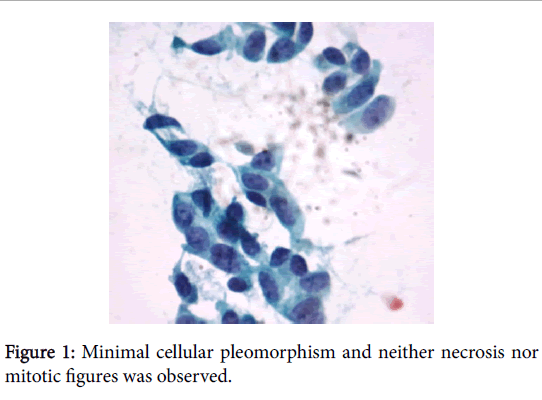
The slide prepared from the FNA revealed a highly cellular aspirate consisting of cohesive or isolated sheets of spindle cells with clusters of polygonal cells with benign features in a background of cyanophilic material which resembles mucin. The spindle cells showed elongated or oval nuclei, finely dispersed chromatin, inconspicuous nucleoli, distinct nuclear membrane and a high nuclear-cytoplasm ratio. There was minimal cellular pleomorphism and neither necrosis nor mitotic figures was observed (Figure 1). Such findings are seen in number of cases illustrated below.
Pathological findings
Macroscopic features: The gross examination of the specimen showed a bosselated, well-circumscribed and firm mass, measuring 2.5 × 2.0 × 1.8 cm. The cut surface was solid, grayishbrown, slightly lobular with residual thyroid at the periphery. There were focal cysts and mucoid areas. Necrosis, calcifications and haemorrhage were observed.
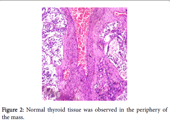
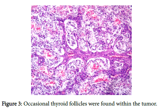
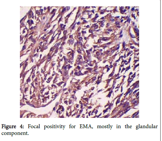
Microscopic Features: Microscopically, the tumor was circumscribed with collagen bands incompletely dividing the tumor in nodules. There was a biphasic pattern composed of a mixture of a predominant component of spindle cells and a minor glandular component with mucinous and respiratory-type epithelium. The mitotic index was low and there were areas with necrosis or hemorrhage. The tumor was highly cellular and formed predominantly by reticulated to compact fascicles of spindle cells with a slightly storiform pattern. The spindle cells displayed scanty eosinophilic cytoplasm and elongated to plump nuclei. The nuclei were minimally pleomorphic with both pointed and blunt ends, distinct nuclear membranes, delicate chromatin and inconspicuous nucleoli. The second component of the tumor consisted mainly of glandular and ductular structures lined by mucinous epithelium and ciliated pseudostratified epithelium with occasional goblet cells. These structures varied from large cystic spaces to smaller structures and cell clusters, merging imperceptibly with the spindle cells. These cells displayed a columnar to cuboidal nuclei. Focal areas with squamous epithelium arising abruptly from the spindle cells were observed. There were interstitial mucin pools, intercellular edema and scant lymphocytes. Normal thyroid tissue was observed in the periphery of the mass, and occasional thyroid follicles were found within the tumor (Figure 2 and 3) Focal positivity for EMA, (Figure 4) mostly in the glandular component, was observed. Calcitotin was negative.
Discussion
SETTLE, a rare primary tumor of the thyroid, is composed of spindle cells of epithelial nature forming fascicles, merging into glandular structures taking the form of tubules, papillae, and cystic spaces. In some cases, cysts or glands lined by mucinous or respiratory epithelium may be present. Rare cases may be predominantly monophasic, with spindle cell predominance. It has been speculated that SETTLE represents a neoplasm arising from brachial pouch remnants or ectopic thymus, showing possible differentiation towards embryonic thymus (thymoblastoma) [3,4]. Up to July, 2010, from the available data in the indexed literature, there were 30 case reports of SETTLE. This rare tumor occurs in young and old individuals with a mean age of 17.9 years at presentation (ranging from 2 to 59 years), with a male-to-female ratio of 1.1. In this case the patient was 74 years old at the time of diagnosis. The generally initial presentation described is a painless mass, within or around the thyroid. In the literature, there are at least 4 patients who had the thyroid mass noticed for four or more years, which suggest a slow tumoral growth. In spite of the disease progression with metastasis development, low mortality rates are observed, demonstrating the indolent course of the tumor. SETTLE may not produce metastasis until many years after the diagnosis (mean of 11 years) and according to case reports with followup information, the overall rate of metastasis is of about 20%, spread is mainly to lung, mediastinum, local lymph node and kidney [2].
Conclusion
In conclusion, this case report showed the first report from our hospital and the possibility of a myofibroblastic differentiation. The SETTLE is among four tumors related to the thyroid, easily differentiated by clinicpathologic correlation. It is a low-grade malignant neoplasm, with epithelial differentiation confirmed by immunohistochemical. Despite all the descriptions in the literature, this tumor remains with uncertain histogenesis and lacking convincing proof of thymic differentiation or branchial pouch derivation. SETTLE can metastasize a long time after the surgical resection. Because of this, the patients need long-term follow-up either radiologically or clinically, with particular attention to monitoring the lungs.
| Diagnosis | Features | Special stain and IHC |
|---|---|---|
| Thymoma | Jigsaw puzzle like lobulation, plump spindle or epithelial cells, which often presents with lymphocytes | CD20++ Tdt+++ thymocytes |
| CASTLE | Malignant tumor resembles thymic carcinoma with lymphoepithelioma like pattern | |
| Teratoma of Thyroid | Mature tissue from all 3 germ layers | |
| Sarcomatoid Anaplastic carcinoma |
Rapid growth, early metastasis, histologically characterized by overt nuclear atypia, necrosis cellular pleomorphism, high mitotic index, vascular invasion, prominent vascularity, giant cells, and wide extrathyroid extension. | Cytokeratin+ CEA + Thyroglobin - |
| Medullary Carcinoma- Spindle cell variant |
Spindle cells, stippled /sand pepper nuclei, granular cytoplasmfibrovascularseptae lack of glandular pattern | Amyloid++ Calcitonin++ Chromogranin++ |
| Synovial Sarcoma | Rapid tumor growth, The spindle cells are usually monomorphic, hyperchromatic, but present a higher mitotic index. The glandular component is well differentiated, but does not display mucinous or goblet cells. | Patchy cytokeratin +/- CD99++ BCL-2++ |
| Ectopic hamartomatousthymoma( BranchialAnalage Mixed Tumor) | Plump spindle cells, delicate fibroblast-like spindled cells, mature adipose tissue, and epithelial elements with squamous, glandular or indeterminate morphology. |
|
| SETTLE | biphasic pattern composed of a predominant component of spindle cells and a minor glandular mucous component, occasionally cystic and collagen stripes |
Congo red - Actin+ Cytokeratin+++ Calcitonin- NSE- |
References
- Rastogi A, Saikia UN, Gupta AK, Bhansali A (2012) Recurrent Thyroid Nodule: Spindle Epithelial Tumor with Thymus Like Differentiation (SETTLE). Indian Pediatrics 49: 482-484.
- Filho LAM, Bordallo MA, Pessoa CH, Corbo R, Bulzico, et al. (2010) Thyroid spindle epithelial tumor with thymus-like differentiation (SETTLE): case report and review. Arq Bras EndocrinolMetab 7: 657-662.
- Cheuk W, Jacobson AA, Chan JK (2000) Spindle Epithelial Tumor with Thymus-Like Differentiation(SETTLE): A Distinctive Malignant Thyroid Neoplasm with Significant Metastatic Potential. Mod Pathol 10: 1150-1155.
- Chan JK, Rosai J (1991) Tumors of the neck showing thymic or related branchial pouch differentiation: A unifying concept. Hum Pathol 22: 349-367.
- Chetty R, Goetsch S, Nayler S, Cooper K (1998) Spindle epithelial tumour with thymus-like element (SETTLE): The predominantly monophasic variant. Histopathology 33: 71-74.
- Abrosimov AY, LiVolsi VA (2005) Spindle epithelial tumor with thymus-like differentiation (SETTLE) of the thyroid with neck lymph node metastasis: a case report. EndocrPathol 16: 139-143.
Citation: Majethia NK (2016) Spindle Epithelial Tumour with Thymus-like Differentiation (Settle): A Distinctive Malignant Thyroid Neoplasm: A Case Report. Otolaryngol (Sunnyvale) 6:222. DOI: 10.4172/2161-119X.1000222
Copyright: © 2016 Majethia NK et al. This is an open-access article distributed under the terms of the Creative Commons Attribution License, which permits unrestricted use, distribution, and reproduction in any medium, provided the original author and source are credited.
Select your language of interest to view the total content in your interested language
Share This Article
Recommended Journals
Open Access Journals
Article Tools
Article Usage
- Total views: 10824
- [From(publication date): 2-2016 - Jul 17, 2025]
- Breakdown by view type
- HTML page views: 9950
- PDF downloads: 874


