State of the Art and Future Trends on the Measurements of the Patient’s Ear in the Audiology and Otologic Anaplastology: Implementation of New Technologies of “3D Inside-the-Canal Auditory Scanners”
Received: 09-May-2019 / Accepted Date: 13-May-2019 / Published Date: 20-May-2019 DOI: 10.4172/2161-119X.1000369
The arrival of 3D scanners to the sanitary prosthesis is not new, it has been used in different fields (audiology, dentistry, etc). However, not until recently there has been a refinement of the scanners to use inside the ear canals in otology and this is stablishing a new era in hearing aids. Guild, which hasn´t brought no major changes in the non-electronic aspects of hearing aids for a long time.
Nowadays, the appearance of inside-duct scanners, the fourth revolution [1] occurs among the manufacturers of hearing aids. The reproduction of the duct is now much more accurate, more precise, and it is performed more conveniently for both, the patient and the technician.
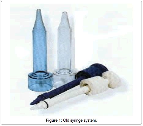
Those who fitted hearing aids before 1988 [2] will remember the handmade molds of those years. In which the molds were only acrylic type, and the measurement was made with two components: a base paste (it was dosed with a small spoon) and a catalyst cream, which was mixed in different proportions and it was then injected by syringe (Figure 1). The Egger A/B two-component formats did not exist yet. This issue has changed a lot, from those first XP peritimpanics that philips launched in 1991 (which, by the way, now returns to the scene of the manufacturers by the hand of DEMANT), in which we made the ear molds with the help of the microscope because the silicone was liquid, instead of dense and viscous as it is nowadays.
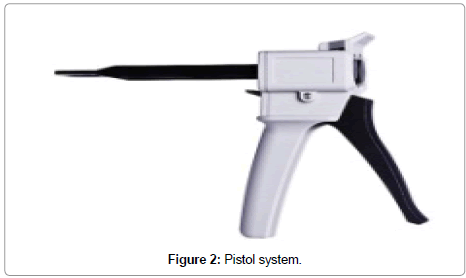
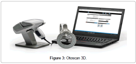
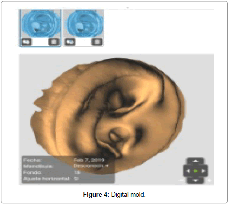
Some years later, our way of reproducing the mold inside the auditory canal changed radically. The “Invisa revolution” and the first Completely-in-the-canal (CIC) came, the arrival of the injection systems with pistols (Figure 2) and the cartridges with automatic automixing cannulas, the micro-molds and Receiver in the ear (RITE). Nowadays, 3D digital scanners (Figure 3) allow us to send the result to the laboratory through the internet (Figure 4).
In the last years several models and prototypes of auditory scanners for use within the ear canal have been disclosed, but we did not obtain good results. Because of this, professionals usually choose to copy the patient’s ear by taking out a silicone mold and then scan that piece externally with a table scanner to obtain a digital file that will be used to manufacture the final mold according to the patient’s exact measures.
Nowadays, digitalization redefines the way we copy ears. The arrival of the new generation of hearing scanners (Otoscan type) represents an advance in technology and also in our daily work in the hearing centers.
Now we take measurements of the patient by scanning his ear canal and three minutes later we are already talking by videoconference with the manufacturing lab. Both the audiologist and the laboratory technician can see the images at the same time to discuss which would be the best way to design the ear mold for our patient. We thus achieve the optimization of manufacturing times.
We access to the smallest details of the internal shape of the ear canal and we can see it easily in 3D models, for the best adaptation of the mold to the anatomy.
We customize our designs much more and improve the final quality of hearing aid fitting.
We reduce manufacturing time, because we save days by sending the package containing the silicone to the laboratory.
We also save money from the cost of materials, shipping and production. And we also help the environment and ecological awareness, because we avoid CO2 from transport and thus improves the carbon footprint of our professional activity (let’s review our environment the international organization of standardization (ISO) 14001 protocols). Neither have we generated the waste from the silicone molds. The whole process is more ecologically efficient and helps sustainability.
The digital files of the images are stored in secure cloud servers, avoiding trips of the patient to take new impressions of the ear, (silicone molds of the analogic audiology deteriorate and they can only be stored for a few months).
On the other hand, the ear canal scanners reproduce it in an identical way, avoiding the modifications that silicone produce, due to the pressure of the silicone paste on the soft area of the ear.
The 3D scanner is less invasive, more comfortable for the patient and also extremely accurate and precise. This is an advantage in elderly patients, with loose tissues, because we take the impression with the tissue in its original position.
Nowadays, working with otoscan has left behind traditional/ conventional silicone impressions, which have more limitations to reach deep areas of the canal, and involve a greater risk. The digital scanner is a tool which helps us making faster, safer and more accurate prints.
Finally, the 3D scanner gives customers a new perspective on the decision to wear hearing aids. As professionals, it is very important to show our patients in real time what their ear canal is like and what their hearing aid will look like.
At this point, let’s also make a brief reminder of an area almost always forgotten: Auditory anaplastology.
Until now, the design of the models for the cases of injuries of the ear was handmade; we had to design the patient’s ear in a very analogic and handmade way. Sometimes, when you were not able to measure it, you had to invent it. Whereas now, we can reproduce ears on the Personal computer (PC) screen and sculpt the anatomy of the user.
Egger®, Philips XP® and INVISA® are registered trademarks.
References
- Barbero A (2018) The fourth Audio-Revolution (The arrival of the CIC –was it the first-, the digital -the second-, the RITE -the third-, and the 3D scanners -the fourth-)", presented at the AUDIOCENTER Summer School, Digital Audio Forum.
- Afers de Comunicació Visual SA (2002) 1988, The year Audiocenter was born. ACV Editors, Barcelona-ESPAÑA, Spain.
Citation: Barbero A (2019) State of the Art and Future Trends on the Measurements of the Patient’s Ear in the Audiology and Otologic Anaplastology: Implementation of New Technologies of “3D Inside-the-Canal Auditory Scanners”. Otolaryngol (Sunnyvale) 9: 369. DOI: 10.4172/2161-119X.1000369
Copyright: © 2019 Barbero A. This is an open-access article distributed under the terms of the Creative Commons Attribution License, which permits unrestricted use, distribution, and reproduction in any medium, provided the original author and source are credited.
Select your language of interest to view the total content in your interested language
Share This Article
Recommended Journals
Open Access Journals
Article Tools
Article Usage
- Total views: 3492
- [From(publication date): 0-2019 - Dec 09, 2025]
- Breakdown by view type
- HTML page views: 2617
- PDF downloads: 875


