Surgical to Interim–Chairside Management of Hemimaxillectomy Patient
Received: 02-Feb-2016 / Accepted Date: 09-Feb-2016 / Published Date: 16-Feb-2017 DOI: 10.4172/2161-119X.1000289
Abstract
Purpose: Fabrication of Interim obturator immediately after surgical procedure may be difficult for the patient due to pain and associated trauma. Method: Use of existing surgical obturator as an impression tray and modifying it into interim obturator is simple and quick procedure Conclusion: Reducing the agony of the patient, maintaining the function and improving the quality of the patient is the primary goal of maxillofacial prosthodontist which is achieved by the technique explained in this article.
Introduction
The principal goal of rehabilitating maxillofacial defects is to reduce the disease and to improve the quality of life for the individuals [1]. Prosthetic intervention should start from the time of surgical resection and will be essential for rest of the patients life [2]. The patient requires immediate care to create an anatomic barrier between the cavities [3,4]. Surgical obturator is created by arbitrary scraping of the cast and it is essential to change it once the surgical pack is removed to preserve the fullness of cheek bone. But, making a new impression with full extension into the depth of surgical site may not be possible on the day of surgical pack removal due to limited mouth opening and associated pain due to surgical trauma. Hence this article presents with technique of converting surgical obturator as an custom impression tray followed by fabrication of closed hollow bulb obturator.
Case Report
A 49 year old female patient was referred from Department of Oral Maxillofacial Surgery. The patient had history of non-healing ulcer in right palatal gingiva adjacent to 16 of the size 1cm × 1 cm diameter and was diagnosed to be squamous cell carcinoma involving floor of maxillary sinus. The patient was planned for hemimaxillectomy and was referred to Department of Oral Maxillofacial Prosthodontics for preoperative surgical obturator fabrication. After surgical procedure the surgical site was wired with the obturator. On the 10th day of surgery, the patient reported back with surgical obturator and the surgical pack. To fabricate the prosthesis to fit the surgical site which would prevent the contraction of surgical site and reduce patient trauma the following methodology was used.
Materials and Method
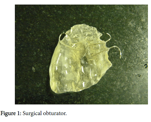
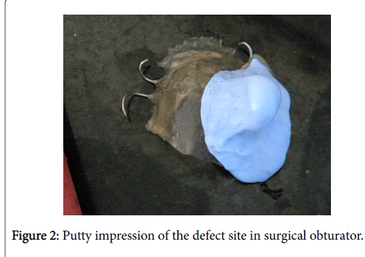
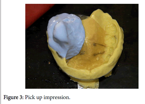
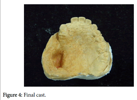
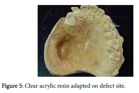
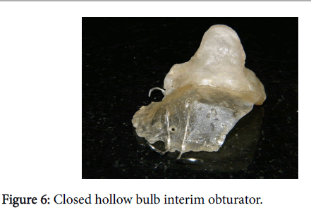
On the day of removal of surgical pack, disinfect the surgical obturator thoroughly (Figure 1) and check for the fit. Apply tray adhesive on the tissue surface of the obturator, and place the putty mix of addition silicone over the defect site in the obturator plate. Make impression of the defect site by placing the obturator in its position on the maxillary arch (Figure 2). Pick up the obturator from the maxillary arch using alginate impression on a perforated stock tray (Figure 3). Apply vaseline over the tissue surface of obturator and pour the cast. Remove the Surgical obturator and paint separating medium on the cast (Figure 4). Adapt the autopolymerising clear acrylic resin on the depth and borders of the defect only, creating a hollow bulb (Figure 5) and place back the surgical obturator on the cast in its previous position. Once set, closed hollow bulb and the plate comes as one unit (Figure 6). Finish, polish and insert the closed hollow interim obturator.
Discussion
Impression procedure after surgical excision is always been traumatic to the patient. Usage of stock impression tray may not well extend into the surgical site and repeated impression will be agonising the patient existing condition. Surgical obturator could be used as custom impression tray as well as could be converted into interim obturator in minimal time, serving to prevent contraction of the soft tissues to maintain facial esthetics [5]. Weight of denture and gravity would affect the retention of maxillary denture [6] and hence closed hollow bulb obturator mentioned in this article would be time effective, simple in fabrication and less traumatic technique.
Conclusion
The conventional impression method could be traumatic to the patient especially when they have reported after a recent surgery. This method described helps in reducing patient trauma and maintains the facial profile and function.
Conflict of Interest
There is no conflict of interest among the authors.
References
- Lethaus B, Lie N, de Beer F, Kessler P, de Baat C, et al. (2010) Surgical and prosthetic reconsiderations in patients with maxillectomy. J Oral Rehabil 37: 138-142.
- Taylor T D (2000) Clinical maxillofacial prosthetics. Quintessence Publishing Co Inc. pp: 85-102.
- Beumer J, Curtis TA, Marunick MT (1996) Maxillofacial rehabilitation prosthodontic and surgical considerations. Journal of Oral and Maxillofacial surgery 55: 786.
- Aramany MA (1978) Basic principles of obturator design for partially edentulous patients: Part II: Design principles. J Prosthet Dent 40: 656–662.
- Lapointe HJ, Lampe HB, Taylor SM (1996) Comparison of maxillectomy patients with immediate versus delayed obturator prosthesis placement. J Otolaryngol 25: 308-312.
- King GE, Gay WD (1979) Application of various removable partial denture design concepts to a maxillary obturator prosthesis. J Prosthet Dent 41: 316-318.
Citation: Raza FB, Choudhary M, Kumar A, Inbarajan A, Kumar M, et al. (2017) Surgical to Interim–Chairside Management of Hemimaxillectomy Patient. Otolaryngol (Sunnyvale) 7:289. DOI: 10.4172/2161-119X.1000289
Copyright: © 2017 Raza FB, et al. This is an open-access article distributed under the terms of the Creative Commons Attribution License, which permits unrestricted use, distribution, and reproduction in any medium, provided the original author and source are credited
Select your language of interest to view the total content in your interested language
Share This Article
Recommended Journals
Open Access Journals
Article Tools
Article Usage
- Total views: 4883
- [From(publication date): 0-2017 - Aug 29, 2025]
- Breakdown by view type
- HTML page views: 3913
- PDF downloads: 970

