Research Article Open Access
Water Flow from Different Solutions During Perfusion of the Functioning Portion of the Small Intestine of the Rats In vivo
Olha V Storchylo*Odessa National Medical University, Medical Chemistry Department, Odessa, Ukraine
- *Corresponding Author:
- Olha V Storchylo
Odessa National Medical University
Medical Chemistry Department, Odessa
Uspenskaya str. 67/69, apt.27, 65012, Ukraine
Tel: 380633085029
E-mail: olha.storchylo@ukr.net
Received date: December 17, 2016; Accepted date: March 17, 2017; Published date: March 24, 2017
Citation: Storchylo OV (2017) Water Flow from Different Solutions During Perfusion of the Functioning Portion of the Small Intestine of the Rats In vivo. J Gastrointest Dig Syst 7:493. doi: 10.4172/2161-069X.1000493
Copyright: © 2017 Storchylo OV. This is an open-access article distributed under the terms of the Creative Commons Attribution License, which permits unrestricted use, distribution, and reproduction in any medium, provided the original author and source are credited.
Visit for more related articles at Journal of Gastrointestinal & Digestive System
Abstract
Objective: The study of the water flow during perfusion of the functioning portion of the small intestine of male rats in vivo by Ringer solution, glucose and glycine. Methods: The experiments were performed on the functioning portion of the small intestine of the rats in the chronic experiments in vivo without narcosis, pain, stress and operation trauma, formed according method. The concentration of PEG was determined based on modified method colorimetrically on CFC-2MP, λ=465nm. The concentration of glycine was determined using method colorimetrically on photoelectrocolorimeter–CFC-2MP, λ=540 nm. The concentration of glucose was determined colorimetrically on photoelectrocolorimeter–CFC-2MP, λ=625 nm. The concentration of bile was determined colorimetrically on CFC-2MP, λ=480 nm. Results: There were no found the dilution of the perfusate with physiological fluids. There were detected an absorption of water from the Ringer solution, from the 25 mmol/L solutions of glucose or glycine, prepared on the Ringer solution. Water absorption from the Ringer's solution was more than from the solutions of 25 mmol/L glycine or glucose, prepared on the same Ringer's solution. It was shown the periodic character of the secretion of bile into the living small intestine of the rats under physiological conditions. Conclusion: Comparison of water flows in the unclosed functioning portion of the small intestine during the thereof perfusion by solutions of Ringer, glucose, or of equimolar concentration glycine showed that in all cases, regardless of the type of the substrate and presence of primordial physiological fluids in the intestine, water absorption is observed from the perfusion solution. Thus, it shows a high rate of water absorption in the small intestine, exceeding the dilution of the perfusion solution.
Keywords
Perfusion; Small intestine; Physiological condition; In vivo ; Absorption; Water; Glucose; Glycine
Introduction
Experimental studies of processes in biological systems typically involve the evaluation of various concentrations of liquid components [1-4]. Changing of the concentration of these components can occur not only due to the transformation and movement of solutes, but also due to the change of the volume of the solvent. It should be borne in mind that the change in the experimental conditions also can affect the content of the solvent (water) in the solution. Therefore, the use of a new method for studying of the functions of small intestine requires an assessment of the water balance in the small intestine during absorption of various nutrients. The value of the study of water flows in the functioning of the small intestine area is particularly high, given the openness of it, as well as the secretion and absorption of water in the intestine.
The following factors and conditions that can affect water balance have been chosen for the experimental work based on the literature analysis:
1) Dynamics of the aqueous composition of the perfusate solution. It reflects the stability of parameter during the experiment (about 60 minutes or more), and the role of the primordial content of the small intestine in absorption, and entering of the secrets of endocrine glands into the intestinal cavity. Dynamics of solvent absorption (especially in the presence of substrate) indirectly reflects the reaction of the loading (temporary or substrate) on the researched portion of intestine.
2) The presence of transported substrates in the intestine. The substrate is able to change the level of functioning of the enterocytes, and also to stimulate the water flow, which conjugates with a substrate transport.
Materials and Methods
The experiments were performed on male rats of Vistar breed weighted 170-180 g that were held out on the standard ration of vivarium and were not fed for 18-24 hours prior to the experiment. There were used rats with functioning part of the small intestine. The functioning portion was prepared according the method described by us previously [5]. 4-5 days after the operation the animals were perfused by peristaltic pump "Zalimp"(Poland). Velocity of perfusion was 0.6 ml/min. For the perfusion we used 25 mmol/L solution of glycine and 25 mmol/L solution of glucose on the Ringer solution (pH=7.4, temperature of the perfusion solution=37°C). We added an unabsorbed marker polyethylene glycol (PEG–400) to the perfusion solution to control possible dilution of perfusion solution with the liquids of digestive tract (saliva, gastric, intestinal and pancreatic juices, bile or reflux from the next part of intestine). The concentration of PEG was determined based on modified method [6] colorimetrically on CFC-2MP, λ=465 nm. The concentration of glycine was determined using method described in ref. [7] colorimetrically on photoelectrocolorimeter–CFC-2MP, λ=540 nm. The concentration of glucose was determined using method described in ref. [8] colorimetrically on photoelectrocolorimeter–CFC-2MP, λ=625 nm. The concentration of bile was determined colorimetrically on CFC-2MP, λ=480 nm.
All experiments were conducted in accordance with scientific/ practical recommendations regarding animal care and work with them [9] and in compliance with the positions of "European convention about defense of the vertebrates used for experimental and scientific aims”. The statistical processing of the obtained data was conducted using “Primer Biostatistics” software.
Results
Absorption of water from the Ringer's solution
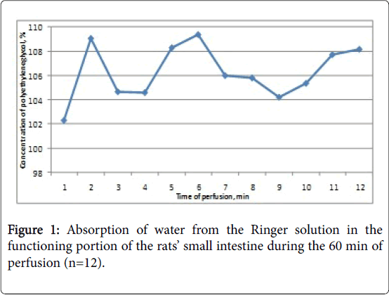
In the first stage of investigation of water absorption Ringer's solution was researched, since in this case to a certain extent eliminated role of ion fluxes. As shown in Figure 1, in the most of the samples of the flowing perfusate during the perfusion takes place the concentration of the perfusion solution, indicating water absorption. In the first sample, corresponding to 5 minutes of perfusion, the concentration of PEG is the minimum for the entire period of perfusion (102.29 ± 4.56%). Apparently, this is due to the presence in the test portion of intestine in the start of perfusion a certain amount of body fluids, which are inconsequential in subsequent samples.
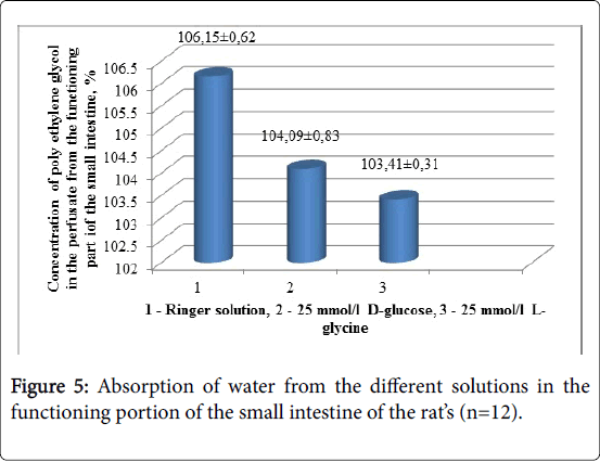
As shown in Figure 1, during the perfusion occurs a concentration of the perfusion solution-this indicates that the water absorption in the intestine prevails over the diluting of perfusate by the physiological fluids. In fact, on average over 60 minutes of perfusion the concentration of PEG in the perfusate from the functioning portion of the small intestine is 106.2 ± 0.6% (p<0.003, n=12). The undulating nature of the curve seems to reflect a periodic delivery of digestive secretions into the analyzed gut fragment. However, this does not result in a significant dilution of the perfusate, since the concentration of PEG in the flowing perfusate does not decrease even up to 100%- index, which corresponds to the concentration of PEG in the perfusion solution at the inlet of the analyzed fragment of intestine. It should be noted that the concentration of PEG in the flowing from the functioning portion perfusate varied within 1 hour of perfusion within 7%. The stability of the water flow over a sufficiently long perfusion combines with substantial variability of this parameter in different experiments: in different animals used in experiments in the same time, the concentrations of PEG in the flowing perfusate were close.
Apparently, the water flow rate is not so much the individual animal feature as an indicator of its functional state. It should also be noted that the experiments conducted on various animals at intervals nearly 1.5 years, gave very similar results. This fact demonstrates the high accuracy of the method of perfusion of the functioning portion of the small intestine of rats and the extremely stable basic properties of intestine under physiological conditions.
Absorption of water from the solution of 25 mmol/L glucose
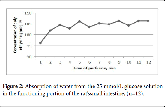
During the perfusion of the functioning portion of the small intestine by the solution of 25 mmol/L of glucose for 60 minutes, the middle concentration of PEG in the perfusate is 104.09 ± 0.83% (Figure 2).
As shown in Figure 2, in the first 10 minutes of perfusion the perfusion solution is diluted, but in subsequent samples is its concentration. If we compare these data with the results of water absorption from the Ringer's solution, we can see that the two curves are virtually close, except for the first 10 minutes of perfusion.
Apparently, removing of the bodily fluids from the functioning fragment of the small intestine during the perfusion with a solution of glucose is slower than during its perfusion with Ringer's solution.
Absorption of water from a solution of 25 mmol/L glycine
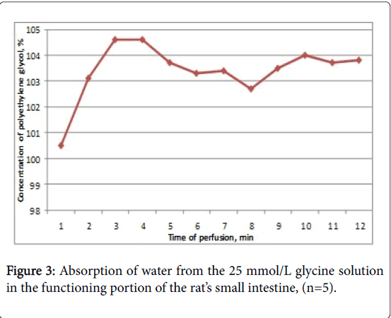
During the perfusion of the functioning portion with the 25 mmol/L solution of glycine for 60 min perfusion the concentration of PEG ranges from 100.50 ± 1.0% in the 1st sample (first 5 minutes of perfusion) to 104.60 ± 0.73 in the 4th sample, which corresponds to the 20th minute of perfusion.
The average concentration of PEG in the perfusate in 1 hour is 103.41 ± 0.31%, and in fact, the curve which explains this process, is the smoothest from all presented in Figure 3.
Secretion of bile into the lumen of the functioning portion of the small intestine of the rats
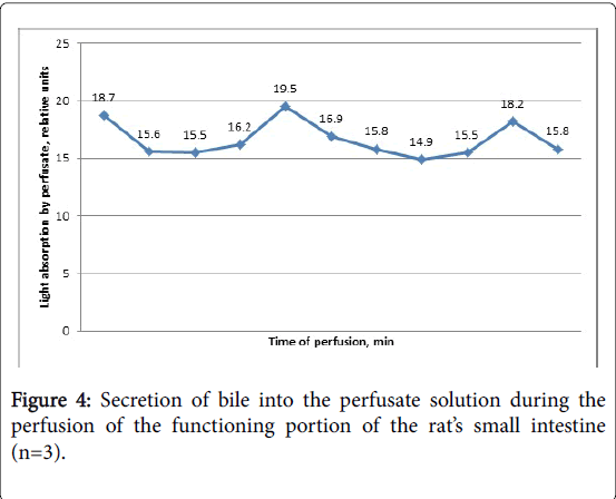
Secretion of the physiological fluids into the small intestine during the perfusion of the functioning portion with Ringer solution was observed by the color of perfusate by bile (Figure 4). The first sample of perfusate contains the maximum concentration of bile, then it declines over time and again rises to the 25th and then in the 50th minute of perfusion. However, if we compare the curves in Figures 1 and 5, it appears that the flow of bile into the perfusion solution has no significant effect on the absorption of water from the Ringer's solution.
Discussion
Normally, in the small intestine there is a certain amount of liquid from adjacent sections, and secreted by the gut. Also here there is a significant absorption of water, and in the proximal fragment this process is more intense than in the rest of the intestine [3,10]. The absorption of water accompanies the transport of monosaccharides (mainly glucose) [4] and amino acids [3,11]. Water absorption in the gut can be of different nature: the transport of water together with the ions, the absorption of water by osmotic pressure of chyme, influenced by various humoral factors or changes in the functional state. The great important in the assessment of the functional activity of the intestine is the study of unstirred aqueous layer near the brush border of enterocytes [12,13]. Therefore, a serious attention is paid to water balance measurement. Accounting water flows can be conducted using the volume-weighted techniques or isotopic labels using no absorbable markers. Isotope methods are useful for determining the unidirectional flow; volume-weighted methods are used only in closed systems. As used in our experiments polyethylene glycol is used as a marker from the 70’s of the last century [14-16]. There is evidence that a small part of the polyethylene glycol can be absorbed in the intestine perfusion [17], but it concerns the polyethylene glycol of low molecular weight (polyethylene glycol 400).
Conclusion
Comparison of water flows in the unclosed functioning portion of the small intestine during the thereof perfusion with solutions of Ringer, glucose, or of equimolar concentration glycine showed that in all cases, regardless of the type of the substrate, water absorption is observed from the perfusion solution. Thus, it shows a high rate of water absorption in the small intestine, exceeding the dilution of the perfusion solution.
References
- Pappenheimer JR (1990) Paracellular intestinal absorption of glucose, creatinine and mannitol in normal animals: relation to body size. Am J Physiol 259: 290-299.
- Gruzdkov AA, Gromova LV, DmitrievaYuV, Alekseeva AS (2015)Free consumption of glucose solution by rats as a criterion for evaluation its absorption in the small intestine. (experimental study and mathematical modeling) Ross FiziolZhIm I M Sechenova101: 708-720.
- Metelsky ST (2009) Physiological mechanisms of intestinal absorption. The main groups of substances. Russian Journal of Gastroenterology, Hepatology and Coloproctology 19: 55-61.
- Zeuthen T, Gorraitz E, Her K, Loo DD, Wright EM (2016) Structural and functional significance of water permeation through cotransporters. ProcNatlAcadSci USA 113: 6887-6894.
- Storchilo OV (2015) Absorption of Glycine in the Small Intestine of Rats underPhysiological Condition. J GastrointestDigSyst 5: 308.
- Malawer SJ, Powell DW (1967) An improved turbidimetric analysis of polyethylene glycol utilizing an emulsifier. Gastroenterology 53: 250-256.
- Ugolev AM, Timofeeva NM (1969) Researching of the peptidase activity. Investigation of the digestive apparatus in the human. Leningrad: Science: 171-178.
- Scott TA, Melvin EH(1953) The determination of hexoses with antrone. AnalytChem25: 1656 -1658.
- Kozhemyakin YM, Chromov YM, Filonenko MA, Saifetdinova GA (2002) Scientifically practical recommendations for the maintenance of the laboratory animals and work with them. Kiev: Avicenna 155.
- Fordtran JS, Ingelfinger FJ (1968) Absorption of water, electrolytes and sugars from the human gut. Handbook of Physiology 3: 1457-1490.
- Powell DW, Malawer SJ (1968) The relationship between water and solute transport from isoosmotic solution by rat intestine in vivo. Amer J Physiol 215: 49-55.
- Winne D (1977) Correction of apparementMichaelis constant, biased by unstirred layer, if a passive transport component is present. BiochimBiophysActa464: 118-126.
- Pappenheimer J R (2001) Role of pre-epithelial «unstirred» layers in absorption of nutrients from the human jejunum. J MembrBiol 179: 185-204.
- Levinson RA, Schedl HP (1966) Absorption of sodium, chloride, water and simple sugars in rat small intestine. Amer J Physiol211: 939-942.
- Turnheim K (1984) Intestinal permeation of water: Handbook of Experimental Pharmacology. Pharmacology of Intestinal Permeation 70: 81-463.
- Cooper H, Levitan R, Fordtran JS (1966) A method of studying of absorption of water and solute from the human small intestine. Gastroenterology 50:1-
- Gardner MLG (1984) Intestinal assimilation of intact peptides and proteins from the diet-a neglected field? Biol Rev 59: 289-331.
Relevant Topics
- Constipation
- Digestive Enzymes
- Endoscopy
- Epigastric Pain
- Gall Bladder
- Gastric Cancer
- Gastrointestinal Bleeding
- Gastrointestinal Hormones
- Gastrointestinal Infections
- Gastrointestinal Inflammation
- Gastrointestinal Pathology
- Gastrointestinal Pharmacology
- Gastrointestinal Radiology
- Gastrointestinal Surgery
- Gastrointestinal Tuberculosis
- GIST Sarcoma
- Intestinal Blockage
- Pancreas
- Salivary Glands
- Stomach Bloating
- Stomach Cramps
- Stomach Disorders
- Stomach Ulcer
Recommended Journals
Article Tools
Article Usage
- Total views: 3445
- [From(publication date):
April-2017 - Aug 30, 2025] - Breakdown by view type
- HTML page views : 2567
- PDF downloads : 878

