The Role of Endoscopic Surgery in the Treatment of Nasal Inverted Papilloma
Received: 02-Sep-2011 / Accepted Date: 29-Jun-2012 / Published Date: 05-Jul-2012 DOI: 10.4172/2161-119X.1000117
Abstract
1.1 Object: To explore the indications for surgical approach of nasal inverted papilloma based on clinical data. 1.2 Methods: In a retrospective study, clinical date of 72 patients with inverted papilloma operated at Anhui Provincial Hospital from 2000 to 2008. A preoperative CT scan was performed in all cases. 1.3 Result: There was no recurrence in any of the two T1 cases. T2--seven recurrence cases among 32 endoscopic cases and one among the four lateral rhinotomy, one among the three combined cases. T3--endoscopic: five recurrences in 22 cases, lateral rhinotomy: one in five cases, combine: one in two. T4--lateral rhinotomy: one case. 1.4 Conclusion: Based on the extensions and sites of the lesions, we should have different approaches to different cases. The endonasal microsurgial approach should be preferred to the cases with tumors localized in the ethmoid, sphenoid, maxilary sinus and frontal sinus ostium.
Keywords: Nose neoplasm, Papilloma, Surgical procedure Endoscopy
251380Introduction
Inverted papilloma is a rare benign neoplasia that, most of the times, originates from the lateral nasal cavity wall, more precisely in the middle meatus region [1]. Inverted papilloma represents from 0.5 to 4% of all nasal cavity tumors and is 25 times less frequent than nasal polyps [2], it is also known as fibromyxoid papilloma, transitional cells papilloma, Ewing papilloma, Schneiderian papilloma or Ringertz’s papilloma [2]. This lesion has an endophytic growth pattern, with epithelium surface inversion to inside the stroma and, although benign, it is locally invasive and tends towards malignant transformation [2]. It has high rates of post-operative recurrence, which emphasizes the importance of an accurate tumor mapping and its complete exeresis [3].
Thus, with the progress of endoscopic surgical technique, this treatment option for the nasosinusal inverted papilloma is under disscussion, being compared to traditional external approaches, in the present study we compared the postoperative results of both surgical techniques, staging patients according to the classification proposed by Krouse [4].
Materials and Methods
We studied the charts of all patients diagnosed with nasosinusal inverted papillomas that were operated upon at the Department of Otorhinolaryngology of the Anhui Provincial Hospital of Anhui Medical University between March, 2000 and August, 2008. All the patients underwent preoperative CT scan and were proved by pathology, and followed up at least for 12 months. They were divided in four staging groups, based on the Krouses’ classification for the nasosinusal inverted papilloma.
The study includes the information on patients’ first symptoms, age, tumor side, gender. All patients’ treatment was surgical, including endonasal, endoscopic, external, or a combined approach. Due to the advances in the endoscopic surgical techniques, there has been a trend towards external approach in the patients operated early on, and more endoscopic in the more recent cases.
Results
Of the 72 studied patients with naosinusal inverted papillomas, 56 cases were males (77.8%), 16 patients were females (22.2%). Age varied from 27 to 78 years, with average 50.4 years. The lesion involved the left side in 36 patients (50%), the right side in 33 (45.8%). and bilateral side in 3 (4.2%). About the first symptoms, 44 patients presented epiataxis, 54 nasal obstructions, 19 rhinorrhea, 9 headache, 14 anosphresia, 1 face anaesthesia and 1 eyesight digression (Table 1).
| Epiataxis | Nasal obstruction | Rhinorrhea | Headache | Ansphresia | Others |
| 44(61%) | 54(75%) | 19(27%) | 9(13%) | 14(19%) | 2(3%) |
Table 1: The first symptoms
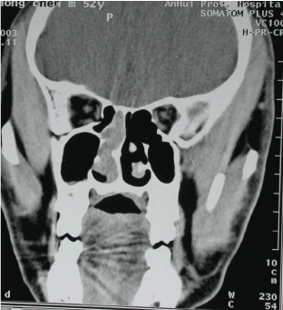
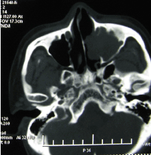
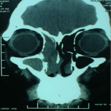
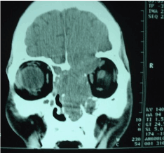
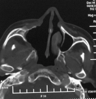
Based on the Krouse’s classification, we had two patients in T1 (3%, Figure 1), 39 cases in T2 (54%, Figure 2), 29 T3 (40%, Figure 3), two T4 (3%, Figure 4). 56 patients were operated upon only endoscopically (78%), 10 cases underwent lateral rhinotomy (14%), five patients underwent endoscopic procedures combined with Caldewell-Luc intervention (7%), and partial resection of maxilla was carried out in one case (1%). The two T1 cases were operated upon endoscopically (100%). Among T2 cases: 32 suffered enodscopic surgery (82%), 4 lateral rhinotomy (10%), 3 combined approach (8%). 29 T3 cases: 22 endoscopic (76%), 5 lateral rhinotomy (17%), 2 combined (7%). One of T4 was operated though lateral rhinotomy, the other though partial resection of maxilla (Table 2).
| Approach | Tumor staging | |||
| T1 | T2 | T3 | T4 | |
| Endoscopic | 2 | 32 | 22 | 0 |
| Lateral rhiontomy | 0 | 4 | 5 | 1 |
| Combined | 0 | 3 | 2 | 0 |
| Partial resection of maxilla | 0 | 0 | 0 | 1 |
Table 2: The approach and tumor staging
There was tumor recurrence in 17 patients (23.6%), 12 of them were operated upon endoscopically, their lateral rhinotomy was combined. There was no recurrence in any of the two T1 cases. Nine of the 39 T2 cases presented recurrence (23.1%). Seven patients among 29 T3 had recurrence (24.1%), one of them was canceration. The T4 patient had a recurrence (50%), one of them was canceration (Table 3).
| Staging of recurrences/of cases | % recurrence | |
| T1 | 0/2 | 0% |
| T2 | 9/39 | 23.1% |
| T3 | 7/29 | 24.1% |
| T4 | 1/2 | 50% |
Table 3: % recurrence by staging
When we consider the number of recurrence cases divided by surgical approach and the staging, we obtained: T2--seven recurrence cases among 32 endoscopic cases and one among the four lateral rhinotomy, one among the three combined cases. T3--endoscopic: five recurrences in 22 cases, lateral rhinotomy: one in five cases, combine: one in two. T4--lateral rhinotomy: one case.
Discussion
The inverted papilloma, also called Schneider papilloma, Ewing papilloma, transitional cells papilloma, epithelial papilloma villus cancer, is a benign sinusal tumor with undefined etiology [5,6].
The clinical aspect of the IP is unilateral nasal obstruction (98%), rhinorrhea (17%), epistaxis (6%), anosmia (4%) and headache [5-7]. Our series had 75% of the patients with nasal obstruction, 61% with epistaxis. From 91% to 99% of the inverted papilloma were unilateral [8]. In our cases, the lesion involved bilateral side in 3 (4.2%), and their first symptom was nasal obstruction and rhinorrhea. It is easy to misdiagnosis as sinusitis.
Although it is a benign lesion, the inverted papilloma is locally aggressive, bears high recurrence rates and is associated to aquamous cell carcinomas in 5 to 15% of the cases [3]; for such reason, many surgeons adopt extra-nasal procedures as treatment of choice for these tumors. However, with the progress in the endonasal endoscopic surgical technique, there has been much debate regarding possible benefits of such surgical approach over traditional ones such as lateral rhinodomy, degloving and sublabial approach [9].
Nasal cavity and paranasal sinuses CT scan suggest IP when there is an image of soft tissue present from the middle meatus all the way to the adjacent maxillary antrum, through an enlarged maxillary ostium, as the one seen in our patient. MRI provides a more accurate assessment of tumor boundaries and implantation site, differentiating it from the adjacent inflammatory tissue and it is also the exam of choice for postoperative follow up [6]. However it is of higher cost for the patients.
Kraft et al. [10] thought the indication of lateral rhinotomy and endoscopic is different. The surgical approach was accord to the position and the range of the lession. If the lession access to nasal cavity, maxillary, ethmoid and sphenoid sinus, it is better to chose the endoscopic approach; if the lesion access to lacrimal sac, nasofrontal duct and the fossa orbitalis, it is better to chose the lateral rhinotomy. In our series, the two T1 cases were approached endoscopically, without recurrence, seven recurrence cases among 32 endoscopic T2 cases and five recurrences in 22 T3 cases. Both of the two approaches have disadvantages. The lateral rhinotomy remove the whole lateral wall of the nasal cavity, expose the tumor thoroughly, especially when the lession reach to the fossa orbitalis and the brain, however, it has not a better visualization of tumor extention and its insertion, and there will be scar on the face. The endoscopic surgery may be beneficial in the anasosinusal papilloma surgery exclusively or in combination with the external access. It has many advantages: 1) It allows better visualization of tumor extension and its insertion, 2) It is less invasive, less bleeding and is necessary to persist structure and function of nasal cavity, allowing for better healing with decreased morbidity and shorter recovery periods. In our cases, we take the endoscopic surgery to T3 cases, the range of the resection can reach the lateral rhinotomy. After surgery six months later, the Figure 5 shows the CT scan. We think whether some benign tumors in the bases of skull like pterygopalatine fossa can be removed by the endoscopic surgery.
Conclusion
Our review with 72 cases of nasosinusal inverted papilloma pointed to a greater trend towards recurrence among the cases with more tumoral extension. Based on the extensions and sites of the lesions, we should have different approaches to different cases. The endonasal microsurgical approach should be preferred to the cases with tumours localized in the ethmoid, sphenoid, maxilary sinus and frontal sinus ostium.
Acknowledgements
We thank Dr. Luo B for the instructions on the manuscript. We also thank everybody who helped us in the study.
References
- Yiotakis I, Psarommatis I, Manolopoulos L, Ferekidis E, Adamopoulos G (2001) Isolated inverted papilloma of the sphenoid sinus . J Laryngol Otol 115: 227-230.
- Alba JR, Armengot M, Diaz A, Perez A, Rausell N, et al. (2002) Inverted papilloma of the sphenoid sinus. Acta Otorhinolaryngol Belg 56: 399-402.
- Lawson W, Ho BT, Shaari CM, Biller HF (1995) Inverted papilloma: a report of 112 cases. Laryngoscope 105: 282-288.
- Krouse JH (2000) Development of a staging system for inverted papilloma. Laryngoscope 110: 965-968.
- Vrabec DP (1994) The inverted Schneiderian papilloma: a 25-year study. Laryngoscope 104: 582-605.
- Myers EN, Fernau JL, Johnson JT, Tabet JC, Barnes EL (1990) Management of inverted papilloma. Laryngoscope 100: 481-490.
- Tsue TT, Bailet JW, Barlow DW, Makielski KH (1997) Bilateral sinonasal papillomas in aplastic maxillary sinuses. Am J Otolaryngol 18: 263-268.
- Oikawa K, Furuta Y, Oridate N, Nagahashi T, Homma A, et al. (2003) Preoperative staging of sinonasal inverted papilloma by magnetic resonance imaging. Laryngoscope 113: 1983-1987.
- Savy L, Lloyd G, Lund VJ, Howard D (2000) Optimum imaging for inverted papilloma. J Laryngol Otol 114: 891-893.
- Kraft M, Simmen D, Kaufmann T, Holzmann D (2003) Long-term results of endonasal sinus surgery in sinonasal papillomas. Laryngoscope 113: 1 541-1 547.
Citation: Liu XW and Sun JW (2012) The Role of Endoscopic Surgery in the Treatment of Nasal Inverted Papilloma. Otolaryngology 2:117. DOI: 10.4172/2161-119X.1000117
Copyright: © 2012 Liu XW, et al. This is an open-access article distributed under the terms of the Creative Commons Attribution License, which permits unrestricted use, distribution, and reproduction in any medium, provided the original author and source are credited.
Select your language of interest to view the total content in your interested language
Share This Article
Recommended Journals
Open Access Journals
Article Tools
Article Usage
- Total views: 15520
- [From(publication date): 8-2012 - Nov 18, 2025]
- Breakdown by view type
- HTML page views: 10725
- PDF downloads: 4795

