A Comprehensive Analysis of the Expression and Prognosis of STATs in Human Breast Invasive Carcinoma
Received: 24-Apr-2023 / Manuscript No. DPO-23-96857 / Editor assigned: 27-Apr-2023 / PreQC No. DPO-23-96857(PQ) / Reviewed: 11-May-2023 / QC No. DPO-23-96857 / Revised: 18-May-2023 / Manuscript No. DPO-23-96857 (R) / Accepted Date: 18-May-2023 / Published Date: 25-May-2023 DOI: 10.4172/2476-2024.8.2.214
Abstract
Background: Multiple cancer types are associated with the Signal Transducer and Activator of Transcription (STAT) family of proteins. The expression and prognostic value of STATs in Breast Invasive Carcinoma (BRCA) remain unclear. Methods: Herein we investigated the clinical data onto 1,222 patients with BRCA based on the Cancer Genome Atlas (TCGA) database, UALCAN, cBio Cancer Genomics Portal (cBioPortal), STRING, and GeneMANIA databases.
Results: The transcriptional levels of STAT4/5A/5B/6 were significantly decreased while the transcriptional levels of STAT1 were elevated in BRCA tissues. A significant correlation exists between STATs expressions and known prognostic factors, e.g., age, pathologic stage, radiation therapy, and Tumor Node Metastasis (TNM) stages. It was discovered that patients with high STAT4 expression had a better prognosis for overall survival (OS) (HR=0.59, p=0.002), Disease-Specific Survival (DSS) (HR=0.59, p=0.018), and Progress Free Interval (PFI) (HR=0.55, p<0.001). STAT4 may be an independent prognostic marker for BRCA through univariate and multivariate Cox regression. In terms of immune infiltrating levels, a correlation between STAT1/2/4/13 expression and immune cell infiltration, including T cells and Th1, has also been noted. Furthermore, the levels of STAT4 were statistically significant correlated with T cells (r=0.822, p<0.001), cytotoxic cells (r=0.746, p<0.001), B cells (r=0.691, p<0.001), Th1 cells (r=0.686, p<0.001), and activated Dendritic Cells (DC).
Conclusion: Based on the findings of this study, STAT4 might serve as a novel prognostic biomarker to predict prognosis and levels of immune infiltration for BRCA.
Keywords: STATs; Expression; Prognosis; Cox regression; Breast invasive carcinoma
Introduction
Worldwide, breast invasive carcinoma is one of the most common malignant tumors in women. The incidence rate is rising each year, which has a significant negative impact on women's health and quality of life [1]. The number of new cases of breast cancer worldwide increased to 2 million 260 thousand in 2020, making it the world's most common cancer [2]. Even though significant advancements have been made in the detection and management of breast cancer, conventional therapy is not the best option for advanced breast cancer. Breast cancer immunotherapy has grown in clinical importance as a result of ongoing research. The earliest immune cells to be identified are Tumor Infiltrating Lymphocytes (TILs) [3]. Numerous studies have shown that the presence of TILs, mainly T cells and B cells, increases the likelihood that a patient would survive their disease [4,5]. Clinical outcomes of breast cancer with HER2 positive TILs have been studied, TILs appear to be able to predict disease-free survival to some extent and identify populations with low recurrence risk [6]. Furthermore, a large number of TILs are reliable prognostic markers for a variety of solid tumors. Early Triple-Negative Breast Cancer (TNBC) patients with high TIL levels have an improved survival rate. A meta-analysis indicated individuals with a large number of interstitial TILs infiltration had a favorable prognosis after adjuvant treatment [7]. Cytotoxic CD8+T cells are the most frequent type of microenvironment in breast cancer. Breast cancer patients with a higher number of CD8+ T cells have a better prognosis [8]. In addition, prognostic and predictive values for B cells [9], Natural Killer (NK) cells [10], and Dendritic Cells (DC) [11] have also been reported in human cancer. Therefore, finding new immune-related therapeutic targets for Breast Invasive Carcinoma (BRCA) is urgently needed.
The Signal Transducer And Activator Of Transcription (STAT) protein family, which is involved in the control of physiological processes like cell division, proliferation, and apoptosis, is both intricate and significant [12]. The human genome has so far been linked to seven stat genes: STAT1, STAT2, STAT3, STAT4, STAT5A, STAT5B, and STAT6, extracellular signal proteins are regulated by them. Numerous researches have so far shown the expression pattern and function of a few STATs in malignancies. There has been evidence that STAT1 is associated with cancer and may have prognostic significance among solid tumor patients [13]. Breast cancer may evade immune monitoring by down-regulating STAT1-mediated interferon signaling, which results in the development of clinically significant tumors, according to Goodman ML [14]. Furthermore, research has identified a possible cancer therapeutic target is STAT3 due to its key role in decreasing the manufacturing of immunosuppressive proteins and lowering the expression of important immune activation regulators [15,16]. In their study on STAT4, Anderson, et al. concentrated on the possibility that activating STAT4 could lessen lymphatic metastases among head and neck squamous cell carcinoma patients [17]. Limited data, however, support STATs' potential prognostic usefulness in BRCA and their connection to immunological infiltrates.
Our research examined how STATs affect the carcinogenesis and prognosis of breast cancer through the clinical data acquired from The Cancer Genome Atlas (TCGA) database, UALCAN, cBioPortal, STRING, and GeneMANIA databases. This is the first in-depth analysis of relationships between STATs family gene expression and clinical, molecular, and immunological traits in BRCA. The research has revealed fundamental processes that contribute to the development of breast cancer and has identified STATs as an important biomarker for diagnosis and prognosis of BRCA.
Materials and Methods
Processing of data and STATs expression analysis
BRCA data (transcripts per million read) and clinic pathological information were downloaded from the TCGA database in high- throughput sequence RNA format. (https://portal.gdc.cancer.gov/). The STATs median value was used to divide the 1,222 BRCA patients into groups with high and low STATs expression. Here, we used the UALCAN portal (http://ualcan.path.uab.edu/analysis-prot.), a tool for online analysis and mining of the TCGA information, to further investigate the degree of STATs protein expression in BRCA [18].
Analysis of genomic alterations and gene and protein networks in the STAT family
Using the cBio Cancer Genomics Portal (http://cbioportal.org), we examined the genomic changes in the STATs family in the TCGA BRCA sample [19]. Next, the STATs family gene and protein network was examined using the GeneMANIA and STRING databases (https://genemania.org/ and https://stringpreview.org/, respectively) [20,21].
STATs expression and clinic pathological characteristics in BRCA
Using the Wilcoxon rank sum test (continuous variables) or Pearson's chi-square test, clinical and pathological traits were compared between groups with high and low STATs expression (rank variables). Based on different classification criteria, including age (≤ 60; >60), pathologic stage (stage I, stage II, stage III, and stage IV), radiation therapy (Yes; No), and tumor node metastasis (TNM) stages (T: T1, T2, T3, T4; N: N0, N1, N2, N3; M: M0, M1) of breast cancer patients were examined using TCGA data, Statistical significance was defined as p<0.05.
Clinical significance of STATs expression in BRCA tumors
An analysis of Kaplan-Meier data was performed on the relationships between STATs expression and Overall Survival (OS), Disease-Specific Survival (DSS), and Progress-Free Interval (PFI) in patients with BRCA. Using the median expression of the gene (high vs. low expression), we divided the patient samples into two cohorts to assess its prognostic potential. To identify independent prognostic variables, both univariate and multivariate Cox regression analyses were performed. The Hazard Ratio (HR) and log-rank P-value with 95% confidence intervals were computed. Random forest regression was carried out using the random Forest R package [22].
Association of STATs and immune cell infiltration in BRCA tumors
Our study examined the relationship between STATs expression and immune cell types, including DCs, immature DCs (iDCs), activated DCs (aDCs), plasmacytoid DCs (pDCs), T cells, T helper (Th) cells, type 1 Th cells (Th1), type 2 Th cells (Th2), type 17 Th cells (Th17), regulatory T cells (Treg), T gamma delta (Tgd), T central memory (Tcm), T effector memory (Tem), T follicular helper (Tfh), CD8+T cells, B cells, neutrophils, macrophages, cytotoxic cells, mast cells, eosinophils, NK cells, NK 56 cells, and NK 56+cells by Spearman’s analysis. By Wilcoxon rank sum analysis, immune cell infiltration levels were compared for high and low STAT expression groups.
Statistical analysis
The median method of gene expression was used as the cut-off value of STATs expression. STATs expression and clinicopathological characteristics were analyzed using logistic regression. Potential prognostic factors can be screened using univariate Cox analysis, and the multivariate Cox analysis can confirm STATs expression's effect on survival when combined with other clinical factors. To perform all of these statistical analyses, R (v4.0.0) was used, and P-values less than 0.05 were considered significant.
Results
STAT4, STAT5A, STAT5B and STAT6 expression are lower and STAT1 expression is higher in BRCA tissues
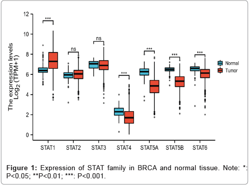
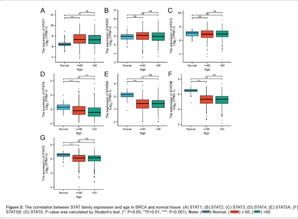
To investigate the difference of STATs expression between BRCA tumor and normal tissue, seven STAT family members have been detected. The levels of STAT expression in cancer and normal samples were compared (Figure 1). The findings demonstrated that STAT4, STAT5A, STAT5B and STAT6 expression levels were decreased in breast cancer tissues while STAT1 expression levels considerably increased (p<0.01). To validate the preceding findings, Figures 2A-2G illustrates a comparison of STAT family expression levels in BRCA and normal tissues. A significant decrease in STAT4, STAT5A, STAT5B, and STAT6 expression levels was observed in BRCA tissues compared to normal tissues (p<0.001), while STAT1 and STAT2 expression levels were remarkably higher in BRCA tissues (p<0.001) and (p<0.05), respectively. As a result, in BRCA tissues, STAT1 expression was higher than in adjacent normal tissue, while STAT4, STAT5A, STAT5B and STAT6 were down-regulated.
Genomic alterations of the STATs family and gene and protein network
In BRCA samples, we assessed the types and frequencies of STATs using the cBioPortal database. It was found that STATs family members rarely mutated (less than five times), indicating that they were highly conserved. GeneMANIA and STRING programs were used to generate the STATs family's gene gene and protein-protein interaction network. We discovered that the STATs family interacted with 20 potential target genes and 11 potential target proteins. Then, we used “correlation analysis” by (bc-GenExMiner v4.7) for seven members of the STATs family, we found STAT1 was positively correlated STAT2 (r=0.41, p<0.0001) and STAT4 (r=0.47, p<0.0001), STAT5A was positively correlated STAT5B (r=0.46, p<0.0001) (Figures 3A-3G).
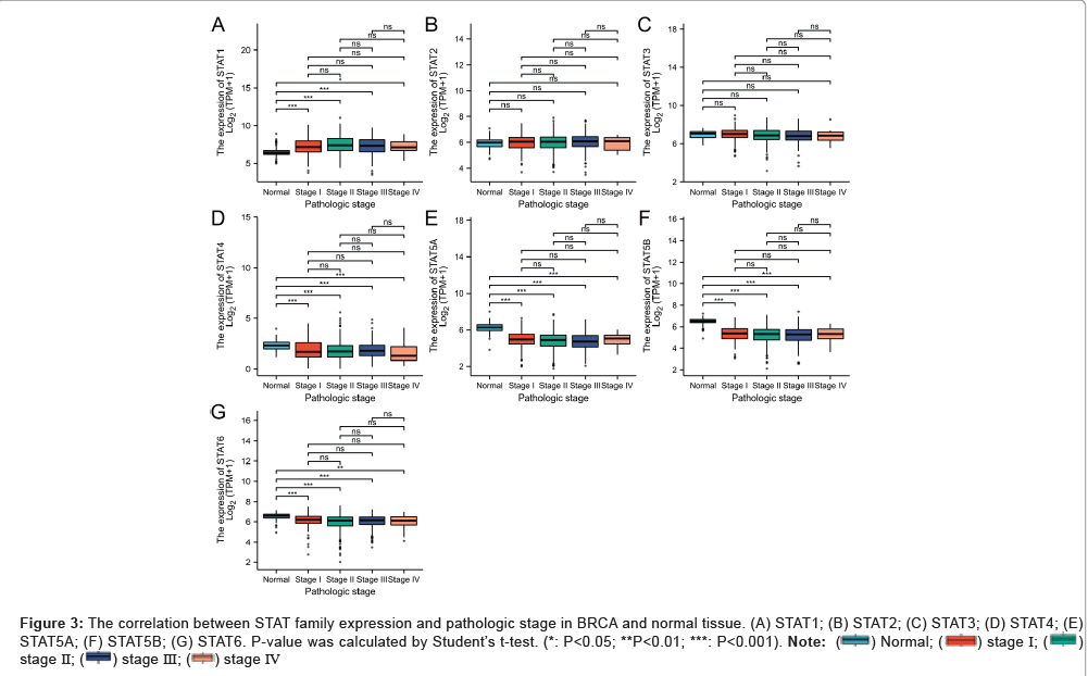
Figure 3: The correlation between STAT family expression and pathologic stage in BRCA and normal tissue. (A) STAT1; (B) STAT2; (C) STAT3; (D) STAT4; (E) STAT5A; (F) STAT5B; (G) STAT6. P-value was calculated by Student’s t-test. (*: P<0.05; **P<0.01; ***: P<0.001). Note: (  ) Normal; (
 ) stage Ⅰ; (
 )
stage Ⅱ; (
  ) stage Ⅲ; (
  ) stage Ⅳ
Association of STATs expression and clinic pathological characteristics in BRCA patients
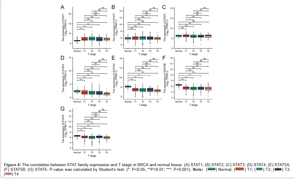
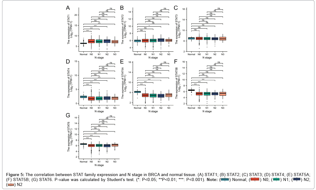
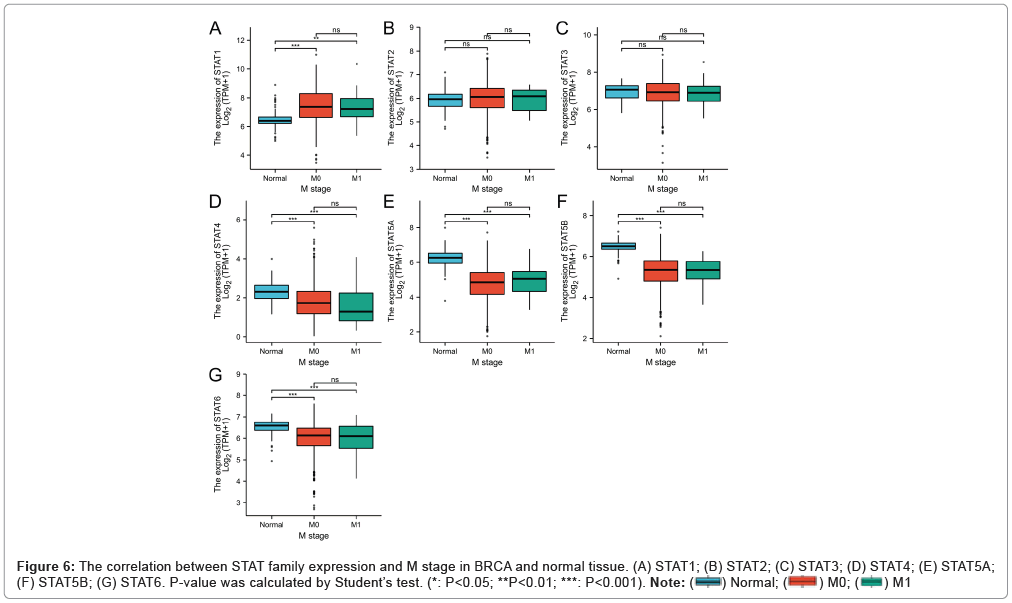
Our study examined the correlation between the expression of STATs and various clinical characteristics of BRCA patients, such as age, pathologic stage, radiation therapy, and TNM stage. The expression of STATs was higher in the ≤ 60-compared with > 60 group, especially STAT4 (p<0.01). The distribution of STAT1, STAT2, STAT3, STAT5A and STAT6 was not significantly different. The expression of STAT family members in patients was also examined in relation to tumor stage. Interestingly, STAT1 was significantly high in stage1/2/3/4 than that in normal. However, the expression of STAT4/5A/5B/6 was significantly low in stage1/2/3/4 than that in normal. In terms of TNM stages, the transcriptional levels of STATs were associated with the TNM stages of the patients. We discovered strongly positive associations between STAT1, STAT4, STAT5A, STAT5B and STAT6 expression and TNM stages. The expression levels of STAT4, STAT5A, STAT5B and STAT6 were marked lower in T1-T4, N0-N3, and M0-M1 than in normal tissues, while the expression level of STAT1 was higher inversely (Figures 4A-4G).
Figure 4: The correlation between STAT family expression and T stage in BRCA and normal tissue. (A) STAT1; (B) STAT2; (C) STAT3; (D) STAT4; (E) STAT5A; (F) STAT5B; (G) STAT6. P-value was calculated by Student’s test. (*: P<0.05; **P<0.01; ***: P<0.001). Note: ( ) Normal; (
 ) T1; (
 ) T2; (
 ) T3; (
 ) T4
) T4
Prognostic value of mRNA expression of STATs in BRCA patients
Next, BRCA prognosis was examined based on STATs family expression. Kaplan-Meier analyses were used to test the effect of STATs on clinical outcomes. Figures 5A-5G shows that high STAT4 expression is related to better OS (H=0.59, p=0.002), DSS (HR=0.59, p=0.018), and PFI (HR=0.55, p<0.001) than those for the low-STAT4 group. However, in terms of OS, DSS, and PFI, STAT1/2/3/5A/5B/6 expression levels showed no significant correlation.
Figure 5: The correlation between STAT family expression and N stage in BRCA and normal tissue. (A) STAT1; (B) STAT2; (C) STAT3; (D) STAT4; (E) STAT5A; (F) STAT5B; (G) STAT6. P-value was calculated by Student’s test. (*: P<0.05; **P<0.01; ***: P<0.001). Note: ( ) Normal; (
  ) N0; (
  ) N1; (
 ) N2; (
  ) N2.
) N2.
For the purpose of comprehending the predictive relevance of STAT4 expression in breast cancer, we used the Cox proportional hazard regression model. Additionally, the outcomes were displayed as forest plots (Figures 6A-6G). As shown in Figures 6A, STAT4 expression were significantly linked with the probability of mortality were patient age (p<0.001), T stage (p=0.012), N stage (p<0.001), M stage (p<0.001), and pathologic stage III and stage IV (p<0.001). An analysis of multivariate Cox regression was performed to determine whether STAT4 expression is an independent predictor of BRCA and to assess STAT4's ability to predict clinical outcomes. STAT4 was a significant good prognostic factor for OS (HR=0.648, p=0.025), with similar findings for and PFI (HR=0.498, p=0.011). Despite the fact that it had no significant predictive ability for DSS. In multivariate Cox regression analysis, the clinical stage, particularly the clinical N and M stages, exhibited predictive advantages for clinical outcomes.
Additionally, we provided Kaplan-Meier analyses for OS, DSS and PFI in male sex, age >60 years, N stages, and M stages. In all of the investigations, the groups with high STAT4 expression experienced noticeably improved clinical outcomes. The prognostic nomogram was created using all statistically relevant prognostic variables in each multivariate Cox regression analysis, and the calibration curve was created to gauge the effectiveness of the nomogram. The nomogram to predict OS, with a C-index of 0.660, contained clinical N and M stages as well as STAT4. Clinical N and M stages, a nomogram was created to predict DSS, STAT4 had a C-index of 0.705. A predictive nomogram for PFI was created using clinical stage, clinical N and M stages, and STAT4, and it had a C-index of 0.631. Except for the 5-year prediction of OS and DSS, all three nomograms of 1, 3 and 5-year clinical results were perfectly predicted by the calibration curves Overall; our findings indicated that STAT4 was a favorable prognostic factor and an independent prognostic marker in BRCA.
Correlation between STATs mRNA expression and immune cell infiltration in BRCA tumors
For a deeper understanding of STAT's suggestive role, it is necessary to investigate the types of immune cells infiltrating BRCA patients. Based on the ssGSEA method, STATs and immune cell infiltration were investigated using Spearman's analysis. As shown in Figures 6B, STAT1 expression correlated strongly with tumor-infiltrating immune cells, including aDCs (r=0.686, p<0.001), Macrophages (r=0.424, p<0.001), T cells (r=0.502, p<0.001), Th1 cells (r=0.549, p<0.001), and Treg cells (r=0.534, p<0.001). aDC and STAT2 expression exhibited a substantial positive association (r=0.403, p<0.001), T helper cells (r=0.392, p<0.001), T central memory cells (r=0.392, p<0.001), and T cells (r=0.366, p<0.001). The expression of STAT3 was inversely correlated with that of aDC, B cells, CD8 T cells, cytotoxic cells, and DCs. More specifically, Statistically, STAT4 expression was positively correlated with T cells (r=0.822, p<0.001), cytotoxic cells (r=0.746, p<0.001), B cells (r=0.691, p<0.001), Th1 cells (r=0.686, p<0.001), and aDC (r=0.642, p<0.001). A moderately positive correlation was found between STAT5A/5B/6 expression and the number of CD8+ T cells, NK cells and T helper cells.
To evaluate the therapeutic potential of STATs-based therapy for breast cancer, the association between STATs mRNA expression and immune checkpoints (PD-1/PD-L1 and CTLA-4) in breast cancer tumors was further investigated in-depth [23]. STAT1 expression was positively correlated with CD274 (PD-1) (r=0.720, p<0.001), PDCD1 (r=0.500, p<0.001) and CTLA-4 (r=0.620, p<0.001), Furthermore, PD-1 and STAT4 expression were significantly correlated (r=0.690, p<0.001), PDCD1 (r=0.760, p<0.001), and CTLA-4 (r=0.770, p<0.001). The expression of STAT3, STAT5A and STAT5B, however, did not correlate with PD-1/PD-L1 and CTLA-4. Based on the combined findings, STATs may influence immune cells in the tumor microenvironment of BRCA.
Discussion
Previous research has demonstrated that the STATs protein family regulates cell proliferation, differentiation, and death in a complex and crucial manner [12,24,25]. The potential roles of STATs in the development of tumors and tumor immunology are still unknown. The immune cells associated with tumors have recently been receiving a great deal of attention [26]. It has been shown that STATs are important predictors of BRCA prognosis in several studies and are currently thought to be biomarkers strongly associated with BRCA [27,28]. More crucially, the possible relevance of STATs with numerous cancer immune checkpoints (PD-1/PD-L1 and CTLA-4) was further examined to fully comprehend theNrelationship between STATs and immune regulation. The immune checkpoint component PD-1/PD-L1 is well known and has been shown to be an effective therapeutic target for invasive breast cancer [29]. TCGA-BRCA data was analyzed for expression profiles, clinicopathological relationships, clinical significance, and immune cell infiltration of STATs. The current work is the first to examine STATs family members' expression, assess their prognostic significance, and forecast the level of immune infiltration in BRCA tumors based on STATs expression.
As a crucial component of IFN signal transduction, STAT1 is a cytoplasmic protein [30]. Studies have revealed that the STAT1 pathway's expression confers cells with resilience to DNA-damaging chemicals and facilitates the formation of aggressive tumors [31]. A potential biomarker for anti-PD-1/anti-PD-L1 immunotherapy in breast cancer is phosphor-STAT1 expression [32]. In our findings, we showed that breast cancer tissues expressed STAT1 at a higher level than healthy tissues. On the basis of age, pathologic stage, and TNM stages, it is interesting to note that STAT1 was much higher in stage1/2/3/4 than in normal. STAT1 expression was not significantly correlated with OS, DSS or PFI in BRCA.
STAT2 has been linked to immune response and carcinogenesis via proteasome degradation [33]. We discovered that the expression of STAT2 in breast cancer tissues and normal tissues did not significantly correlate with one another. no association was seen between STAT2 expression and age, pathologic stage, or TNM stage in BRCA. Additionally, there was no discernible relationship between OS, DSS, and PFI in BRCA and STAT2 expression. The findings suggest that STAT2 is ineffective for predicting BRCA. STAT3 has been identified as a prognostic biomarker in head and neck cancer and TNBC [34,35]. The study provides evidence that STAT3 is related to immunosuppression and drug response in clinical treatment [36]. There was no significant link between STAT3 expression in breast cancer tissues and normal tissues in our study, and it was unrelated to clinicopathological factors such age, pathologic stage, or TNM stage. Furthermore, STAT3 expression was not shown to be significantly associated to OS, DSS and PFI, demonstrating that STAT3 is not suitable for the prognosis of BRCA.
A key mediator in the development of tumors and inflammation is STAT4 [37]. However, it is unclear how STAT4 is expressed and what its prognostic significance is in breast cancer. To definitively address this question, we determined the predictive value of STAT4 in BRCA, high expression levels of STAT4 indicated better OS, DSS and PFI, indicating that STAT4 was a good prognostic factor in BRCA. After controlling for potential confounding variables, using a multivariate Cox regression analysis, we attempted to determine whether STAT4 expression is an independent predictor of BRCA, taking into account the effect of STAT4 on clinicopathological features of cancer. All of the results showed that high-STAT4 expression groups had considerably superior clinical outcomes, indicating STAT4 may be an independent prognostic factor in BRCA. The most important discovery was the statistically significant positive correlation between STAT4 expression and T cells, cytotoxic cells, B cells, Th1 cells, and activated DCs. The clinical response potential of cancer immunotherapy is determined by T cell infiltration created in human cancer [38]. In the occurrence and growth of malignancies, B lymphocytes are crucial [39]. DCs are essential for T cell-mediated cancer immunity because they are the primary modulator of the adaptive immune response [40]. Additional research is necessary to confirm the use of STAT4 as a prognostic biomarker and independent prognostic marker for BRCA. The highly conserved Janus kinase (JAK)/STAT signaling system, which includes STAT5A and STAT5B, is thought to be the cause of the disease because its signal is amplified in a number of hematological malignancies [41]. In our study, A lower expression of STAT5A/B was found in breast cancer tissues than in normal tissues, and this expression was not correlated with age, pathologic stages, or TNM stage in BRCA.
In acute myeloid leukemia, STAT6 has been discovered as an important biological target [42]. The immunoreactivity of nuclear STAT6 is highly sensitive and specific for the detection of solitary fibrous tumors [43]. Additionally, it has been noted that STAT6 signaling is frequently active in cancerous cells and controls a number of genes necessary for immune response, inflammation, and cell growth [44]. Up until recently, little was understood about STAT6's expression and function in breast cancer. Our findings indicated that STAT6 expression was noticeably low in BRCA patients. Age, pathological stages, TNM stages, and prognosis were all clinicopathological parameters that did not significantly correlate with each other.
Conclusion
Based on our findings, a significant down-regulation in STAT4, STAT5A, STAT5B, and STAT6 was observed in BRCA, while an up- regulation of STAT1 was observed. Another intriguing point is that in BRCA, elevated STAT4 expression was an excellent predictor of outcome and may perhaps be a standalone prognostic diagnostic. Conversely, STAT4 expression was significantly correlated with immune infiltration. Our research offers a promising new biomarker and practical knowledge for the creation of an immunotherapy for BRCA. We anticipate that our research will establish STAT4 as a major biomarker for human BRCA prognosis.
Declarations
Availability of data and materials
The data that support the findings of this study are openly available in the TCGA database (https://portal.gdc.cancer.gov/), the UALCAN portal (http: //ualcan.path.uab.edu/analysis-prot.), the cBioPortal (http://cbioportal.org), the GeneMANIA (https://genemania.org/), and the STRING databases (https://string-preview.org/).
Funding
This project was supported by the National Natural Science Foundation of China, China (No. 82172356, No. 81972003), the Natural Science Foundation of Guangdong, China (No. 2021A1515012144).
Autours’ Contributions
QH: designed the work, writing-original draft preparation, performed the statistical analysis, participated in the discussion and language editing. NX: writing-review and editing.
Competing Interests
The authors declare no competing interests.
Ethics Approval and Consent to Participate
Not applicable.
Consent for Publication
All authors have seen and agreed to publish.
References
- Siegel RL, Miller KD, Jemal A (2019) Breast cancer statistics, 2019. CA: A Cancer Journal for Clinicians 69:438-51.
[Crossref] [Google Scholar] [PubMed]
- Xu B, Zhang Q, Zhang P, Hu X, Li W, et al. (2021) DAWNA-1 Study Consortium. Dalpiciclib or placebo plus fulvestrant in hormone receptor-positive and HER2-negative advanced breast cancer: a randomized, phase 3 trial. Nat Med 27:1904-1909.
[Crossref] [Google Scholar] [PubMed]
- Dieci MV, Radosevic-Robin N, Fineberg S, van den Eynden G, Ternes N, et al. (2018) Update on tumor-infltrating lymphocytes (TILs) in breast cancer, including recommendations to assess TILs in residual disease after neoadjuvant therapy and in carcinoma in situ: A report of the International Immuno-Oncology Biomarker Working Group on Breast Cancer. Semin Cancer Biol 52:16-25.
[Crossref] [Google Scholar] [PubMed]
- Baxevanis CN, Sofopoulos M, Fortis SP, Perez SA (2019) The role of immune inftrates as prognostic biomarkers in patients with breast cancer. Cancer Immunol Immunother 68:1671–1680.
[Crossref] [Google Scholar] [PubMed]
- Savas P, Virassamy B, Ye C, Salim A, Mintoff CP, Caramia F, et al. (2018) Single-cell profling of breast cancer T cells reveals a tissue-resident memory subset associated with improved prognosis. Nat Med 24:986-993.
[Crossref] [Google Scholar] [PubMed]
- Kim RS, Song N, Gavin PG, Salgado R, Bandos H, et al. (2019) Stromal Tumor-infltrating Lymphocytes in NRG Oncology/NSABP B-31 Adjuvant Trial for Early-Stage HER2-Positive Breast Cancer. J Natl Cancer Inst 111:867-871.
[Crossref] [Google Scholar] [PubMed]
- Byrne A, Savas P, Sant S, Li R, Virassamy B, et al. (2020) Tissue-resident memory T cells in breast cancer control and immunotherapy responses. Nat Rev Clin Oncol 17:341-348.
[Crossref] [Google Scholar] [PubMed]
- Tiainen S, Tumelius R, Rilla K, Hamalainen K, Tammi M, et al. (2015) High numbers of macrophages, especially M2-like (CD163-positive), correlate with hyaluronan accumulation and poor outcome in breast cancer. Histopathology 66:873-83.
[Crossref] [Google Scholar] [PubMed]
- Garaud S, Buisseret L, Solinas C, Gu-Trantien C, de Wind A, et al. (2019) Tumor infltrating B-cells signal functional humoral immune responses in breast cancer. JCI Insight 5:e129641.
[Crossref] [Google Scholar] [PubMed]
- Terrén I, Orrantia A, Vitallé J, Zenarruzabeitia O, Borrego F (2019) NK Cell Metabolism and Tumor Microenvironment. Front Immunol 10:2278.
[Crossref] [Google Scholar] [PubMed]
- Patente TA, Pinho MP, Oliveira AA, Evangelista GCM, Bergami-Santos PC, et al. (2019) Human Dendritic Cells: Their Heterogeneity and Clinical Application Potential in Cancer Immunotherapy. Front Immunol 9:3176.
[Crossref] [Google Scholar] [PubMed]
- Verhoeven Y, Tilborghs S, Jacobs J, De Waele J, Quatannens D, et al. (2020) The potential and controversy of targeting STAT family members in cancer. Semin Cancer Biol 60:41-56.
[Crossref] [Google Scholar] [PubMed]
- Zhang J, Wang F, Liu F, Xu G (2020) Predicting STAT1 as a prognostic marker in patients with solid cancer. Ther Adv Med Oncol 12:1758.
[Crossref] [Google Scholar] [PubMed]
- Goodman ML, Trinca GM, Walter KR, Papachristou EK, D'Santos CS, et al. (2019) Progesterone Receptor Attenuates STAT1-Mediated IFN Signaling in Breast Cancer. J Immunol 202:3076-3086.
[Crossref] [Google Scholar] [PubMed]
- Zou S, Tong Q, Liu B, Huang W, Tian Y, et al. (2020) Targeting STAT3 in Cancer Immunotherapy. Mol Cancer 19:145.
[Crossref] [Google Scholar] [PubMed]
- Lee H, Jeong AJ, Ye SK (2019) Highlighted STAT3 as a potential drug target for cancer therapy. BMB Rep 52:415-423.
[Crossref] [Google Scholar] [PubMed]
- Anderson K, Ryan N, Volpedo G, Varikuti S, Satoskar AR, et al. (2020) Immune Suppression Mediated by STAT4 Defciency Promotes Lymphatic Metastasis in HNSCC. Front Immunol 10:3095.
[Crossref] [Google Scholar] [PubMed]
- Chandrashekar DS, Bashel B, Balasubramanya SAH, Creighton CJ, Ponce-Rodriguez I, et al. (2017) UALCAN: A Portal for Facilitating Tumor Subgroup Gene Expression and Survival Analyses. Neoplasia 19:649-658.
[Crossref] [Google Scholar] [PubMed]
- Gao J, Aksoy BA, Dogrusoz U, Dresdner G, Gross B, et al. (2013) Integrative analysis of complex cancer genomics and clinical profles using the cBioPortal. Sci Signal 6:pl1.
[Crossref] [Google Scholar] [PubMed]
- Warde-Farley D, Donaldson SL, Comes O, Zuberi K, Badrawi R, et al. (2010) The GeneMANIA prediction server: biological network integration for gene prioritization and predicting gene function. Nucleic Acids Res 38:214-220.
[Crossref] [Google Scholar] [PubMed]
- Szklarczyk D, Gable AL, Nastou KC, Lyon D, Kirsch R, et al. (2021) The STRING database in 2021: customizable protein-protein networks, and functional characterization of user-uploaded gene/measurement sets. Nucleic Acids Res 8:D605-D612.
[Crossref] [Google Scholar] [PubMed]
- Svetnik V, Liaw A, Tong C, Culberson JC, Sheridan RP, et al. (2003) Random forest: a classifcation and regression tool for compound classification and QSAR modelling. J Chem Inf Comput Sci 43:1947-1958.
[Crossref] [Google Scholar] [PubMed]
- Huang X, Tang T, Wang X, Bai X, Liang T (2020) Calreticulin couples with immune checkpoints in pancreatic cancer. Clin Transl Med 10:36-44.
[Crossref] [Google Scholar] [PubMed]
- Gutiérrez-Hoya A, Soto-Cruz I (2020) Role of the JAK/STAT Pathway in Cervical Cancer: Its Relationship with HPV E6/E7 Oncoproteins. Cells 9:2297.
[Crossref] [Google Scholar] [PubMed]
- Loh CY, Arya A, Naema AF, Wong WF, Sethi G, et al. (2019) Signal Transducer and Activator of Transcription (STATs) Proteins in Cancer and Infammation: Functions and Therapeutic Implication. Front Oncol 9:48.
[Crossref] [Google Scholar] [PubMed]
- Wang B, Chen D, Hua H (2021) TBC1D3 family is a prognostic biomarker and correlates with immune infltration in kidney renal clear cell carcinoma. Mol Ther Oncolytics 22:528-538.
[Crossref] [Google Scholar] [PubMed]
- Huo Q, Li Z, Cheng L, Yang F, Xie N (2020) SIRT7 Is a Prognostic Biomarker Associated With Immune Infltration in Luminal Breast Cancer. Front Oncol 10:621.
[Crossref] [Google Scholar] [PubMed]
- Loi S, Drubay D, Adams S, Pruneri G, Francis PA, et al. (2019) Tumor-Infltrating Lymphocytes and Prognosis: A Pooled Individual Patient Analysis of Early-Stage Triple-Negative Breast Cancers. J Clin Oncol 37(7):559-569.
[Crossref] [Google Scholar] [PubMed]
- Dong H, Strome SE, Salomao DR, Tamura H, Hirano F, et al. (2002) Tumor-associated B7-H1 promotes T-cell apoptosis: a potential mechanism of immune evasion. Nat Med 8:793-800.
[Crossref] [Google Scholar] [PubMed]
- Zhang Y, Liu Z (2017) STAT1 in cancer: friend or foe?. Discov Med 24:19-29.
[Google Scholar] [PubMed]
- Khodarev NN, Roizman B, Weichselbaum RR (2012) Molecular pathways: interferon/stat1 pathway: role in the tumor resistance to genotoxic stress and aggressive growth. Clin Cancer Res 18:3015-3021.
[Crossref] [Google Scholar] [PubMed]
- Nakayama Y, Mimura K, Tamaki T, Shiraishi K, Kua LF, et al. (2019) Phospho‑STAT1 expression as a potential biomarker for anti‑PD‑1/anti‑PD‑L1 immunotherapy for breast cancer. Int J Oncol 54:2030-2038.
[Crossref] [Google Scholar] [PubMed]
- Lee CJ, An HJ, Cho ES, Kang HC, Lee JY, et al. (2020) Stat2 stability regulation: an intersection between immunity and carcinogenesis. Exp Mol Med 52:1526-1536.
[Crossref] [Google Scholar] [PubMed]
- Van Ruitenbeek NJ, van der Woude LL, van Krieken JH, van Engen-van Grunsven ACH, Willemsen AECAB, et al. (2020) STAT3 as a predictive biomarker in head and neck cancer: A validation study. Pathol Res Pract 216:153172.
[Crossref] [Google Scholar] [PubMed]
- Qin JJ, Yan L, Zhang J, Zhang WD (2019) STAT3 as a potential therapeutic target in triple negative breast cancer: a systematic review. J Exp Clin Cancer Res 38:195.
[Crossref] [Google Scholar] [PubMed]
- Chen W, Dai X, Chen Y, Tian F, Zhang Y, et al. (2020) Significance of STAT3 in Immune Infiltration and Drug Response in Cancer. Biomolecules 10:834.
[Crossref] [Google Scholar] [PubMed]
- Yang C, Mai H, Peng J, Zhou B, Hou J, et al. (2020) STAT4: an immunoregulator contributing to diverse human diseases. Int J Biol Sci 16:1575-1585.
[Crossref] [Google Scholar] [PubMed]
- Van der Leun AM, Thommen DS, Schumacher TN (2020) CD8+ T cell states in human cancer: insights from single-cell analysis. Nat Rev Cancer 20:218-232.
[Crossref] [Google Scholar] [PubMed]
- Wang SS, Liu W, Ly D, Xu H, Qu L, (2019) Tumor-infltrating B cells: their role and application in anti-tumor immunity in lung cancer. Cell Mol Immunol 16:6-18.
[Crossref] [Google Scholar] [PubMed]
- Gardner A, Ruffell B (2016) Dendritic Cells and Cancer Immunity. Trends Immunol 37:855-865.
[Crossref] [Google Scholar] [PubMed]
- Maurer B, Kollmann S, Pickem J, Hoelbl-Kovacic A, Sexl V (2019) STAT5A and STAT5B-Twins with Different Personalities in Hematopoiesis and Leukemia. Cancers (Basel) 11:1726.
[Crossref] [Google Scholar] [PubMed]
- Liu W, Zhu F, Yan J, Liu Y, Chen C, et al. (2020) Identification and Validation of STAT6 as a Prognostic and Predictive Biomarker in Acute Myeloid Leukaemia. Onco Targets Ther 13:11165-11176.
[Crossref] [Google Scholar] [PubMed]
- Yoshida A, Tsuta K, Ohno M, Yoshida M, Narita Y, et al. (2014) STAT6 immunohistochemistry is helpful in the diagnosis of solitary fibrous tumors. Am J Surg Pathol 38:552-559.
[Crossref] [Google Scholar] [PubMed]
- Hebenstreit D, Wirnsberger G, Horejs-Hoeck J, Duschl A (2006) Signaling mechanisms, interaction partners, and target genes of STAT6. Cytokine Growth Factor Rev 17:173–188.
[Crossref] [Google Scholar] [PubMed]
Citation: Huo Q, Xie N (2023) A Comprehensive Analysis of the Expression and Prognosis of STATs in Human Breast Invasive Carcinoma. Diagnos Pathol Open 8:214 DOI: 10.4172/2476-2024.8.2.214
Copyright: © 2023 Huo Q, et al. This is an open-access article distributed under the terms of the Creative Commons Attribution License, which permits unrestricted use, distribution, and reproduction in any medium, provided the original author and source are credited.
Select your language of interest to view the total content in your interested language
Share This Article
Open Access Journals
Article Tools
Article Usage
- Total views: 3130
- [From(publication date): 0-2023 - Dec 10, 2025]
- Breakdown by view type
- HTML page views: 2701
- PDF downloads: 429

