Case Report Open Access
A Pediatric Case of Cardiobacterium Hominis Endocarditis after Right Ventricular Outflow Tract Reconstruction
| Mehdi Slim*, Rym Gribaa, Elies Neffati, Sana Ouali, Fehmi Remadi and Essia Boughzela | ||
| Department of Cardiology, Sahloul Hospital, Sousse, Tunisia | ||
| Corresponding Author : | Mehdi Slim Hôpital Sahloul, Route de la ceinture Hammam Sousse 4054, Sousse, Tunisia Tel: +216 98696847 Fax: +216 73 367 451 Email: mehdislim_fms@yahoo.fr |
|
| Received January 30, 2015, Accepted April 11, 2015, Published April 18, 2015 | ||
| Citation: Gribaa R, Slim M, Neffati E, Ouali S, Remadi F, et al. (2015) A Pediatric Case of Cardiobacterium Hominis Endocarditis after Right Ventricular Outflow Tract Reconstruction. J Infect Dis Ther 3:210. doi:10.4172/2332-0877.1000210 | ||
| Copyright: © 2015 Slim M, et al. This is an open-access article distributed under the terms of the Creative Commons Attribution License, which permits unrestricted use, distribution, and reproduction in any medium, provided the original author and source are credited. | ||
Related article at Pubmed Pubmed  Scholar Google Scholar Google |
||
Visit for more related articles at Journal of Infectious Diseases & Therapy
Abstract
Cardiobacterium hominis, a member of the HACEK group of organisms, is a rare cause of endocarditis and is even rarer in pediatric population. In this report, we present a case of infective endocarditis caused by C. hominis in a 16-year-old Tunisian girl who had undergone right ventricular outflow tract reconstruction using a Hancock® heterograft for double outlet right ventricle with pulmonary stenosis. Two weeks before admission, the patient suffered from worsened shortness of breath and fever. Tranthoracic echocardiography revealed right ventricular outflow tract stenosis and vegetation attached to the leaflet conduit. The Subsequent blood cultures grew Cardiobacterium hominis and the patient was treated successfully with 6 weeks of intravenous ceftriaxone therapy. Conduit replacement was performed after appropriate antibiotic therapy with favourable course.
| Abstract |
| Cardiobacterium hominis, a member of the HACEK group of organisms, is a rare cause of endocarditis and is even rarer in pediatric population. In this report, we present a case of infective endocarditis caused by C. hominis in a 16-year-old Tunisian girl who had undergone right ventricular outflow tract reconstruction using a Hancock® heterograft for double outlet right ventricle with pulmonary stenosis. Two weeks before admission, the patient suffered from worsened shortness of breath and fever. Tranthoracic echocardiography revealed right ventricular outflow tract stenosis and vegetation attached to the leaflet conduit. The Subsequent blood cultures grew Cardiobacterium hominis and the patient was treated successfully with 6 weeks of intravenous ceftriaxone therapy. Conduit replacement was performed after appropriate antibiotic therapy with favourable course. |
| Keywords |
| HACEK organisms; Cardiobacterium hominis; Endocarditis in Children; Gram-negative endocarditis |
| Abbreviations |
| RVOT: Right Ventricular Outflow Tract; DORV/PS: Double Outlet Right Ventricle with Pulmonary Stenosis, RV: Right Ventricle |
| Introduction |
| Cardiobacterium hominis, a fastidious Gram-negative bacillus, is a member of the HACEK group of microorganisms (Hemophilus influenza, Actinobacillus actinomycetemcomitans, C. hominis, Eikenella corrodens, Kingella kingae) [1]. Part of the normal human oropharyngeal flora, the organism is an unusual cause of human disease, but as its identification requires special media and prolonged incubation, it is notorious for causing apparently culture negative endocarditis. There have been approximately 100 cases of Cardiobacterium hominis endocarditis reported in the literature, but only few have been reported in children [2]. We report a case of infective endocarditis caused by C. hominis in a female child who had undergone right ventricular outflow tract (RVOT) reconstruction for double outlet right ventricle with pulmonary stenosis (DORV/PS). To our knowledge this is the fifth case of C. Hominis endocarditis reported in children. |
| Case Report |
| A 16-year-old female child, who had RVOT reconstruction for DORV/PS and subsequent replacements of right ventricle to pulmonary artery conduit for conduit stenosis using a Hancock® heterograft when she was 10 years old, was referred to our faculty because of two weeks history of worsened shortness of breath, fever, intermittent cough and decreased appetite. One month before admission, echocardiogram performed by his attending cardiologist revealed right ventricle (RV) hypertension, RV dysfunction and conduit stenosis. Conduit replacement was scheduled. The patient had a history of poor dentition, hypothyroidism under replacement therapy and hemolytic anemia which led to cholecystectomy for pigment stones two years prior to current presentation. The patient had no recent dental work or skin injury. Physical findings on presentation included temperature of 36.8°C, a heart rate of 70 beats/min, respiratory rate of 16 breaths/min, a blood pressure of 120/80 mmHg and oxygen saturation of 95% in room air. Cardiovascular examination demonstrated a grade 3 (of 6) systolic murmur at the left sternal border and right sided heart failure. Poor dentitions with dental caries were found at oral examination. The remainder of the physical examination was unremarkable. |
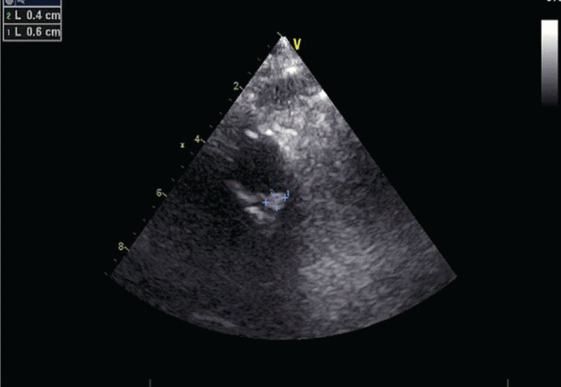
| A complete blood count revealed a white blood cell count of 3.57 cells/mm3, with 63.4% neutrophils and 30.4% lymphocytes. The C-reactive protein level was 140 µg/l, and ESR was 128 mm/hour. 48 hour after admission, there was fever onset with petechiae (Figure 1). Because infective endocarditis was suspected, a series of 7 aerobic and anaerobic blood cultures was taken before starting empirical treatment with intravenous vancomycin, rifampicin and gentamicin. A trans-thoracic echocardiogram performed on the third day after admission demonstrated evidence of a new oscillating mass on a pulmonary valve leaflet concerning for a possible vegetation (Figure 2). This was not present on the previous echocardiogram (approximately 1 month prior). |
| Two of the seven blood culture drawn at admission was reported positive at 72 hours (Bactec 9240 System) for small gram-negative bacilli. The isolate was subsequently identified as C. hominis. All subsequent blood cultures were reported as negative. The susceptibility of the bacteria is shown in Table 1. |
| Because third-generation cephalosporin’s are regarded as the drugs of choice for treating C. hominis endocarditis [3], vancomycin-rifampicin-gentamicin was switched to cefotaxime (1 g q 8 h intravenously). In conjunction with antibiotherapy, dental care was performed. The patient’s symptoms improved quickly and she remained without fever throughout with repeatedly normal ESR, CRP, complement fractions, and white cell counts. Elective replacement of the RVOT stenotic conduit was decided. At surgery, after six weeks of antibiotic treatment, the heterograft was found to be friable with fibrous degenerescence of the valve conduit, although no vegetations were identified. The RVOT conduit was replaced by a Carpentier-Edwards® valved conduit. The diameter of the valved conduit was 20 mm. The excised graft underwent prolonged culture but subsequent bacteriological examination showed no growth. The patient made an uneventful recovery and was discharged ten days after surgery. |
| Discussion |
| C. hominis is a fastidious gram negative bacillus [1]. It is a member of the HACEK (Haemophilus parainfluenzae, Haemophilus aphrophilus, and Haemophilus paraphrophilus, Actinobacillus actinomycetemcomitans, C. hominis, Eikenella corrodens, and Kingella kingae) group of microorganisms and is differentiated from them by a positive oxidase reaction and the production of indole [3]. Endocarditis caused by C. hominis, is rare in adults and extremely rare in children [4-6]. Reports in children have been recently reviewed [2]. To our knowledge this is the fifth report of C. Hominis endocarditis in pediatric population (Table 2). The presentation of endocarditis with one of the HACEK microorganisms is often insidious in onset. Patients often experience malaise with or without low grade fevers which can last weeks to months before seeking medical attention or a diagnosis is confirmed [2]. The patient presented here was afebrile on admission but had some intermittent fevers two weeks before. The patient therefore met modified Duke Criteria for definite infective endocarditis (echocardiographic findings, fever, predisposition, and positive blood cultures). C. Hominis is found as part of normal respiratory flora, the likely source of the organism in our patient, but it is seldom identified in that setting due to its slow growth characteristics compared with other resident bacteria [6]. The 72-hour interval between the initial blood draw and the report of the positive culture result in our patient is typical of this slow-growing organism [4-6]. Initial isolates of C. hominis were penicillin and ampicillin sensitive. However, beta-lactamase producing strains of C. hominis have been identified and since antimicrobial susceptibility testing may be difficult to perform on HACEK microorganisms [6], the American Heart association (AHA) now recommend that all HACEK microorganisms should be considered ampicillin resistant and third generation cephalosporins should be the treatment of choice [1]. For patients who cannot tolerate betalactams, trimethoprim/sulfamethoxazole, fluroquinolones and aztreonam are appropriate alternatives [1]. This is the third case demonstrating beta-lactamase-producing C. hominis in a pediatric patient. The first pediatric case was described in a 7-year-old girl with a history of Tetralogy of Fallot who was treated with aztreonam [7]. The second one was reported by Priyanka et al. [2] in a 12 year old boy with a history of RVOT reconstruction using a valved conduit for tetralogy of Fallot with pulmonary atresia. The patient was treated with 6 week antibiotic course of Levofloxacin due to an allergic response to ceftriaxone and ampicillin/sulbactam. In our case, as soon as Gram-negative bacilli were identified in cultures, antibiotherapy was switched to ceftriaxone. Intravenous antibiotherapy was administered for 6 weeks according to the AHA guidelines [1]. |
| Underlying heart disease is the major risk factor for developing endocarditis with approximately 76% of cases occurring in patients with surgically repaired structural cardiac abnormalities [2,6]. Because HACEK microorganisms are part of the normal oral flora, recent dental procedures are a risk factor for developing endocarditis with this group of microorganisms, especially for patients with underlying heart disease. Wormser et al. noted that 12 of 27 patients with endocarditis caused by one of the HACEK microorganisms had recent dental work or an oral infection prior to presentation [10]. Additionally children with congenital heart disease harbor HACEK microorganisms to a greater extent and have more gingival inflammation than healthy children [11]. In this case, the patient did not undergo a dental procedure but was noted to have poor dentition. |
References
- Wilson WR, Karchmer AW, Dajani AS, Taubert KA, Bayer A, et al. (1995) Antibiotic treatment of adults with infective endocarditis due to streptococci, enterococci, staphylococci, and HACEK microorganisms: American Heart Association. JAMA274: 1706-1713.
- Suresh P, Blackwood RA (2013) A pediatric case of cardiobacterium hominis endocarditis.Infect Dis Rep 5: e7.
- Geraci JE, Greipp PR, Wilkowske CJ, Wilson WR, Washington JA 2nd (1978) Cardiobacterium hominis endocarditis. Four cases with clinical and laboratory observations.Mayo Clin Proc 53: 49-53.
- Mylonakis E, Calderwood SB (2001) Infective endocarditis in adults.N Engl J Med 345: 1318-1330.
- Ferrieri P, Gewitz MH, Gerber MA, Newburger JW, Dajani AS, et al. (2002) Unique features of infective endocarditis in childhood.Pediatrics 109: 931-943.
- Feder HM Jr, Roberts JC, Salazar J, Leopold HB, Toro-Salazar O (2003) HACEK endocarditis in infants and children: two cases and a literature review.Pediatr Infect Dis J 22: 557-562.
- Currie PF, Codispoti M, Mankad PS, Godman MJ (2000) Late aortic homograft valve endocarditis caused by Cardiobacterium hominis: a case report and review of the literature.Heart 83: 579-581.
- Groner A, Kowalsky S, Arnon R, Tosi MF (2012) Endocarditis Due to Cardiobacterium hominis in a 4-Year-Old Boy, Complicated by Right Lower Lobe Pulmonary Artery Mycotic Aneurysm. J Ped Infect Dis 2: 278-280.
- Maurissen W, Eyskens B, Gewillig M, Verhaegen J (2008) Beta-lactamase-positive Cardiobacterium hominis strain causing endocarditis in a pediatric patient with Tetralogy of Fallot. Clin Microbiol News30:132-133.
- Wormser GP, Bottone EJ (1983) Cardiobacterium hominis: review of microbiologic and clinical features.Rev Infect Dis 5: 680-691.
- Steelman R, Einzig S, Balian A, Thomas J, Rosen D, et al. (2000) Increased susceptibility to gingival colonization by specific HACEK microbes in children with congenital heart disease.J Clin Pediatr Dent 25: 91-94.
Tables and Figures at a glance
| Table 1 | Table 2 |
Figures at a glance
 |
 |
|
| Figure 1 | Figure 2 |
Relevant Topics
- Advanced Therapies
- Chicken Pox
- Ciprofloxacin
- Colon Infection
- Conjunctivitis
- Herpes Virus
- HIV and AIDS Research
- Human Papilloma Virus
- Infection
- Infection in Blood
- Infections Prevention
- Infectious Diseases in Children
- Influenza
- Liver Diseases
- Respiratory Tract Infections
- T Cell Lymphomatic Virus
- Treatment for Infectious Diseases
- Viral Encephalitis
- Yeast Infection
Recommended Journals
Article Tools
Article Usage
- Total views: 13909
- [From(publication date):
April-2015 - Aug 07, 2025] - Breakdown by view type
- HTML page views : 9309
- PDF downloads : 4600
