Accessory Pinna with Accessory Tragus–A Rare Entity
Received: 07-Oct-2016 / Accepted Date: 24-Oct-2016 / Published Date: 31-Oct-2016 DOI: 10.4172/2161-119X.1000272
Abstract
The objective is to report a rare case of Accessory Pinna with Accessory Tragus. We present a case of a 13 year old boy who presented with swelling in the left cheek and left pre-auricular region since childhood. We diagnosed the case to be Accessory pinna with accessory tragus. Thorough evaluation was done to rule to presence of associated anomalies. The child was being ridiculed by his peers, hence surgical correction of the accessory pinna and accessory tragus was done
Keywords: Accessory pinna; Accessory tragus; Congenital anomaly of ear
253445Introduction
The pinna develops from the first branchial cleft at 6th week of intra-uterine life by the fusion of six hillocks. Initially the pinna begins to develop in the base of the neck but as the mandible develops it migrates to the normal anatomical position by 20th week of intrauterine life. Improper development of the external ear leads to congenital anomalies such as Polyotia, which is defined as an accessory pinna that is large enough to resemble an additional pinna. It is a rare congenital anomaly of the external ear. Accessory tragus may be found anywhere along the line of ascent-an imaginary line from tragus to angle of mouth or along anterior border of sternocleidomastoid. Accessory Pinna and Accessory Tragus are caused by failure of proper fusion of the six Hillocks of His during embryological development. It consists of skin, subcutaneous fat and may also contain elastic cartilage. There have been less than 30 reported cases of accessory pinna. Incidence of accessory tragus is approximately 5-10/1000 live births [1,2]. Management consists of surgical correction only for cosmetic purpose. They are also more prone to have associated renal and cardiac anomalies.
Case Report
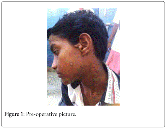
A 13 year old boy presented to the ENT OPD with a large Accessory Pinna near his left ear along with Accessory Tragus in the left cheek midway between angle of mouth and tragus (Figure 1). His accessory pinna was approximately 2.5 × 2.5 × 1 cm in size with cartilaginous component. His accessory tragus was approximately 1.0 cm × 0.5 cm in size. Right ear was within normal limits. Rest of ENT examination was within normal limits.
Pure Tone Audiometry, Ultrasound Abdomen, CT Temporal Bone were normal.
ECHO showed mild Mitral Valve Prolapse without significant Mitral Regurgitation.
The child was being ridiculed by his peers hence wanted surgical correction.
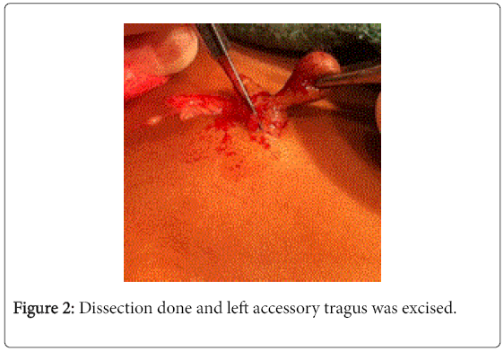
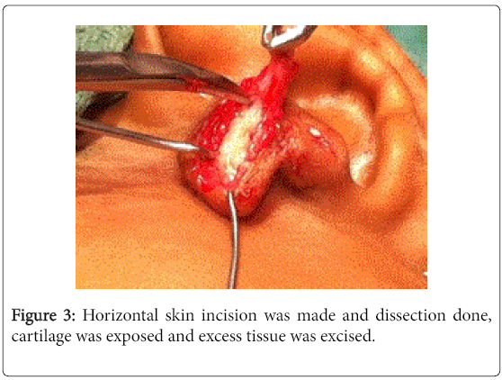
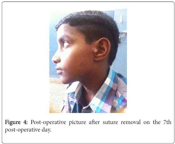
Surgical Correction-Under general anesthesia, both the Accessory Tragus and Accessory Pinna were excised (Figures 2 and 3) was uneventful. Patient was followed by at 1st week, 3rd week, 1st month and at 3rd months (Figure 4).
Discussion
Accessory pinna or polyotia is an extremely rare condition. It occurs due to failure of proper fusion of the six Hillocks of his during embryological development of the ear. Recent studies suggest that it results from extraordinary migration of neural crest cells in the branchial arch during development [3-8]. According to Lammer, fetal exposure to retinoic acid affects the migration of the neural crest cells. It may be single or multiple, unilateral or bilateral.
The first report of polyotia was by von Bol et al. in 1918. Due to rarity of Accessory Pinna, surgical techniques have not yet been established. Yung Moon et al reported a case of polyotia in 2014 where 9 year old boy presented with a large accessory pinna of size 2.3 × 2 cm. Surgical correction was done in two procedures [3]. The patient had a cartilaginous concave bowl that resembled a conchal hollow behind the accessory pinna. Surgical correction was done in two procedures. In the first procedure, the accessory pinna was resected in a wedge shape. The inferior chondrocutaneous flap was used to construct a new tragus. In the second procedure they eliminated the concha-resembling cartilaginous bowl. Raw et al. reported a case of polyotia where surgical correction was done using extraneous tissue and dermal substitute [3].
Accessory pinna has been reported to co-exist with syndromes such as Goldenhar Syndrome, and Treacher Collins Syndrome. Children with accessory tragus are also at a higher risk of renal anomalies. Durakbasa et al. reported that 4% of the children with Accessory Tragi were found to have cardiac and renal anomalies [1]. Thus ultrasound abdomen and ECHO should be mandatorily done.
Conclusion
In conclusion, patients presenting with Accessory Pinna and Accessory Tragus should be evaluated thoroughly to rule out presence of any syndromes. Parents should be advised to receive genetic counselling. Surgical correction is done only for cosmetic purpose.
References
- Durakbasa, Cigdem (2004) Should children with isolated preauricular tags be routinely evaluated for associated renal and cardiac malformations. Marmara Medical Journal 17: 109-112.
- Moon IY, Oh KS (2014) Surgical correction of an accessory auricle, polyotia.Arch PlastSurg 41: 427-429.
- EunYR (2015) Surgical Treatment of Polyotia. Archives of Craniofacial Surgery 16: 84-87.
- Kono T, Ro H, Murakami M (2015) Accessory auricles affecting the tragus and cheek occurring with cervical chondrocutaneousbranchial remnants: A Case Report. JPRAS Open 6: 20-24.
- Chander B, Dogra SS, Raina R, Sharma C, Sharma R (2014) Chondrocutaneousbranchial remnants or cartilaginous choristoma: terminology, biological behavior and salience of bilateral cervical lesions.Turk PatolojiDerg 30: 195-200.
- Aboud, Daifullah (2012) Syndromes which may feature accessory tragus : An Overview. Journal of Pakistan Association of Dermatologists 22: 262-263.
- Kar, Sumit (2013) Multiple Accessory Tragi: A Case Report. International Journal of Basic and Applied Medical Sciences3: 1-3.
- Kohelet D, Arbel E (2000) A prospective search for urinary tract abnormalities in infants with isolated preauricular tags.Pediatrics 105: E61.
Citation: Shanmugam R, Ravindra V, Shanmugam VU, Mariappan RG, Swaminathan B, et al. (2016) Accessory Pinna with Accessory Tragus–A Rare Entity. Otolaryngol (Sunnyvale) 6:272. DOI: 10.4172/2161-119X.1000272
Copyright: © 2016 Shanmugam R, et al. This is an open-access article distributed under the terms of the Creative Commons Attribution License, which permits unrestricted use, distribution, and reproduction in any medium, provided the original author and source are credited.
Select your language of interest to view the total content in your interested language
Share This Article
Recommended Journals
Open Access Journals
Article Tools
Article Usage
- Total views: 13932
- [From(publication date): 10-2016 - Aug 30, 2025]
- Breakdown by view type
- HTML page views: 13000
- PDF downloads: 932


