Make the best use of Scientific Research and information from our 700+ peer reviewed, Open Access Journals that operates with the help of 50,000+ Editorial Board Members and esteemed reviewers and 1000+ Scientific associations in Medical, Clinical, Pharmaceutical, Engineering, Technology and Management Fields.
Meet Inspiring Speakers and Experts at our 3000+ Global Conferenceseries Events with over 600+ Conferences, 1200+ Symposiums and 1200+ Workshops on Medical, Pharma, Engineering, Science, Technology and Business
Case Report Open Access
Bilateral Glenoid Osteochondritis Dissecance Detected on MR Arthrogram
| Abdullah Al–Mulhim1*, Mishal Al-Shalan2 and Alia Al-Barwan3 | |
| 1Radiology Demonstrator, Dammam University, Saudi Arabia | |
| 2Head section of MSK radiology, King Faisal Specialist Hospital and Research Center, Saudi Arabia | |
| 3Khoula Hospital, Sultanate of Oman, Saudi Arabia | |
| Corresponding Author : | Abdullah Al–Mulhim Radiology Demonstrator Dammam University and Musculoskeletal Radiology Fellow King Faisal Specialist Hospital and Research Centre, Saudi Arabia Tel: +966569908288 E-mail: dr.aalmulhim@gmail.com |
| Received October 29, 2012; Accepted December 04, 2012; Published December 08, 2012 | |
| Citation: Al-Mulhim A, Al-Shalan M, Al-Barwan A (2013) Bilateral Glenoid Osteochondritis Dissecance Detected on MR Arthrogram. OMICS J Radiology. 2:110. doi: 10.4172/2167-7964.1000110 | |
| Copyright: © 2013 Al-Mulhim A, et al. This is an open-access article distributed under the terms of the Creative Commons Attribution License, which permits unrestricted use, distribution, and reproduction in any medium, provided the original author and source are credited. | |
Visit for more related articles at Journal of Radiology
Abstract
OCD classically occur in convex articular surfaces. The femoral condyles, humeral capitulum and humeral head are the most common affected sites. It rarely involves the glenoid and the diagnosis requires a high index of suspicion. The pathological process and imaging findings are almost similar in all sites but most of the literature focused in the knee joint. The radiographic diagnosis might not be straight forward, and the use of CT scan with or without intra articular contrast and magnetic resonance imaging helps to establish the stability status. The mostly used treatment of stable lesions is conservative treatment and arthroscopic surgical management is preserved for unstable lesions.
| Introduction |
| True osteochondrosis must be differentiated from ossification variants. Normal variation in epiphyseal ossification centers are incidental cases [1], bilateral symmetrical and asymptomatic most of the time however, this normal variant eventually progress to OCD especially if the fragmentation is extensive. |
| Glenoid OCD is a rare entity with few case reports isolated in the literature. Unlike other locations, glenoid OCD [2] bony involvement is theorized to have less traumatic effect because most of the energy is being absorbed in the adjacent muscles and ligaments. In this report we documented a case of bilateral OCD of glenoid with different staging. |
| Objectives |
| To report the rare case of bilateral Osteochondritis Dissecance (OCD) in young non athletic boy and to review the latest literature, updates for similar findings in the glenoid articular surface. |
| Case Report |
| An 18 years old male presented to the orthopedic clinic complaining of bilateral, occasionally painful shoulder clicking that is aggravated by repetitive movement. He presented to the radiology department at King Faisal Specialist Hospital and Research Center and a Magnetic Resonant Arthrogram of both shoulders was performed. Consent was obtained from the patient for publication of this report. |
| Methods |
| Fluoroscopic guided injection of diluted Gadolinium contrast was performed. The mixer contains 0.1 ml of Gadolinium (Dotarem), 2 ml of xylocaine containing 1% epinephrine and 17 ml of sterile water. Then an MR arthrogram was obtained with coronal T2WI, axial, sagittal and coronal T1WI with fat suppression. Axial oblique T1WI fat saturated sequence in Abduction- External Rotation (ABER) was also performed [3]. |
| The right shoulder exam was performed in Trio Tim 3 Tesla Siemens and using axial T1WI with fat saturation (TE11/TR315, ST3.5, Matrix 320×240, FOV 16). Coronal T1WI (TE12/TR354 ST3), Sagittal T1WI with fat sat (TE11/TR368 ST3), Coronal T2WI (TE84/ TR3940 ST3 Matrix 384×257). |
| The left shoulder exam was performed in 1.5 Tesla GE Medical Systems And using axial T1WI fat saturation (TE9/TR483, ST3 Slice Spacing 4 mm, Matrix 320×244, FOV16), [4,5] Coronal T1WI with fat sat (TE8/TR633), Sagittal T1WI with fat sat (TE9/TR500), Coronal T2WI (TE97/TR3583, ST3, Slice Spacing 3.3 Matrix 384×256), Axial oblique T1WI with fat sat in ABER position (TE7.7/TR683 Spacing 3.3, ST3, FOV 16 Matrix 320×224). |
| The images were evaluated by three musculoskeletal radiologists using GE healthcare PACS (Centricity Radiology RA1000). |
| Results |
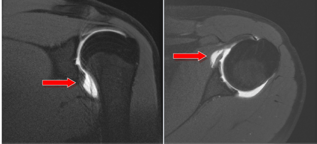
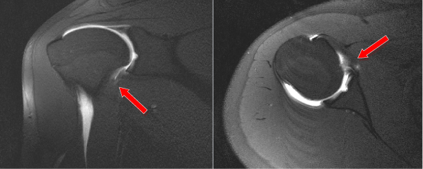
| The left shoulder showed an osteochondral defect measuring 3×5 mm in the central portion of the glenoid articular surface and containing tiny low signal intensity lose body which is surrounded by intra articular contrast [6]. The right shoulder showed an osteochondral defect with similar dimensions that is filled completely with intra articular contrast. |
| These findings are consistant with grade 3 OCD of the left shoulder (Figure 1) (detached in situ fragment) and stage 4 OCD (Figure 2) in the right shoulder (completely detached fragment) [7]. No definite rotator and non rotator cuff abnormality was depicted. The glenohumeral ligaments and other supporting structures were intact and there were no signs of capsular injury. |
| Discussion |
| Typically an osteochondrosis dissecans occurs in the region of a convex articular surface, with the medial femoral condyle being the most common location. Two theories help to explain the etiology of OCD. The first suggests the presence of poor vascular supply to the concave articular surfaces as compared to the convex surfaces. The other theory claims that the basic instability of the shoulder joint protects the bone against the occurrence of the lesion through the function of the muscle-ligament structures, softening much the traumatic forces thereby saving the glenoid from injuries. However, the exact of an OCD has not been clearly established. |
| An important mimicker is the presence of accessory ossification center of the glenoid. At birth most of the scapula is ossified. There are two separate centers for the glenoid; an upper glenoid center which gives rise to the base of the coracoid and the upper third of the glenoid, and a lower glenoid center which is horseshoe-shaped, giving rise to the lower two thirds of the glenoid. Both the upper and lower centers typically fuse by the age of ten to twelve years. After they fuse a small depression is noted mainly at the articular cartilage coverage which represents the central gelnoid region [8]. |
| A focal thickening of the subchondral bone in the mid aspect of the glenoid with thinning of overlying cartilage is considered a normal variant and named tubercle of Assaki. |
| The radiological appearance and classification is similar in most of the involved joints and different imaging modalities can be used to convey the abnormality to the clinician. Investigation of the cartilage and subchondral bone in young patients with mechanical pain should not be confined to the convex articular surfaces, although these are more likely to sustain trauma-related damage. |
| A wide spectrum of clinical signs may be evident including joint effusion, painful articular movements, clicking or locking. And among most of the cases repetitive movements elicits and exaggerates the pain yet, the condition might be painless. A spectrum is seen ranging from subchondral edema, subchondral fracture and fragmentation with or without displacement of an osteochondral fragment. Nevertheless, healing may occur initially. |
| Conventional radiographs are often diagnostic and the covering articular cartilage can be assessed by CT scan and MRI especially with use of intra articular contrast. It is important to assess the viability of the overlying cartilage and wither a loose osteochondral fragment is present and addressing its size and location. |
| Classically, the non-weight-bearing medial femoral condyle is the location in 85% of cases of OCD of the knee. OCD must be ruled out in the contra lateral joint, because 20–30% of cases are bilateral [3]. |
| We have not come across any papers that mention the presence of bilateral glenoid OCD in the literature of radiology. Until 2006 only 13 cases were mentioned in the English literature and none presented with bilateral disease. |
References |
|
Figures at a glance
 |
 |
| Figure 1 | Figure 2 |
Post your comment
Relevant Topics
- Abdominal Radiology
- AI in Radiology
- Breast Imaging
- Cardiovascular Radiology
- Chest Radiology
- Clinical Radiology
- CT Imaging
- Diagnostic Radiology
- Emergency Radiology
- Fluoroscopy Radiology
- General Radiology
- Genitourinary Radiology
- Interventional Radiology Techniques
- Mammography
- Minimal Invasive surgery
- Musculoskeletal Radiology
- Neuroradiology
- Neuroradiology Advances
- Oral and Maxillofacial Radiology
- Radiography
- Radiology Imaging
- Surgical Radiology
- Tele Radiology
- Therapeutic Radiology
Recommended Journals
Article Tools
Article Usage
- Total views: 13808
- [From(publication date):
March-2013 - Jul 07, 2025] - Breakdown by view type
- HTML page views : 9208
- PDF downloads : 4600
