Case Report Open Access
Bilateral Uveitis in Rickettsial Disease
| Thanuja G Pradeep*, Sindhu Satyanarayanan and Malavika Krishnaswamy | ||
| Department of Ophthalmology, M.S. Ramaiah Medical College and Hospital, Bangalore, India | ||
| Corresponding Author : | Thanuja G. Pradeep Department of Ophthalmology MS Ramaiah Medical College Bangalore, Karnataka, India Tel: 9611600888 E-mail: thanugopal@yahoo.co.in |
|
| Received October 27, 2014; Accepted January 03, 2015; Published January 07, 2015 | ||
| Citation: Pradeep TG, Satyanarayanan S, Krishnaswamy M (2015) Bilateral Uveitis in Rickettsial Disease. J Infect Dis Ther 3:199. doi: 10.4172/2332-0877.1000199 | ||
| Copyright: © 2015 Pradeep TG, et al. This is an open-access article distributed under the terms of the Creative Commons Attribution License, which permits unrestricted use, distribution, and reproduction in any medium, provided the original author and source are credited. | ||
Related article at Pubmed Pubmed  Scholar Google Scholar Google |
||
Visit for more related articles at Journal of Infectious Diseases & Therapy
Abstract
We report a case of bilateral uveitis in a 52-year-old male patient who was admitted to the intensive care unit with fever and skin rashes. He was diagnosed to have rickettsial fever and treated with antibiotics. He was referred to the ophthalmologist for red eye, which on examination revealed an exudative membrane over the iris in both eyes. The patient also had associated vitritis. The patient responded to topical and oral steroids but had sectoral iris atrophy both eyes and poor visual recovery. Ocular manifestations are rare in rickettsial disease and easily overlooked by the treating physician. Early referral of these cases helps in initiation of appropriate treatment that will reduce the visual morbidity in these individuals.
| Introduction | |
| Rickettsial diseases are the most covert re-emerging infections of present times, generally incapacitating and notoriously difficult to diagnose [1]. Under diagnosed or misdiagnosed rickettsial infection are public health problems in recent times with untreated cases having a poor prognosis. | |
| Rickettsial infections are characterized by a triad of high fever, headache, general malaise and skin rashes. They are transmitted by arthropod to humans. Narendra et al. [1] have reported about the rickettsial infection in the Indian scenario and they have concentrated on systemic features where in ocular manifestations are easily overlooked. There have been case reports of ocular affection in rickettsioses but most of them concentrated on posterior segment manifestation [2-5] with very few reports on anterior segment affection [6-8]. | |
| Diagnostic dilemma of rickettsial infection usually delays the recognition. The laboratory tests are also not widely available making the diagnosis difficult, thus delaying the treatment. | |
| Case Report | |
| A 52 years old male was admitted to our hospital with 4 days of fever with chills and rigors. He was a known alcoholic and a case of chronic liver disease. Initially he was treated as viral fever with thrombocytopenia after which he had altered sensorium and developed petechial rashes all over the body involving palms and soles for which he was admitted to ICU. Investigations for dengue serology, leptospirosis, widal and malarial parasite, were negative. He had altered LFT with elevated liver enzymes (AST, AGT and ALP). Patient had thrombocytopenia (Platelet count-26000/cubic mL) and low lymphocyte count (5%). He had hyponateremia (Na-122mEq) and hypoalbuminemia (Serum Albumin-1.7 g/dl). He was diagnosed to have sepsis with procalcitonin level of 6.46 ng/ml (greater than 2 - significant of sepsis) and treated symptomatically. A dermatology opinion was taken on the third day for the petechial rashes and a differential diagnosis of rickettsia was considered. | |
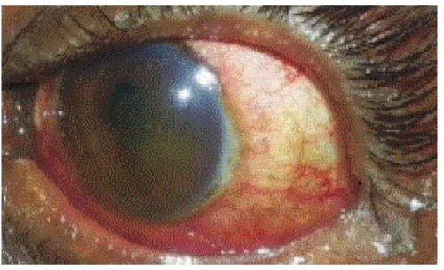
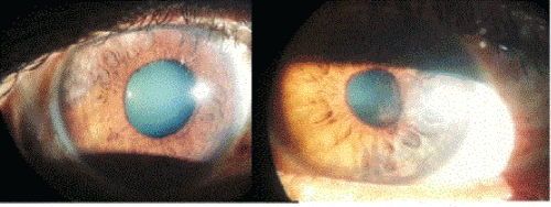
| An ophthalmology reference was sent for red eye one day after admission. On examination patient had altered sensorium, visual acuity could not be assessed. He had ciliary congestion in both eyes. The right eye (RE) showed exudative reaction involving superotemporal quadrant of the iris and small non-reacting pupils. In the left eye (LE) cornea was hazy with exudative membrane involving temporal half of the iris (Figure 1). Pupils were small and not reacting to light. Fundus could not be examined. Patient was started on mydriatics and topical steroids. Pupils were dilated after one day to reveal posterior synechiae. Posterior segment showed significant vitreous haze obscuring the retinal details. | |
| On the seventh day the laboratory tests showed a positive Weil Felix reaction of 1:320 titres with OX-19, which was considered significant for rickettsia. Immunofluorescence was not performed due to nonavailability. Patient was started on T. Doxycycline 100 mg b.d. Patient showed defervescence after 24 hours and had an improvement in the general condition thus confirming the possibility of rickettsial infection. Ocular examination showed resolving exudative reaction in the anterior chamber with iris atrophic patches, which were sectoral, but the patient continued to have visual acuity of CF -2 m right eye and 6/60 left eye due to vitreous inflammation. Fundus examination did not show any retinal involvement. | |
| Patient was started on oral steroids prednisolone 1mg/kg body weight after 2 weeks of oral antibiotics once his systemic condition stabilized. Patient showed resolution in vitreous inflammation after a week of oral steroids and was discharged after 28 days of hospital admission with a tapering dose of steroids. Follow up after 2 months showed poor vision of 6/36 OD and 6/60 OS. Patient had sectoral iris atrophy and an altered foveal reflex. At 6 month follow up patient had BCVA of 6/18 in the RE and 6/24 in the LE. | |
| Discussion | |
| Rickettsial infections are emerging public health problem in India. Batra [9] has reported a high magnitude of scrub typhus, spotted fever and India tick typhus caused by R.conorii. | |
| The course of the rickettsial infections is usually mild and benign. The prognosis varies [1,9] depending on the virulence of the bacterial strain and the host co-morbidities and early diagnosis. The disease usually mimics viral fevers and non-availability of laboratory investigations results in the delayed diagnosis. The Weil Felix test used in diagnosis has poor sensitivity and specificity and usually requires immunofluorescence to diagnose which is not widely available. | |
| Ocular manifestations are rare and easily overlooked in comparison to systemic manifestations. The review of literature revealed two case reports of non-granulomatous uveitis [6,7] that were reported as mild to moderate resolving with steroids and mydriatics. These cases showed resolution in 48 hours after treatment but in our case uveitis was severe with posterior segment inflammation and showed slow response to topical treatment and required systemic steroids to control inflammation. | |
| Ocular manifestations with rickettsial infections have been reported with predominantly posterior segment manifestations [2,3,5], there have been scanty reports of anterior segment manifestations. Anterior segment manifestation [4] included conjunctivitis, conjunctival nodule, subconjunctival hemorrhage, marginal corneal ulcer and keratitis. We report a case of iridocyclitis with extensive exudative reaction in anterior chamber and associated vitreous haze. On resolution of inflammation, patient had sectoral iris atrophy. | |
| Angiotropism makes vessels a potential target. The rickettsial organism usually affects the vascular endothelium that could explain the iris atrophy, which was sectoral in this case. Review of literature revealed case reports showing sectoral iris atrophy following herpes zoster infection [10-12] and sickle cell disease [13] but no reports are suggestive of iris atrophy following rickettsial infection. Therefore we must consider rickettsial infection as a differential diagnosis in a case of sectoral iris atrophy. The patient also had significant vitritis that resolved after starting systemic steroids under the cover of antibiotics. The delay in diagnosis of rickettsial infection resulted in late commencement of doxycycline and also his systemic condition did not allow us in starting the oral steroids immediately. All these could have contributed to poor visual recovery in our patient. Dilated ocular examination is essential in all cases to rule out posterior segment involvement and early institution of treatment to prevent visual morbidity. | |
| The treating physician needs to be aware of the ocular affections in rickettsial diseases. Early referral will help in recognition and timely treatment of the ocular inflammation and thus helps in preserving vision in these patients. | |
References
- Rathi N Rathi A (2010) Rickettsial infections: Indian perspective. Indian Pediatr 47: 157-164.
- Khairallah M Ladjimi A, Chakroun M, Messaoud R, Yahia SB, et al. (2004) Posterior segment manifestations of Rickettsia conorii infection. Ophthalmology 111: 529-534.
- Lukas JR, Egger S, Parschalk B, Stur M (1998) Bilateral small retinal infiltrates during rickettsial infection. Br J Ophthalmol 82: 1217-1218.
- Pinna A (2009) Ocular manifestations of rickettsiosis: 1. Mediterranean spotted fever: laboratory analysis and case reports. Int J Med Sci 6: 126-127.
- Beselga D Campos A Castro M Mendes S Campos J et al. (2014) A rare case of retinal artery occlusion in the context of mediterranean spotted Fever. Case Rep Ophthalmol 5: 22-27.
- Cherubini TD, Spaeth GL (1969) Anterior nongranulomatous uveitis associated with Rocky Mountain spotted fever. First report of a case. Arch Ophthalmol 81: 363-365.
- DEDUIT Y (1963) IRIDOCYCLITIS AND FOCUS OF RETINITIS IN THE COURSE OF A THERAPEUTIC RICKETTSIOSIS WITH RICKETTSIA QUINTANA. Ann Ocul (Paris) 196: 934-938.
- Beltrán LM, García S, Vallejo AJ, Bernabeu-Wittel M (2011) Bilateral anterior uveitis and Rickettsia typhi infection. EnfermInfeccMicrobiolClin 29: 235-236.
- Batra HV (2007) Spotted fevers & typhus fever in Tamil Nadu. Indian J Med Res 126: 101-103.
- Van der Lelij A Ooijman FM, Kijlstra A, Rothova A (2000) Anterior uveitis with sectoral iris atrophy in the absence of keratitis: a distinct clinical entity among herpetic eye diseases. Ophthalmology 107: 1164-1170.
- Lightman S, Marsh RJ, Powell D (1981) Herpes zoster ophthalmicus: a medical review. Br J Ophthalmol 65: 539-541.
- Doran M.Understanding and treating viral anterior uveitis.AAO Sept 2009
- Acheson RW, Ford SM, Maude GH, Lyness RW, Serjeant GR (1986) Iris atrophy in sickle cell disease. Br J Ophthalmol 70: 516-521.
Figures at a glance
 |
 |
|
| Figure 1 | Figure 2 |
Relevant Topics
- Advanced Therapies
- Chicken Pox
- Ciprofloxacin
- Colon Infection
- Conjunctivitis
- Herpes Virus
- HIV and AIDS Research
- Human Papilloma Virus
- Infection
- Infection in Blood
- Infections Prevention
- Infectious Diseases in Children
- Influenza
- Liver Diseases
- Respiratory Tract Infections
- T Cell Lymphomatic Virus
- Treatment for Infectious Diseases
- Viral Encephalitis
- Yeast Infection
Recommended Journals
Article Tools
Article Usage
- Total views: 14650
- [From(publication date):
February-2015 - Aug 17, 2025] - Breakdown by view type
- HTML page views : 9997
- PDF downloads : 4653
