Research Article Open Access
Black Stains in Primary Teeth: Overview
Fawzi Rachid1* and Hariri El Mehdi2
1Department Head in Pediatric Dentistry, Faculty of Dentistry, center of dental treatment and consultation, Mohammed V University in Rabat, Military Hospital Mohammed V, Rabat, Morocco
2Pediatric Dentistry, Faculty of Dentistry, Center of Consultation and Dental Treatment, Mohammed V University In Rabat, Military Hospital Mohammed V, Rabat, Morocco
- *Corresponding Author:
- Rachid F
Faculty of dentistry, Center of Dental Treatment And Consultation
Pediatric Dentistry Department, Mohammed V University In Rabat
Military Hospital Mohammed V, P.O: 6212 - Rabat Instituts, Rabat, Morocco
Tel: +212661208830
E-mail: fawzirachid@gmail.com
Received date: October 15, 2016; Accepted date: November 08, 2016; Published date: November 16, 2016
Citation: Rachid F, Mehdi HE (2016) Black Stains in Primary Teeth: Overview. Pediatr Dent Care 1:123.
Copyright: © 2016 Rachid F, et al. This is an open-access article distributed under the terms of the Creative Commons Attribution License, which permits unrestricted use, distribution, and reproduction in any medium, provided the original author and source are credited.
Visit for more related articles at Neonatal and Pediatric Medicine
Abstract
Black stain (BS) is a specific type of external discoloration observed at 2.4-21% of all children, on the buccal or lingual surfaces along the gingival margin and firmly attached to the tooth surface. According to some investigators, these stains are related with a lower frequency of caries. However, the cause of the black stain is still unknown. The purpose of this article is to summarize the fundamentals about black stain in primary teeth, its diagnosis and possible differential diagnoses as well as its microbiology and therapy.
Keywords
Black stains; Primary teeth; Caries
Introduction
Black tooth stain is a characteristic extrinsic discoloration, which occurs along the third cervical line of the buccal and/or lingual surfaces of teeth, particularly in the primary dentition. However, the permanent teeth may be also concerned. They appear early on the tooth enamel, at the age of 2 or 3. Its negative effect on the dental aesthetic perception causes concerns in parents and can have significant effects on the personality and self-confidence of the child.
Mechanism of Constitution
Black stain is a form of dental plaque. It’s different from other types by insoluble iron salt and high calcium and phosphate composition.
The black material is a ferric compound, most likely a ferric sulfide, which arises from the interaction in the saliva or gingival fluid betwen hydrogen sulfide (produced by bacteria in the periodontal environment) and iron. Some hypothesis concerning the association between black stains and some bacterial strains (actinomyces, lactobacillus sp, prevotella melaninogenica) has been reported [1,2]. The attraction of materials to the tooth surface is important to the formation of extrinsic dental stain. These attractive forces include electrostatic, van der Waals, hydration forces, hydrophobic interactions and hydrogen bonds. However, the mechanisms that determine the strength of adhesion are not perfectly understood.
Diagnosis and Classification
Criteria for the diagnosis of black stains are not well established. Some authors propose different criteria to establish a diagnosis; Koch et al. [3] considered that the presence of dark dots (diameter less than 0.5 mm) forming linear discoloration (parallel to the gingival margin) at dental smooth surfaces of at least two different teeth without cavitation of the enamel surface. As standardised classification systems are generally useful for clear communication between practitioners, numerical indices have also been introduced.
Shourie [3] recorded the presence or absence of pigmented plaque as: (1) no line; (2) incomplete coalescence of pigmented dots; (3) continuous line formed by pigmented spots. Gasparetto et al. [4] added another criteria based on the area of the tooth surface affected: First degree corresponds to the presence of pigmented dots or thin lines with incomplete coalescence parallel to the gingival margin; second degree indicates the presence of continuous pigmented lines, which are easily observed and limited to half of the cervical third of the tooth surface; third one equals the presence of pigmented stains extending beyond half of the cervical third of the tooth surface. A similar classification was used by Leung who used four scores: (1) correspond on thin line of 1 mm or less in width across the surface, (2) and (3) equalling staining of one third and two thirds respectively and (4) involving all dental surface.
Differential Diagnosis
Tooth discoloration is a frequently found condition that can be associated with medical problems and might also lead to psychological challenges of the individual affected.
The etiology of tooth discoloration are classified according to the location of the stain and are divided into extrinsic, intrinsic, or internalized.
Black stain is part of this group, but there also exist brown, green or orange stain, metallic stain, or stains caused by tobacco, restorative materials and pharmaceuticals. Tannins found in tea, coffee and other beverages are the presumed cause of the thin, bacteria free and pellicle like brown stain. Green stain appears as a heavy grey green, soft and ‘furry’ film, and has been attributed to fluorescent bacteria and fungi [5]. This condition is more prevalent in males than in females and has been associated with poor oral hygiene [6]. Orange stain is a light, thin deposit with a red to orange color. It is supposedly caused by chromogenic bacteria and again is found in a population with poor oral hygiene [5]. Metallic stains, which form after industrial exposure to iron, manganese and silver are also part of the extrinsic, stain group, as well as chlorhexidine stains. The latter are caused by the interaction of the dicationic antiseptic chlorhexidine with dietary chromogens. Intrinsic stains, in contrast, have a completely different appearance and can hardly be mistaken for black stain. They result from the incorporation of pigments into the dental tissues, which can happen either during odontogenesis or after eruption [7-11].
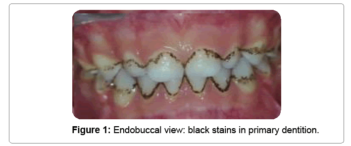
Frequent reasons for this condition are infections (e.g. rubella, measles), drug interactions (tetracycline, fluorides), malnutrition, hematopoietic disorders (sickle cell anaemia, thalassemia), or developmental disorders (amelogenesis and dentinogenesis imperfecta) [12-15]. Another important differential diagnosis is dental caries. However, while caries manifests with an irreversible local enamel or dentin defect, black stain is a deposit that can be removed through instrumentation and polishing. Also, the dental surface is left intact without signs of decalcification [7,8]. Its dotted appearance and characteristic localisation close to the gingival margin also facilitate the diagnosis (Figure 1).
Treatment
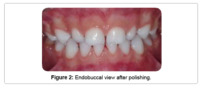
The black stain particularly poses an aesthetic problem. Daily Tooth brushing is not enough to remove this external stain. The professional cleaning is necessary to remove stains and resolve this aesthetic problem. Although a simple scaling and tooth brushing with pumice powder are usually sufficient, frequently black stain is recurrent. The ultrasonic cleaning is not recommended, this modality can lead to enamel removal; therefore, their repeated use is undesirable. Nevertheless, it can challenge the dentist, especially when it is deposited on roughened or pitted areas of the tooth. In a professional dental hygiene appointment, removal through polishing with rubber cup and fluoride pumice is possible. If the staining is resistant, the excess water can be blotted from the pumice and the tooth should be dried before the polishing procedure is performed [12-15]. Also, sharp scaling instruments are of use against firmly attached deposit. Black stain tends to reform again despite good personal oral care, but quantity may be less when biofilm control procedures are meticulous [12] (Figure 2).
Conclusion
Black stain is an extrinsic pigmented deposit that is generally observed in primary dentition. The mechanisms of constitution of these stains are not perfectly understood. In contrast, individuals with this condition seem to present with lower caries prevalence, explained by the presence of different oral bacterial strain in association with this discoloration.
References
- Shah HN, Bonnett R, Mateen B, Williams RA (1979) The porphyrin pigmentation of subspecies of Bacteroides melaninogenicus. Biochem J 180: 45-50.
- Stenudd C, Nordlund A, Ryberg M, Johansson I, Kallestal C, Stromberg N (2001) The association of bacterial adhesion with dental caries. J Dent Res 80: 2005-2010.
- Koch MJ, Bove M, Schroff J, Perlea P, Garcia-Godoy F, Staehle HJ (2001) Black stain and dental caries in schoolchildren in Potenza, Italy. ASDC J Dent Child 68: 353-355
- Gasparetto A, Conrado CA, Maciel SM, Miyamoto EY, Chi- carelli M et.al. (2003) Prevalence of black tooth stains and dental caries in Brazilian schoolchildren. Braz Dent J 14: 157-161.
- Hattab FN, Qudeimat MA, al-Rimawi HS (1999) Dental discolora- tion: an overview. J Esthet Dent 11: 291-310.
- Sarkonen N, Kononen E, Summanen P, Kanervo A, Takala A et.al (2000) Oral colonization with Actinomyces species in infants by two years of age. J Dent Res 79: 864-867.
- Sarkonen N, Kononen E, Summanen P, Kononen M, Jousi- mies-Somer H (2001) Phenotypic identification of Actinomyces and related species isolated from human sources. J Clin Microbiol 39: 3955-3961.
- Theilade J, Pang KM (1987) Scanning electron microscopy of black stain on human permanent teeth. Scanning Microsc 1: 1983-1989.
- Heinrich-Weltzien R, Monse B, Helderman W (2009) Black stain and dental caries in Filipino schoolchildren. Community Dent Oral Epidemiol 37: 182-187.
- Paredes Gallardo V, Paredes Cencillo C (2005) Black stain: a common problem in pediatrics. An Pediatr (Barc) 62: 258-260.
- Reid JS, Beeley JA, MacFarlane TW (1976) A study of the pigment produced by Bacteroides melaninogenicus. J Dent Res 55: 1130.
- Saba C, Solidani M, Berlutti F, Vestri A, Ottolenghi L, et al. (2006) Black stains in the mixed dentition: a PCR micro-biological study of the etiopathogenic bacteria. J Clin Pediatric Dent 30: 219-224.
- Saba C, Solidani M, Berlutti F, Vestri A, Ottolenghi L, Polimeni A (2006) Black stains in the mixed dentition: a PCR micro- biological study of the etiopathogenic bacteria. J Clin Pediatric Dent 30: 219-224.
- Theilade J (1977) Development of bacterial plaque in the oral cavity. J Clin Periodontol 4: 11-12.
- Wilkins EM( 2005) Clinical practice of the dental hygienist (9th Edn). Philadelphia: Lippincott Williams & Wilkins 316- 317.
- Bonecker M, Cleaton-Jones P (2003) Trends in dental caries in Latin American and Caribbean 5-6 and 11-13-year-old children: a systematic review. Community Dent Oral Epidemiol 31: 152-157.
Relevant Topics
- About the Journal
- Birth Complications
- Breastfeeding
- Bronchopulmonary Dysplasia
- Feeding Disorders
- Gestational diabetes
- Neonatal Anemia
- Neonatal Breastfeeding
- Neonatal Care
- Neonatal Disease
- Neonatal Drugs
- Neonatal Health
- Neonatal Infections
- Neonatal Intensive Care
- Neonatal Seizure
- Neonatal Sepsis
- Neonatal Stroke
- Newborn Jaundice
- Newborns Screening
- Premature Infants
- Sepsis in Neonatal
- Vaccines and Immunity for Newborns
Recommended Journals
Article Tools
Article Usage
- Total views: 133044
- [From(publication date):
specialissue-2016 - Aug 16, 2025] - Breakdown by view type
- HTML page views : 130660
- PDF downloads : 2384


