Research Article Open Access
Changes in Muscle Coordination Following Robot-assisted Gait Training in Hemiparetic Stroke
| Thrasher TA1* and Fisher S2 | |
| 1Center for Neuromotor and Biomechanics Research, University of Houston, Houston, USA | |
| 2The Methodist Neurological Institute, Houston, USA | |
| Corresponding Author : | Adam Thrasher 3855 Holman Street, Garrison Room 104 Houston, TX 77204-6015, USA Tel: 713-743-5276 Fax: 713-743-9860 E-mail: athrasher3@uh.edu |
| Received April 22, 2014; Accepted June 27, 2014; Published June 30, 2014 | |
| Citation: Thrasher TA, Fisher S (2014) Changes in Muscle Coordination Following Robot-assisted Gait Training in Hemiparetic Stroke. J Nov Physiother 4:217. doi: 10.4172/2165-7025.1000217 | |
| Copyright: © 2014 Thrasher TA, et al. This is an open-access article distributed under the terms of the Creative Commons Attribution License, which permits unrestricted use, distribution, and reproduction in any medium, provided the original author and source are credited. | |
Visit for more related articles at Journal of Novel Physiotherapies
Abstract
Robot-Assisted Gait Training (RAGT) has been shown to improve walking function in hemiparetic stroke. It is assumed, but unproven, that these improvements are associated with enhanced muscle coordination resulting from neurological changes in locomotor control. The goal of this study is to assess changes in muscle coordination in the lower extremities before and after an RAGT intervention using the AutoambulatorTM. Four individuals with subacute stroke participated in this prospective case series. All participants had hemiparesis and were able to walk with supervision or minimal contact assistance. Each participant received 18 one-hour sessions of RAGT over an 8-week period. Before and after the RAGT intervention, gait was assessed using a Timed Up-and-Go and a 10 m Walk Test. Participants also underwent a muscle coordination evaluation based on surface electromyography (SEMG) recorded from lower extremity muscles. SEMG electrodes were placed to record the following major muscle groups bilaterally: vastus lateralis, biceps femoris, tibialis anterior and lateral gastrocnemius. A Symmetry Index (SI) was defined to represent the similarity of SEMG signals from both sides of the body during gait. As well, an Index of Rhythmicity (IR) was defined to represent the degree to which the composite SEMG signals could be characterized by a basic recurring pattern. Following the RAGT intervention, participants demonstrated a 16 to 60% increase in walking speed and a 14 to 37% decrease in time to complete the Timed Up-and-Go test. Similarly, all four participants showed improvements in SI and IR. These results indicate that in addition to significant improvements in walking function following RAGT, muscle activation patterns were more rhythmic and more coordinated between both sides of the body. These findings suggest that the improvements in gait function following RAGT are associated with improvements in muscle coordination. These changes are likely due to positive adaptations in the central nervous system.
| Keywords |
| Surface electromyography (SEMG); Gait; Rehabilitation; Coordination; Locomotor training |
| Introduction |
| Recovery of ambulation is a key goal for many stroke patients with hemiparesis. Hemiparetic gait is typically characterized by slow walking speed, asymmetrical limb movements, short step length and reduced range of motion on the affected side. Long-term improvements in gait are possible through intensive locomotor therapy [1]. Robot-Assisted Gait Training (RAGT) is an alternative to manually-assisted treadmill training that is designed to alleviate the burden on therapists. RAGT provides external forces to the limbs of patients to mimic normal kinematics during locomotor training [2]. Learning occurs via high doses of repetitive, strictly-guided movements. Randomized controlled trials have shown that RAGT is as effective as conventional physical therapy and body-weight supported treadmill training [3]. To date, there have been several studies of the efficacy of RAGT in stroke [4], but few have investigated its specific effects on coordination of muscle activation patterns. |
| The idea that human locomotion is driven by oscillating neural circuits located in the spinal cord has been advanced for decades [5]. Locomotor training focuses on adaptation of the Central Pattern Generator (CPG), which is a largely autonomous neural circuit that produces cyclical bursts of pre-determined muscle activation signals [6,7]. The output of the CPG can be measured in the extremities using surface electromyography (SEMG), from which rhythmic patterns can be identified using a statistical classification technique [8]. This way, we can evaluate the degree to which multiple muscles act in a stable, rhythmic, coordinated manner. Furthermore, we can infer changes that take place in the central nervous system by repeating this evaluation at different points during rehabilitation (i.e., before and after an intervention). |
| To date, most studies on RAGT in stroke evaluated locomotion using standard clinical scores, such as the 10 m Walk Test and the Timed-Up-and-Go test. Ostensibly, the improvements in these scores are due to enhanced locomotor control governed by the central nervous system. However, it has never been demonstrated that lower extremity muscle activation patterns do change fundamentally during training. This study is a first attempt to confirm that the muscle activation signals after training are more symmetrical and rhythmic (i.e., coordinated) than before training. |
| Materials and Methods |
| Four (n=4) individuals with hemiparesis due to stroke were recruited from an outpatient clinic in a rehabilitation hospital located in Humble, TX. To qualify, participants must have had a stroke in the previous 6 months, and it must have been their first stroke. Furthermore, they must have scored 4 or 5 on the locomotion component of the Functional Independence Measure (FIM) indicating moderately impaired gait requiring supervision or minimal assistance. Descriptive statistics of the participants are provided in Table 1. All participants provided informed written consent. The study procedures were approved by the University of Houston’s Committee for Protection of Human Subjects. |
| Each participant received 24 one-hour sessions of RAGT using the AutoAmbulator at a schedule of three sessions per week for eight weeks. Each session consisted of 30 minutes of stretching and strengthening exercises administered by a highly-trained neurorehabilitation team consisting of a Physical Therapist and two Physical Therapy Assistants who applied appropriate manual assistance as necessary. This was followed by 30 minutes of RAGT using the Autoambulatora (HealthSouth, Birmingham, AL) [9]. It is mounted over a treadmill and includes body-weight support. The device settings, including walking speed and percentage of bodyweight support, were custom-fitted to each participant each session by a therapist (the same therapist for all four subjects). |
| Gait function was assessed before and after treatment using the following clinical tests: Timed Up-and-Go, and 10 m Walk Test. Both tests were performed in a wide, empty hallway. Participants were allowed to use a 4-point cane. |
| During the 10 m Walk Test, participants’ lower extremities were instrumented with an 8-channel SEMG system (Biometrics DataLOG, Biometrics Ltd, Ladysmith, VA, USA). SEMG was recorded from four major muscle groups bilaterally: vastus lateralis, long head of biceps femoris, tibialis anterior and gastrocnemius lateralis. The skin was cleaned and lightly abraded before electrodes were attached over the muscle bellies. SEMG signals were amplified, filtered (bandpass: 15-450 Hz), and A/D converted at a sampling rate of 2000 Hz. The signals were then rectified and low-pass filtered at a cut-off frequency of 10 Hz. The beginning of each step was determined by initial contact of the heels, which was detected by the activation of a foot switch placed on the sole of each shoe directly under the heel. |
| These signals were then separated into n individual gait cycles marked by heel contact, and time-normalized relative to the gait cycle using linear interpolation. Each gait cycle was normalized to exactly 100 points with a constant time interval. A Symmetry Index (SIm) was then determined for each muscle group using the following equation. |
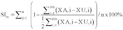 |
| Where m refers to the muscle in question, XA,i and XU,i refer to the amplitudes of the normalized SEMG signal for the affected and unaffected legs, respectively, at the i th time point in the j th gait cycle. By this definition, two muscles that produce the same time-normalized signal starting from respective heel contact (i.e., 180 degrees out of phase) have perfect alternating symmetry, SIm =100%. An unweighted average of all four muscle groups (m =1 ... 4) was used to determine overall SI, which was used in the following analysis. |
| Rhythmicity was assessed by processing and coding SEMG signals using fuzzy sets according to a classification procedure previously described [8]. This method was designed to represent multiple muscle activation signals as a recurrent sequence of four basic burst patterns in the manner of the CPG. The relative amount of SEMG signal that fits this model can be quantified using the statistical variable, R2. We interpreted R2 as the Index of Rhythmicity (IR); it represents the proportion of the muscle activation signals that recur with observed regularity. All data processing was performed using custom-written software in the MATLAB programming language (The Mathworks, Inc., Natick, MA, USA). |
| Results |
| All participants successfully completed the RAGT regimen, as well as the baseline and post-RAGT tests. All participants improved in terms of the Timed Up-and-Go and 10m Walk Tests. The initial and final values of the functional mobility tests are shown for all participants in Figure 1. Participants demonstrated a 16 to 60% increase in walking speed and a 14 to 37% decrease in time to complete the Timed Up-and-Go test. High variability can be seen between participants in terms of both mobility tests, especially at baseline. This variability decreased notably after RAGT. In general, the improvements in these scores were greater among participants with more severe impairment. Our analysis of the SEMG data revealed that all participants exhibited increases in SI and IR after RAGT. These data are shown in Figure 2. |
| Discussion |
| Four individuals with hemiparesis due to stroke underwent an 8- week intervention of RAGT administered using a powered exoskeleton with partial body-weight support. All participants improved in terms of functional mobility assessments. These improvements were accompanied by increased symmetry of muscle activation during gait. That is, the muscle activation signals in the affected leg were more similar to those in the unaffected leg (180 degrees out of phase) after RAGT. Also, the combined muscle activation patterns of the lower extremities exhibited increased rhythmicity during gait. We can infer from these results that alterations occurred within the central nervous system such that locomotor activity is more coordinated and more consistent with a model of CPG control. This supports the idea that RAGT may affect positive adaptations in the central nervous system in hemiparetic stroke. |
| We assessed rhythmicity using a statistical model of CPG control that yielded values of IR between zero and one. In our previous work, we measured values of IR in normal, healthy adults between 0.70 and 0.80 [8]. Perfect rhythmicity (IR=1.00) represents a set of perfectly periodic SEMG signals with no deviations. Normal, healthy gait involves some variations from periodic muscle activity. According to the optimal variability hypothesis [10], a certain amount of variability is desirable in physiological systems to deal with perturbations and remain within a state of dynamic equilibrium. Three of the four hemiparetic subjects in this study had subnormal IR (less than 0.70), indicating some aberrant, “noisy” muscle activity during gait. Following RAGT, the aberrations were reduced, resulting in IR scores closer to the normal range. In our analysis, we applied the simplest interpretation of “improvement” as any change in IR greater than zero. We acknowledge that small changes that are greater than zero may not be clinically significant. Further study is needed to establish a threshold value that correlates to significance in this context. |
| The present study is one of the few analyses that deal with changes in coordination (synchronization and ryhthmicity) of multiple muscles following locomotor training. |
| Future work should involve experiments to compare changes in muscle coordination between patients who have undergone RAGT and other forms of locomotor training, such as conventional physical therapy and body-weight supported treadmill training with manual assistance. |
| Conclusion |
| Our data showed that muscle coordination improved following 8 weeks (24 sessions) of RAGT. This signifies the possibility that RAGT may affect fundamental alterations in locomotor control, a hypothesis that should be investigated via an experimental design. In our case series, we utilized a non-invasive method to identify how the central nervous system controls locomotion before and after a rehabilitation intervention. We believe that this is a practical method to measure changes in the central nervous system for any intervention for locomotor training. |
| Acknowledgements |
| All financial support for this study was provided by HealthSouth Corporation (Humble, TX). |
References |
|
Tables and Figures at a glance
| Table 1 |
Figures at a glance
 |
 |
| Figure 1 | Figure 2 |
Relevant Topics
- Electrical stimulation
- High Intensity Exercise
- Muscle Movements
- Musculoskeletal Physical Therapy
- Musculoskeletal Physiotherapy
- Neurophysiotherapy
- Neuroplasticity
- Neuropsychiatric drugs
- Physical Activity
- Physical Fitness
- Physical Medicine
- Physical Therapy
- Precision Rehabilitation
- Scapular Mobilization
- Sleep Disorders
- Sports and Physical Activity
- Sports Physical Therapy
Recommended Journals
Article Tools
Article Usage
- Total views: 14179
- [From(publication date):
September-2014 - Aug 20, 2025] - Breakdown by view type
- HTML page views : 9546
- PDF downloads : 4633