Make the best use of Scientific Research and information from our 700+ peer reviewed, Open Access Journals that operates with the help of 50,000+ Editorial Board Members and esteemed reviewers and 1000+ Scientific associations in Medical, Clinical, Pharmaceutical, Engineering, Technology and Management Fields.
Meet Inspiring Speakers and Experts at our 3000+ Global Conferenceseries Events with over 600+ Conferences, 1200+ Symposiums and 1200+ Workshops on Medical, Pharma, Engineering, Science, Technology and Business
Case Report Open Access
Correlation of MRI Pattern and Histological Features in a Schwannoma of the Soft Palate in a 13-Year-Old Girl
| Claudio Sicca1, Angelina Cistaro2,3,4*, Natale Quartuccio5, Eleonora Sardo6, Mattia Berrone7, Ubaldo Familiari8, Sergio Gandolfo9 and Monica Pentenero9 | |
| 1Indipendent Clinical Dentistry, Forno Canavese and Bruino, Turin, Italy | |
| 2Positron Emission Tomography Centre IRMET S.p.A., Euromedic inc., Turin, Italy | |
| 3Co-ordinator of PET Pediatric AIMN InterGroup, Italy | |
| 4Associate researcher of Institute of Cognitive Sciences and Technologies, National Research Council, Rome, Italy | |
| 5Nuclear Medicine Unit, Department of Biomedical Sciences and of the Morphological and Functional Images, University of Messina, Messina, Italy | |
| 6Department of Radiology, AOU S. Luigi Gonzaga, Orbassano, Italy | |
| 7University of Turin, Department of Oncology, Oral Medicine and Oral Oncology Unit, Italy | |
| 8Pathology Division, AOU S. Luigi Gonzaga, Orbassano, Italy | |
| 9Department of Clinical and Biological Sciences, Oral Medicine and Oral Oncology Section, University of Turin, Turin, Italy. | |
| Corresponding Author : | Angelina Cistaro Positron Emission Tomography Centre IRMET S.p.A. Euromedic Inc. V. O. Vigliani 89/A, 10138 Turin, Italy Tel : +390113160158 Fax : +390113160828 E-mail: a.cistaro@irmet.com |
| Received November 24, 2014; Accepted January 13, 2015; Published January 16, 2015 | |
| Citation: Sicca C, Cistaro A, Quartuccio N, Sardo E, Berrone M (2015) Correlation of MRI Pattern and Histological Features in a Schwannoma of the Soft Palate in a 13-Year-Old Girl. OMICS J Radiol 4:177. doi: 10.4172/2167-7964.1000177 | |
| Copyright: ©2015 Sicca C et al. This is an open-access article distributed under the terms of the Creative Commons Attribution License, which permits unrestricted use, distribution, and reproduction in any medium, provided the original author and source are credited. | |
Visit for more related articles at Journal of Radiology
Abstract
Schawnnoma or neurilemmoma is a benign slow-growing neoplasm of Schwann cells that can arise from any cranial, peripheral or autonomic nerve. The involvement of the oral cavity is extremely rare. We present a case of a schwannoma lesion in the soft palate in a young girl. This report introduces the clinical aspects of schwannoma of the soft palate, and the correlation between the histopathological features and the Magnetic Resonance Imaging (MRI) features of this lesion.
| Keywords |
| Schwannoma; Magnetic Resonance Imaging; Soft palate |
| Introduction |
| Schwannoma is an uncommon benign tumor involving the head and neck region in about 25-45 % of cases. Only 1% of cases arise from the oral cavity [1-3]. The involvement of the soft palate is rare [4,5]. This tumor is typical of the adult age [5,6]. Malignant changes have been reported rarely and in long-lasting lesions [4]. |
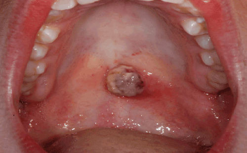
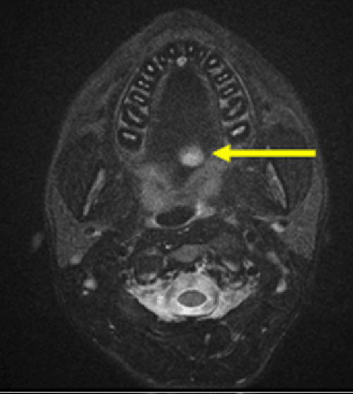
| We report a particular case of schwannoma arising in the soft palate of a 13 years old girl, referred by the pediatrician for the presence of a swelling rapidly growing on the border between hard and soft palate since a couple of weeks ago. The pediatrician administered antibiotic therapy without any results. The lesion continued to enlarge. On examination, a purplish round-shaped sessile soft and friable mass of about 15 mm with superficially ulcerated surface was observed (Figure 1). Given the fast growing of the lesion and the uncertain diagnosis, an incisional biopsy was performed. The pathological report described a mass, mainly formed by proliferating neural cells with granulation tissue, consistent with schwannoma and suggesting a complete excision for more accurate analyses. A MRI exam was obtained with a General Electric MRI of 1,5 Tesla using a dedicated maxillofacial ‘s protocol consisted by sagittal, coronal and axial images T1 and T2 - weighted images before and after injection of paramagnetic contrast media. MRI showed a little oval lesion of 10 mm with slightly irregular margins in the median-paramedian left side region of the soft palate (Figure 2). The lesion showed a hypointense MRI signal on T1-weighted images and a hyperintense MR signal on T2-weighted and in FLAIR sequences. After endovenous injection of paramagnetic contrast media, this solid lesion had a strong and uniform enhancement on T1- weighted images as well as on T2, with no evidence of cystic or necrotic area. Therefore, the MRI features suggested a possible diagnosis of schwannoma, showing a mass leaning on the overlying nasal mucosa without involving it and without any bone erosion in the near hard palate. |
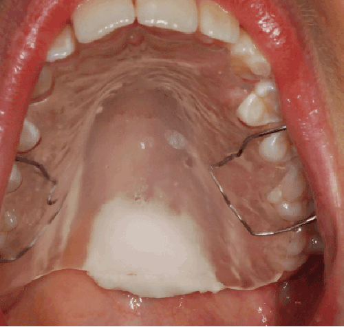
| Surgery was performed under general anesthesia with nasotracheal intubation via an intra-oral approach, aiming in order to achieve a complete enucleation. The mass was enucleated leaving intact the overlying nasal mucosa (Figure 3); having obtained a proper hemostasis by bipolar diathermy, the wound was packed by a collagen membrane [7,8]. At histology, the lesion showed well-defined borders but without fibrous capsule formation and with ulceration of the oral mucosa. At the histopathological study, performed by an experienced pathologist, cytological atypia was mild, with small oval nuclei. Nucleoli were absent. Mitotic figures were very infrequent, and atypical mitotic figures were absent. Antoni A pattern was prevalent: tumor cells were spindle-shaped, uniform and arranged in sheets interspersed in a dense collagenous stroma, with prominent nuclear palisading. Antoni patterns (A and B) are specific cellular pattern which have been recognized in the peripheral nerve sheath tumors and first described by Prof. Nils Ragnar Eugene Antoni [5]. The lesion stained intensively in both nuclei and cytoplasm for S-100 protein. Ki-67 index was low (5%). No degenerative changes suggestive for ancient schwannoma were found. No other stains were done. |
| A removable custom made resin plate was used to protect the wound (Figure 3); the postoperative course was free of complications and a complete second intention healing was achieved in about 5 weeks. After a 6-months follow-up the patient is free from disease. Usually schwannoma presents as submucosal nodule, nevertheless our patient showed a purplish lesion with superficially ulcerated surface. Generally, the treatment of choice for schwannomas is a conservative surgical excision [9] and only rarely malignant changes may arise in untreated schwannomas [1]. |
| The prevalence of Antoni types A or B morphological patterns has been described as having a potential impact on the neuroimaging features [10]. The present case was mainly characterized by a histological Antoni types A pattern and showed a regular MRI signal and a homogeneous enhancement after injection of paramagnetic contrast media. MRI had a key role in the definition of the extent of the lesion in order to plan a proper surgical approach. The use of a resin plate to guide and protect the wound healing allowed a less invasive and shorter surgical approach, as the defect was not covered by any pedicle flap. Moreover, the protection given by the plate almost immediately allowed feeding and swallowing. |
| To the best of our knowledge, a few cases of schwannoma in the oral cavity are present in literature [3,4,11,12] with only four cases of schwannoma of the soft palate reported in adults and three in pediatric patients [4,13,14]. MRI appears to be an additional and valuable tool in supporting the diagnosis of schwannoma of the soft palate. This case elucidates that a correct preoperative diagnostic protocol comprehending incisional biopsy and MRI imaging is mandatory to properly plan surgical excision of lesions with non-univocal clinical features. |
References |
|
--
Figures at a glance
 |
 |
 |
| Figure 1 | Figure 2 | Figure 3 |
Post your comment
Relevant Topics
- Abdominal Radiology
- AI in Radiology
- Breast Imaging
- Cardiovascular Radiology
- Chest Radiology
- Clinical Radiology
- CT Imaging
- Diagnostic Radiology
- Emergency Radiology
- Fluoroscopy Radiology
- General Radiology
- Genitourinary Radiology
- Interventional Radiology Techniques
- Mammography
- Minimal Invasive surgery
- Musculoskeletal Radiology
- Neuroradiology
- Neuroradiology Advances
- Oral and Maxillofacial Radiology
- Radiography
- Radiology Imaging
- Surgical Radiology
- Tele Radiology
- Therapeutic Radiology
Recommended Journals
Article Tools
Article Usage
- Total views: 14487
- [From(publication date):
February-2015 - Aug 29, 2025] - Breakdown by view type
- HTML page views : 9904
- PDF downloads : 4583
