Dendrimer-Trastuzumab Contrast Agent Improves Imaging of Magnetic Resonance Imaging (MRI) On Her2 Positive Breast Cancer Cell Line HCC-1954 in Apple Scaffold
Received: 11-Nov-2017 / Accepted Date: 24-Nov-2017 / Published Date: 30-Nov-2017 DOI: 10.4172/2167-7964.1000281
Abstract
Background: Gd-DOTA is a micromolecular compound that is not specific for HER2+ breast cancer cells. It is not retained in the tissues and easily excreted via kidney and stool leading to suboptimal imaging. Conjugation of Gd-DOTA with trastuzumab by dendrimer will create a macromolecule and specifically bind to HER2 receptor. This study aimed to evaluate enhancing intensity of Gd-Dota-Dendrimer-Trastuzumab by Magnetic Resonance Imaging (MRI) in HER2+ breast cancer cells HCC1954 in apple scaffold.
Methods: HER2+ breast cancer cell, HCC-1954 was culture in 3 dimensional culture system using apple scaffold. Intensity of contrast agents were evaluated using MRI. Cytotoxic activity of Gd-Dota-Dendrimer-Trastuzumab was evaluated in HER2+ breast cancer cells (HCC-1954 and SKBR-3) and HER2- breast cancer cell (MCF-7) using MTT assay. This study was an experimental test using Generalized Estimating Equation (GEE) method with statistical test of Generalized Linear Model (GLM) with exchangeable matrix correlation structure.
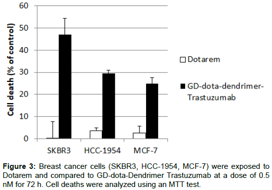
Result: We confirmed that HCC-1954 cells were grown in apple scaffold and increased intensity of MRI image. Interestingly, Gd-Dota-Dendrimer-Trastuzumab tended to increase intensity of MRI image compared to Gd-Dota using HCC-1954 cells in apple scaffold, although the difference was not significant (p>0.05). Importantly, the HER2+ sensitive to trastuzumab cells, SKBR3 cells were still sensitive to Gd-Dota-Dendrimer-Trastuzumab but not the HCC-1954 and MCF-7 cells.
Conclusion: Gd-Dota-Dendrimer-Trastuzumab was potentially increase MRI intensity in HER2+ breast cancer cells.
Keywords: Apple scaffold; HER2+ breast cancer; Gd-DOTADendrimer-Trastuzumab; Magnetic Resonance Imaging (MRI)
Introduction
Magnetic Resonance Imaging (MRI) is a radiological imaging examinations that exploits proton elements (hydrogen ion) residing in human tissue. The advantage of MRI imaging compared to CT scan are a non-ionizing radiation imaging, multiple image/multiplanar drawing images, good spatial resolution, no bone artifact, noninvasive and superior able to assess soft tissues such as breast cancer [1]. The general approach to MRI data analysis involves the extraction of signals from a given area. The use of ROI analysis becomes standard practice across all MRI imaging [1,2].
One way to improve imaging quality of MRI is administration of contrast agent. MRI contrast agent is a paramagnetic compound having considerable moments and able to shorten the relaxation time in T1 and T2 weighing mode, which will result in a more vivid and clearly discernible tissue image [3,4].
Gadolinium chelate is a widely used as MRI micromolecular contrast agent, acting on extracellular and non-specific. To overcome nonspecific trait to become specific agent, another compound is needed. The target is that the contrast compound would be able to be retained in intracellular and a clearer image. Hardiani et al. have successfully performed the synthesis and stability test of Radiogadolinium (III) Gadolinium-DOTA-PAMAM (Poliamidoamine) Generation 3.0-Trastuzumab as SPECT contrast agent (Single Photon Emission Computed Tomography) - MRI contrast agent for breast cancer diagnostics [5-7]. Previous result successfully demonstrated a strong signal intensity increase on MRI examination with the Gd-DOTADendrimer-Trastuzumab contrast compound against the SKOV3 cell line. SKOV3 is a positive HER-2 cell line in ovarian cancer that is sensitive to trastuzumab.
HCC cell line 1954 breast cancer which is a cell line of breast cancer of HER-2 positive expression resistant to Trastuzumab. Approximately 25-30% of breast cancers express HER-2 (Human epidermal growth factor receptor 2), although the incidence is low but has poor biological behavior in the context of aggressiveness, thus affecting its prognosis [8-10].
Cell culture is a technique associated with a complex process of cell isolation from the original environment (in vivo) as well as in controlled and aseptic (in vitro) environmental conditions. The cell is taken from the original tissue, primary culture or cell line in the form of suspension of cells isolated for days to weeks in sterile conditions with an environment adapted to the in vivo environment of the cell so as to survive and controlled proliferation occurs [11-15].
Cell cultures can be planted in apple scaffolds. Characteristics of apples used are apples of fuji type. The apple scaffold after peeling is frozen in temperature -80°C, and then incorporated into the methanol solution resulting in deselularization of the organelles within the apple cytoplasm. Then cell line culture is planted in the framework of apples that have experienced the deselularization [16-20].
The purpose of this study was to evaluate Dendrimer-Trastuzumab in enhancing of imaging using MRI in cell line HCC 1954 on breast cancer HER-2 positive expressions.
Materials And Methods
Chemicals and reagents
Roswell Park Memorial Institute (RPMI) 1640 medium, fetal bovine serum (FBS), and penicillin streptomycin were purchased from GIBCO, USA. Dimethylsulfoxide (DMSO), and 3-[4,5-dimethylthiazol- 2-yl]-2,5 diphenyl tetrazolium bromide (MTT) were purchased from Sigma-Aldrich, USA. All the other chemicals were of analytical grade purchased from Merck, USA.
Gd-DOTA-Dendrimer-Trastuzumab was developed in-house by supervision of Prof Mutakin from Department of Chemistry, Faculty of Math and Science, Universitas Padjadjaran. In the synthesis conjugat Gd-DOTA-Dendrimer-Trastuzumab required stages of DOTA NHS ester conjugation on dendrimer PAMAM G3.0, then activation of conjugate DOTA NHS ester-PAMAM G 3.0 using Traut’s reagent, conjugated with trastuzumab then marked with 153Gd then tested pharmaceutical. The conjugate synthesis of 153Gd-DOTA-Dendrimer-Trastuzumab and pharmaceutical test was performed by P2RR Batan.
Cell culture conditions
Human breast cancer cell lines were used in this study including HCC-1954, SKBR-3 and MCF-7 cells. HCC-1954 was a gift from Prof. Stefan Wiemann (DKFZ, Heidelberg, Germany), SKBR-3 was from Prof Andreas Trummp (DKFZ, Heidelberg, Germany) and MCF- 7 cells were a gift from Dr. Ahmad Faried (Universitas Padjadjaran, Bandung, Indonesia). The cell lines were cultured using RPMI, supplemented with 10% FBS and 1% penicillin streptomycin (GIBCO, USA), incubated with a controlled temperature of 37°C and 5% CO2 [2].
Apple scaffold for 3D cell culture
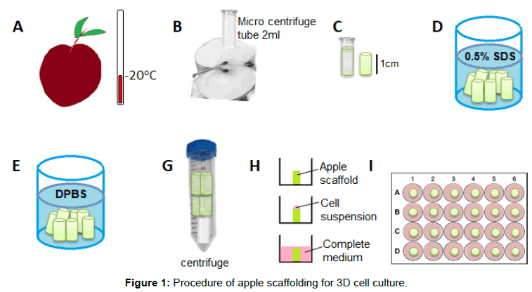
Apple scaffold was made and modified according to previous study (Figure 1). Briefly, Fuji apple was put in a -20°C freezer for 5 min before punched with micro-centrifuge tube 2 ml and cut to a uniform tube form with 1 cm height. Tissue was decellularized from apple scaffold using 0.5% sodium dodecyl sulphate (SDS) (Sigma-Aldrich) solution for 24 h then washed and incubated with PBS (GIBCO) for 24 h.
For culture, apple scaffold was centrifuged at 1000 rpm for 10 s to drain remaining PBS then placed on 24-well-plate followed by 0.5 ml cell suspension of 1 x 10⁶ breast cancer cells. Sample was then incubated in temperature of 37°C and 5% CO2 for an hour before added 1.5 ml complete medium.
Experiments were conducted in triplicate and two repeats. Breast cancer cell line was cultured in apple scaffold for 8 weeks inside a 12 well plate with RPMI medium. Culture condition was monitored and medium was changed twice a week.
HE staining and microscopy
Fixation of cell inside apple scaffolding was done using cold methanol 100% for 10 min. The sample were then washed with pH balanced PBS 3x and then stored for one day before embedded in paraffin with standard method. Paraffin block then sliced into 5 μm slice using microtome and mounted on glass slide. Hematoxylin and Eosin stain was done to visualize structure of interest.
Image was acquired using Olympus CX-51 inverted microscope with mounted digital camera.
Gd-DOTA trastuzumab treatment
To evaluate enhancing MRI imaging of GD-dota-dendrimer trastuzumab in HER2+ breast cancer cells, HCC-1954 cells in apple scaffolds were treated with 0.5 nM of Gd-DOTA-Dendrimer- Trastuzumab or Gd-DOTA for 24 h prior to scanning with MRI.
Magnetic Resonance Imaging (MRI) scanning and Image analysis
MRI is an imaging modality that has the magnetic properties that each proton has in producing the image. Every human body is made up of atoms. These atoms will form a water molecule consisting of 2 hydrogen atoms and 1 oxygen atom. Conventional MRI generally empowers the magnetic properties of protons. MRI imaging is focused on T2 differences between tissues so the sequence is called T2-weighted imaging (T2 WI). If the contrast difference in the image is not focused on the difference between T1 and T2 between tissues, but more towards the difference in proton density, then the sequence is called Proton Density Weighted Imaging (PDWI).
Treated cell lines were scanned using Magnetom Essenza 1.5T (Siemens, Germany). Five planes were scanned using T1 weighted. At T1WI the TR is given a short enough so that the fat and water tissue is not enough time to recover the initial magnetization value, so there is a considerable difference between water and fat. In T1WI water has a weak signal that has a dark picture or hypointens, while fat has a stronger signal that has a brighter picture or hyperintens. In the T2WI sequence water has a stronger signal that has a lighter or hyperhensive image whereas fat has a weaker signal resulting in a less light, dark or hypointens picture. Fat sequences are usually used in MRI imaging to suppress signals from adipose tissue or detect adipose tissue. This can be applied to T1WI and T2WI. Due to the short relaxation time, fat has a high signal on MRI. This high signal, easily recognizable on MRI, may be useful for marking lesions. The 3 middle plane were then use for image analysis.
Region of Interest (ROI) was selected which covered all scaffold regions. Three planes from every sample were selected. Average intensity and area were analysed using RadiAnt Dicom Viewer v 3.4.1 software (Medixant, Poland). ROI selection and calculation were done triplicate.
Cytotoxicity assay
The cytotoxic effect of Gd-DOTA-Dendrimer-Trastuzumab as well as Gd-DOTA contrast ware evaluated in breast cancer cells using MTT assay, modified from our previous study [20,21]. Briefly, cells were seeded on 96-well plate then treated with indicated concentrations on the next day followed by incubation for 72 h in an incubator with 5% CO2 at the temperature of 37°C. Cells with 1% DMSO were used as control. The MTT solution was added and incubated for 4 h before DMSO was added to dissolve the formazan crystal. Absorbance was measured at the wavelength of 450 nm for finding percentages of viable cells on treated cells compared to the control cells. (Figures 2 and 3)
Statistical analysis
Generalized Estimating Equation (GEE) method with the exchangeable matrix correlation structure was used to calculate the difference in signal intensity in the ROI of the Gd-DOTA treatment group with Gd-DOTA-Dendrimer-Trastuzumab. Analysis was done using SPSS for Windows version 13.0 with 95% confidence degree with value p ≤ 0.05%.
Result and Discussion
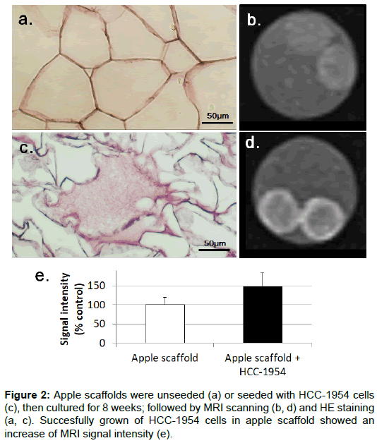
Before we evaluated enhancing MRI imaging of GD-dotadendrimer trastuzumab in HER2+ breast cancer cells, we verified that the HER2+ breast cancer cells were grown in apple scaffold. Interestingly, HCC-1954 cells in apple scaffold were not only detected by HE staining but also enhanced intensity of MRI (Figure 2).
Experiments were performed using HCC-1954 cell line of HER-2 positive IHC-proven HER-2 breast cancer grown within the framework of cultured cells of apple (scaffold). MRI examination was performed on apple scaffold without and with HCC cell line 1954 cancer of HER-2 positive expression of breast to obtain enhanced imaging by measuring signal intensity at 6 areas (ROI). The Dendrimer Trastuzumab Gd- DOTA contrast compound specifically causes death in breast cancer cells with positive HER-2.
The result of MRI on apple scaffold preparation without and with cell line by measuring ROI and statistical analysis using Generalized Estimating Equation (GEE) Method with exchangeable matrix correlation structure is used to overcome the correlation observation obtained p value <0.001. The Gd-DOTA Dendrimer Trastuzumab contrast compound increases the intensity of the MRI signal.
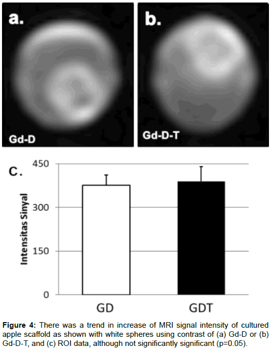
There is a tendency of increased imaging in HCC cell line 1954 of HER-2 positive expression breast cancer after contrast administration of Gd-DOTA-Dendrimer-Trastuzumab when compared with the administration of Gd-DOTA contrast compound using ROI (Figure 4). Although the results of statistical analysis with p value 0.06 (p>0.05) but not necessarily reduce the benefits of clinical aspects. HCC cell line 1954 cancer of HER-2 positive expression is a cell line that is resistant to trastuzumab, so at some internalization does not occur endocytosis. The intensity of the signal resulting from the bonding of the Gd-DOTA and dendrimer contrast compounds forms the macromolecules to enhance the imaging.
This study has several limitations among others; no trials of trastuzumab in HER-2 cell line HCC 1954 cancer of positive HER-2 breast cancer were performed.
Conclusion
There is a trend of signal intensity increase between apple scaffold and apple scaffold with cell line; i.e. there was a binding of contrast compound Gd-DOTA-Dendrimer-Trastuzumab in cell line HCC 1954, although the increase of signal intensity on the average MRI examination of the Gd-Dotrim-Dendrimer- Trastuzumab contrast group was not significantly significant (p 0.06). Further tests are needed to prove trastuzumab attached to the HER-2 receptor of breast cancer.
Acknowledgement
Authors would like to thanks Dr. Kurnia Wahyudi for statistical analysis suggestion and Staff of Advanced Biomedic Laboratory for all the technical assistance.
References
- Grover VP, Tognarelli JM, Crossey MM, Cox IJ, Taylor-Robinson SD, et al. (2015) Magnetic resonance imaging: principles and techniques: lessons for clinicians. J Clin Exp Hepatol 5: 246-255.
- Weishaupt D, Köchli VD, Marincek B (2008) How does MRI work?: an introduction to the physics and function of magnetic resonance imaging. Springer Science & Business Media, pp: 103-122.
- Maiti PK, Çaǧın T, Wang G, Goddard WA (2004) Structure of PAMAM dendrimers: Generations 1 through 11. Macromolecules 37: 6236-6254.
- Adang H, Soetikno R, Mutalib, Yono S (2010) Preparasi, biodistribusi dan clearance senyawa pengontras MRI Gd-DTPA-PAMAM G4-Nimotuzumab. 712-17.
- Fiddiyanti I (2012) Afinitas Pengikatan Antibodi Monoklonal Trastuzumab Terhadap Reseptor HER2 yang Diekspresikan Tumor Ganas Ovarium. Fakultas Kedokteran Unpad 46-51.
- El Khouli RH, Macura KJ, Kamel IR, Jacobs MA, Bluemke DA (2011) 3 Tesla dynamic contrast enhanced magnetic resonance imaging of the breast: pharmacokinetic parameters versus conventional kinetic curve analysis. AJR 197: 1498-1505.
- Rahmania H, Mutalib A, Ramli M, Levita J (2015) Synthesis and stability test of radiogadolinium (III)-DOTA-PAMAM G3. 0-trastuzumab as SPECT-MRI molecular imaging agent for diagnosis of HER-2 positive breast cancer. J Rad Res Appl Sci 8: 91-99.
- Jeyakumar A, Younis T (2012) Trastuzumab for HER2-positive metastatic breast cancer: clinical and economic considerations. Clin Med Insights Oncol 6: 179.
- Kuhl CK, Schrading S, Leutner CC, Morakkabati-Spitz N, Wardelmann E, et al. (2005) Mammography, breast ultrasound, and magnetic resonance imaging for surveillance of women at high familial risk for breast cancer. J Clin Oncol 23: 8469-8476.
- Shukla R, Thomas TP, Peters JL, Desai AM, Kukowska-Latallo J, et al. (2006) HER2 specific tumor targeting with dendrimer conjugated anti-HER2 mAb. Bioconjugate Chem 17: 1109-1115.
- Swanson SD, Kukowska-Latallo JF, Patri AK, Chen C, Ge S, et al. (2008) Targeted gadolinium-loaded dendrimer nanoparticles for tumor-specific magnetic resonance contrast enhancement. Int J Nanomed 3: 201.
- Hashemi RH, Bradley WG, Lisanti CJ (2012) MRI: the basics. Lippincott Williams Wilkins pp: 41-47.
- Bellin MF (2006) MR contrast agents, the old and the new. Eur J Rad 60: 314-323.
- Sinn HP, Kreipe H (2013) A brief overview of the WHO classification of breast tumors. Breast Care 8: 149-154.
- Hengerer A, Grimm J (2006) Molecular magnetic resonance imaging. Biomed Imag Interven J 2: e8.
- Modulevsky DJ, Lefebvre C, Haase K, Al-Rekabi Z, Pelling AE (2014) Apple derived cellulose scaffolds for 3D mammalian cell culture. PloS one 9: e97835.
- Herborn CU, Honold E, Wolf M, Kemper J, Kinner S, et al. (2007) Clinical safety and diagnostic value of the gadolinium chelate gadoterate meglumine (Gd-DOTA). Investigative Radiol 42: 58-62.
- Rosner B (2011) Fundamentals of Biostatistics. (7th edn) Harvard University, Cambridge pp: 213 -222.
- Liu J, Colditz GA (2017) Optimal design of longitudinal data analysis using generalized estimating equation models. Biometrical J 59: 315-330.
- Rezano A, Kuwahara K, Yamamoto-Ibusuki M, Kitabatake M, Moolthiya P, et al. (2013) Breast cancers with high DSS1 expression that potentially maintains BRCA2 stability have poor prognosis in the relapse-free survival. BMC Cancer 13: 562.
- Vallet S, Bashari MH, Fan FJ, Malvestiti S, Schneeweiss A, et al. (2016) Pre-Osteoblasts Stimulate Migration of Breast Cancer Cells via the HGF/MET Pathway. PloS one 11: e0150507.
Citation: Soekersi H, Bashari MH, Huda F, Rudiman R, Sahiratmadja E, et al. (2017) Dendrimer-Trastuzumab Contrast Agent Improves Imaging of Magnetic Resonance Imaging (MRI) On Her2 Positive Breast Cancer Cell Line HCC-1954 in Apple Scaffold. OMICS J Radiol 6: 281. DOI: 10.4172/2167-7964.1000281
Copyright: ©2017 Soekersi H, et al. This is an open-access article distributed under the terms of the Creative Commons Attribution License, which permits unrestricted use, distribution, and reproduction in any medium, provided the original author and source are credited.
Select your language of interest to view the total content in your interested language
Share This Article
Open Access Journals
Article Tools
Article Usage
- Total views: 5405
- [From(publication date): 0-2017 - Nov 28, 2025]
- Breakdown by view type
- HTML page views: 4427
- PDF downloads: 978


