Make the best use of Scientific Research and information from our 700+ peer reviewed, Open Access Journals that operates with the help of 50,000+ Editorial Board Members and esteemed reviewers and 1000+ Scientific associations in Medical, Clinical, Pharmaceutical, Engineering, Technology and Management Fields.
Meet Inspiring Speakers and Experts at our 3000+ Global Conferenceseries Events with over 600+ Conferences, 1200+ Symposiums and 1200+ Workshops on Medical, Pharma, Engineering, Science, Technology and Business
Case Report Open Access
Diagnosis of Osteoporosis and Radiotherapy Induced Fracture by F-18 FDG PET/CT in A Case with Colon Cancer
| T KürÃ?Â?at Dabak1, Funda Aydin2*, Güzide AyÃ?Â?e Ocak3, Fatma Yalçin Müsri4, Gökhan Arslan5, Adil Boz2 and H Ã?Â?enol CoÃ?Â?kun4 | |
| 1Department of Orthopaedics and Traumatology, School of Medicine, Akdeniz University, Antalya, Turkey | |
| 2Department of Nuclear Medicine, School of Medicine, Akdeniz University, Antalya, Turkey | |
| 3Department of Pathology, School of Medicine, Akdeniz University, Antalya, Turkey | |
| 4Department of Medical Oncology, School of Medicine, Akdeniz University, Antalya, Turkey | |
| 5Department of Radiology, School of Medicine, Akdeniz University, Antalya, Turkey | |
| Corresponding Author : | Funda Aydin Department of Nuclear Medicine School of Medicine, Akdeniz University Antalya, Turkey Tel: +902422496490 E-mail: afunda@akdeniz.edu.tr |
| Received: December 31, 2015; Accepted: January 05, 2016; Published: January 08, 2016 | |
| Citation: Dabak TK, Aydin F, Ocak GA, Musri FY, Arslan G, et al. (2016) Diagnosis of Osteoporosis and Radiotherapy Induced Fracture by F-18 FDG PET/CT in A Case with Colon Cancer. OMICS J Radiol 5:212. doi:10.4172/2167-7964.1000212 | |
| Copyright: © 2016 Dabak TK, et al. This is an open-access article distributed under the terms of the Creative Commons Attribution License, which permits unrestricted use, distribution, and reproduction in any medium, provided the original author and source are credited. | |
Visit for more related articles at Journal of Radiology
Abstract
Pelvic Insufficiency Fractures (PIF) is caused by the effect of normal or physiologic stresses on bone with reduced mineral content or elastic resistance. Positron Emission Tomography (PET)/Computed Tomography (CT) is widely used in the work-up of patients with various types of cancer. In our case, a patient with colon cancer who was investigated by many forms of conventional imaging for pelvic pain. We found to have osteoporotic fractures using PET/CT.
| Keywords |
| Positron emission tomography (PET)/Computed tomography (CT); Colon cancer; Osteoporosis; Pelvic irradiation |
| Introduction |
| Pelvic Insufficiency Fractures (PIF) is caused by the effect of normal or physiologic stresses on bone with reduced mineral content or elastic resistance [1]. The sacral ala adjacent to the sacroiliac joint is the most commonly involved site because it is a weight-bearing site [2]. The pubic rami adjacent to the symphysis pubis and acetabulum can also be affected. Postmenopausal osteoporosis, use of high-dose steroids, rheumatoid arthritis, and pelvic irradiation are risk factors for PIF [3]. PIF causes lower back pain which has a mild to severe intensity and which sometimes mimics bony metastases of cancer. In CT, PIF appears as linear sclerotic lesions with or without cortical discontinuity in the sacral body parallel to sacroiliac joints. MRI shows reduced T1 and T2 signal intensity in the same locations. This distribution results in the characteristic “H” or “Honda” sign in the sacrum in bone scintigraphy [4]. |
| F-18 fluorodeoxyglucose (FDG) Positron Emission Tomography (PET)/Computed Tomography (CT) is widely used in the work-up of patients with various types of cancer [5]. The value of this tool in the diagnosis and staging of colorectal carcinoma has been well established [6]. It is sensitive in the diagnosis of metastatic disease, as well as the identification of benign metabolic processes such as osteoporotic fractures [7]. PET/CT scanner permits simultaneous acquisition of anatomic (CT) and functional (PET) data, as well as accurately fused images. It has demonstrated great utility in the detection and staging of malignancy [8]. Specifically related to malignant skeletal disease, PET/CT has demonstrated high sensitivity and specificity in detecting lytic and sclerotic malignant bone lesions [9]. |
| In this case report, PET/CT demonstrated PIF and osteoporotic fractures and excluded malignant skeletal disease in a patient with known colorectal cancer and pelvic pain. |
| Case Report |
| A 70-year old female with a history of colorectal cancer treated with surgery followed by 5 courses of chemotherapy and postoperative irradiation for 30 days presented to our clinic. At the time of surgery, the patient had been categorized as stage III rectal adenocarcinoma. |
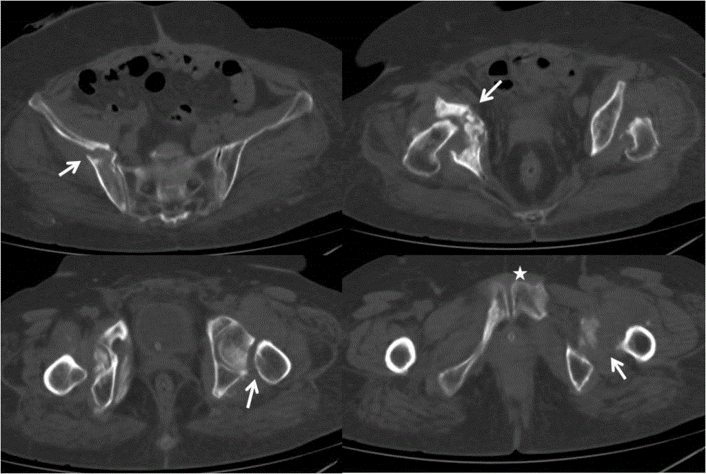
| She also had a history of postmenopausal osteoporosis. A DXA scan showed osteoporosis. Two years after treatment she began experiencing pelvic-hip pain when sitting on the toilet. The CT imaging of the pelvic area was performed, which detected multiplepart displaced fractures in right iliac bone, right acetabulum, and both femoral heads (Figure 1). |
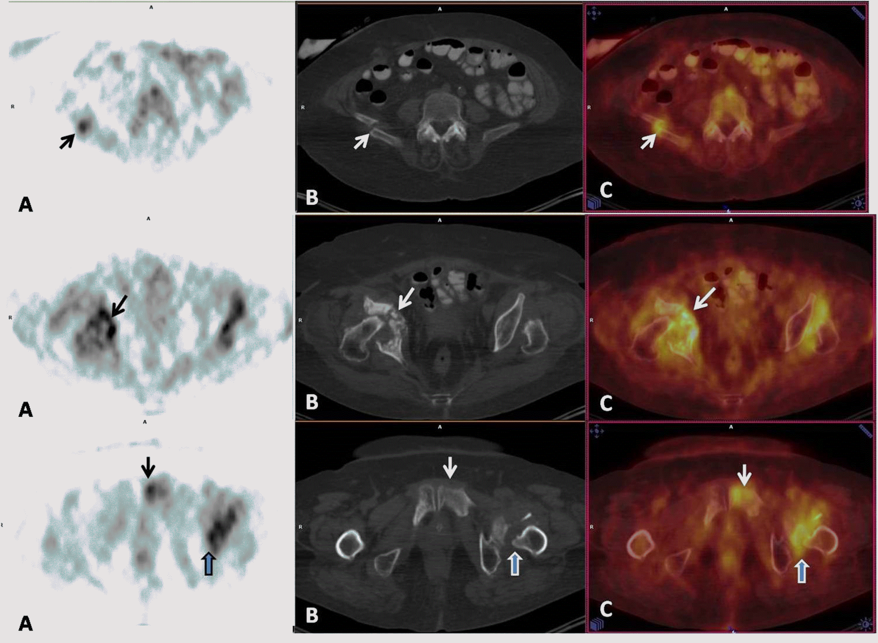
| A PET/CT scan was performed to confirm the lesions and to search for other possible metastatic disease. In a 6-hour fasting state (serum glucose level 95 mg/dL), the patient received an intravenous injection of 12.2 mCi (451MBq) F-18 FDG. After a 1-hour waiting period, the patient was imaged using an integrated PET/CT camera (Biograph-16 True Point PET/CT; Siemens). The CT part of the study was done without intravenous contrast medium. PET/CT scan revealed uniformly increased FDG uptake in right scapula which fused with multiple lytic fractures in the CT scan. The scan also showed increased FDG uptake around the right shoulder joint, primarily suggestive of inflammation. There was an increased FDG accumulation in the 3rd and 4th ribs on the left side and bone window images of the CT scan showed the fracture line. Mild to diffuse FDG uptake was observed in right iliac bone and acetabulum, femoral necks, ischium, and pubis (Figure 2). |
| The corresponding CT slices clearly revealed fracture lines on pelvic bones. Furthermore, an increased FDG uptake was shown around both hip joints, primarily suggestive of inflammation. A maximum standard uptake value (SUVmax) was variable and ranged from 3.4 to 12.1. Normal FDG uptake was seen in other parts of the body, and there was no metastasis. These findings were suggestive of fractures related to osteoporosis and pelvic irradiation. |
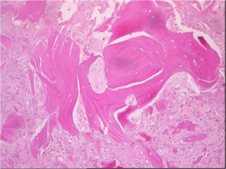
| The broken left femoral head and neck were resected. Pathological investigation supported the diagnosis of PIF by demonstrating bone and fat necrosis, callus formation, and absent malignant tissue (Figure 3). No evidence of recurrent disease or metastasis were found at follow-up. |
| Discussion |
| The development of PIF associated with radiation or osteoporosis is not a rare situation in patients with cancer; some authors have found an incidence of 8.2-19.7% [1,10]. Bone scintigraphy and MRI scans help differentiate PIF from bony metastases. Only a few reports have been described the findings of PIF in FDG-PET scanning, although FDG-PET has been recognized as an important imaging tool for the evaluation of patients with cancer [1]. |
| In this case, a patient with colorectal carcinoma, osteoporosis, and a history of pelvic irradiation who had pelvic and back pain was found to have multiple areas of osteoporosis and radiation-induced fractures. The diagnosis was confirmed and fracture sites were accurately localized using PET/CT. Increased FDG uptake is experienced in both acute and sub-acute phases of benign fractures [7]. The FDG activity in these fractures may subside as they heal. With the integration of PET/CT, the diagnosis of benign fracture is more accurate and the age of the fracture may also be determined. At the same time, malignant disease can be accurately excluded. PET/CT offers a greater sensitivity as well as a greater specificity than PET scan alone in the diagnosis of malignant bone disease. Integrated PET/CT scanners are able to produce the identical corresponding PET and CT slices of the body within a single examination. Thus, morphologic characteristics of a PET positive lesion can be easily examined and by doing so some falsepositive studies encountered on PET imaging, including benign bony fractures, can be accurately differentiated, as occurred in our case. On CT scans, sacral insufficiency fractures generally appear as fracture lines, sclerotic lines or both, or areas of sclerosis within the sacral alae parallel to the sacroiliac joints [11]. |
| Whole-body FDG PET/CT imaging has gained wide clinical acceptance in both cancer diagnosis and staging, as well as in followup. FDG has already been established as a good tumor-seeking agent for various malignancies [12]. Although there is a growing body of evidence for the use of FDG in differentiating malignant from benign disease, it is well known that FDG uptake is not specific for malignant cells [13]. Numerous benign causes with abnormal FDG uptake have been described in the literature. From a practical point of view, all granulomatous and inflammatory processes would be the source of false-positive results on FDG PET, mainly due to their content of active alveolar macrophages, cells with a well-known high affinity for the FDG molecule, comparable to malignant tissue [14]. Possibly for the same reason, benign bone fractures may accumulate a considerable amount of FDG and cause false-positive PET results as described many times before [15]. Although FDG accumulation in fractures decreases with time, little is known regarding the exact time duration of FDG uptake in fracture regions and the relation of the degree of uptake with the fracture stage. There may be important differences in the duration and intensity of uptake depending on the site of fracture. Benign fractures may become FDG-negative 8 weeks after fracture [7], and acute fractures may show no FDG uptake [16]. |
| The utility of SUVs remains unclear, although a trend toward lower uptake in benign fractures compared to uptake in malignant lesions has been noted [16]. Considerable overlapping regarding the intensity of FDG uptake between malignant and benign conditions is very well known. Fayad et al. [15] calculated SUVs ranging from 2.2 to 3.6 in 3 cases with sacral fractures. In our case, SUVs were found ranging from 3.4 to 12.1 and strongly increased uptake was seen in both femoral necks, and acetabulum was more prominent on the right ilium. Fracture zones typically observed in sacrum in cases with PIF were not observed in our case. There were, however, fracture zones with increased FDG uptake in right scapula and ribs. |
| Recognition of osteoporotic fractures was important in the care of this patient and led to the implementation of appropriate therapy. Osteoporosis is a common consequence of chemotherapy. For these patients, fractures often result in chronic pain, loss of mobility, and loss of independence [17]. In this case, CT alone was less helpful in defining the cause of this patient’s symptoms. FDG PET/CT showed pelvic bone fractures as well as fractures in other bones. Furthermore, the absence of metastasis in extra-osseous areas was also demonstrated by PET/CT. |
| Our case is important with respect to being the first to have fractures that were shown by PET/CT, and confirmed by histopathological examination, to have occurred secondary to osteoporosis and radiotherapy. |
| Moreover, there are only a few case reports and studies in which only PIF has been shown by PET/CT performed for follow-up of colorectal carcinoma [1,18]. In that case report and study, only fractures of pelvic bones were reported. In the present case, PET/CT showed zones of increased FDG uptake considered to be secondary to osteoporosis in right scapula and ribs in addition to pelvic bones, which were accompanied by fracture zones in CT. The non-metastatic nature of these foci was confirmed at the follow-up without administration of any metastasis-directed therapy. With that respect, our case is the first in the literature. |
| With this case report, we aimed to re-emphasize the role of FDG PET/CT imaging in showing metastases in patients with cancer. We also aimed to stress that not every pathological fracture is necessarily metastasis-related, but it may also result from other benign conditions (such as osteoporosis, radiotherapy). Besides, increased FDG uptake takes place not only in malignant and/or metastatic pathologies, but also in benign pathologies; we therefore aimed to underscore the importance of the evaluation of CT along with PET in PET-positive cases and simultaneous assessment of PET/CT fusion imaging for more accuracy in diagnostic process. PET/CT may be utilized in the differentiation of malignant and benign conditions as a non-invasive, simple diagnostic tool. |
References
- Oh D, Huh SJ, Lee SJ, Kwon JW (2009) Variation in FDG uptake on PET in patients with radiation-induced pelvic insufficiency fractures: a review of 10 cases. Ann Nucl Med 23: 511-516.
- Cooper KL, Beabout JW, Swee RG (1985) Insufficiency fractures of the sacrum. Radiology 156: 15-20.
- Peh WC, Gough AK, Sheeran T, Evans NS, Emery P (1993) Pelvic insufficiency fractures in rheumatoid arthritis. Br J Rheumatol 32: 319-324.
- Salavati A, Shah V, Wang ZJ, Yeh BM, Costouros NG, et al. (2011) F-18 FDG PET/CT findings in postradiation pelvic insufficiency fracture. Clin Imaging 35: 139-142.
- Wahl RL, Hutchins GD, Buchsbaum DJ, Liebert M, Grossman HB, et al. (1991) 18F-2-Deoxy-2-Fluoro-D-Glucose uptake into human tumor xenografts. Cancer 67: 1544-1550.
- Ruers TJ, Langenhoff BS, Neeleman N, Jager GJ, Strijk S, et al. (2002) Value of positron emission tomography with _F-18_fluorodeoxyglucose in patients with colorectal liver metastases: a prospective study. J Clin Oncol 20: 388-395.
- Shon IH, Fogelman I (2003) F-18 positron emission tomography and benign fractures. Clin Nuclear Med 28: 171-175.
- Bar-Shalom R, Yefremov N, Guralnik L, Gaitini D, Frenkel A, et al. (2003) Clinical performance of PET/CT in evaluation of cancer: additional value for diagnostic imaging and patient management. J Nucl Med 44: 1200-1209.
- Even-Sapir E, Metser U, Flusser G, Kollender Y, Lerman H, et al. (2004) Assessment of malignant skeletal disease: initial experience with 18F-Fluoride PET/CT and comparison between 18F-Fluoride PET and 18F-Fluroide PET/CT. J Nucl Med 45: 272-278.
- Baxter NN, Habermann EB, Tepper JE, Durham SB, Virnig BA (2005) Risk of pelvic fractures in older women following pelvic irradiation. JAMA 294: 2587-2593.
- Peh WC, Khong PL, Yin Y, Ho WY, Evans NS, et al. (1996) Imaging of pelvic insufficiency fractures. Radiographics 16: 335-338.
- Yasuda S, Ide M, Fujii H, Nakahara T, Mochizuki Y, et al. (2000) Application of positron emission tomography imaging to cancer screening. Br J Cancer 83: 1607-1611.
- Schelbert HR, Hoh CK, Royal HD, Brown M, Dahlbom MN, et al. (1998) Procedure guideline for tumor imagi ng using fluorine-18-FDG. Society of Nuclear Medicine. J Nucl Med 39: 1302-1315.
- Bakheet SM, Powe J (1998) Benign causes of 18-FDG uptake on whole body imaging. Semin Nucl Med 28: 352-358.
- Fayad LM, Cohade C, Wahl RL, Fishman EK (2003) Sacral fractures: a potential pitfall of FDG positron emission tomography. AJR Am J Roentgenol 181:1239 -1243.
- Kato K, Aoki J, Endo K (2003) Utility of FDG-PET in differential diagnosis of benign and malignant fractures in acute to subacute phase. Ann Nucl Med 17: 41-46.
- Pfeilschifter J, Diel IJ (2000) Osteoporosis due to cancer treatment: pathogenesis and management. J Clin Oncol 18: 1570-1583.
- Katzel JA, Heiba SI (2005) PET/CT F-18 FDG scan accurately identifies osteoporotic fractures in a patient with known metastatic colorectal cancer. Clin Nucl Med 30: 651-654.
Figures at a glance
 |
 |
 |
| Figure 1 | Figure 2 | Figure 3 |
Post your comment
Relevant Topics
- Abdominal Radiology
- AI in Radiology
- Breast Imaging
- Cardiovascular Radiology
- Chest Radiology
- Clinical Radiology
- CT Imaging
- Diagnostic Radiology
- Emergency Radiology
- Fluoroscopy Radiology
- General Radiology
- Genitourinary Radiology
- Interventional Radiology Techniques
- Mammography
- Minimal Invasive surgery
- Musculoskeletal Radiology
- Neuroradiology
- Neuroradiology Advances
- Oral and Maxillofacial Radiology
- Radiography
- Radiology Imaging
- Surgical Radiology
- Tele Radiology
- Therapeutic Radiology
Recommended Journals
Article Tools
Article Usage
- Total views: 12744
- [From(publication date):
February-2016 - Aug 30, 2025] - Breakdown by view type
- HTML page views : 11796
- PDF downloads : 948
