Make the best use of Scientific Research and information from our 700+ peer reviewed, Open Access Journals that operates with the help of 50,000+ Editorial Board Members and esteemed reviewers and 1000+ Scientific associations in Medical, Clinical, Pharmaceutical, Engineering, Technology and Management Fields.
Meet Inspiring Speakers and Experts at our 3000+ Global Conferenceseries Events with over 600+ Conferences, 1200+ Symposiums and 1200+ Workshops on Medical, Pharma, Engineering, Science, Technology and Business
Case Report Open Access
Dual Thyroid Ectopy, Sublingual and Pretracheal
| Tariq Alam, Hidayatullah Hamidi* and Ahmad Reshad Faizi | |
| Radiology department of French medical institute for children (FMIC), Kabul, Afghanistan | |
| Corresponding Author : | Hidayatullah Hamidi Radiology department of French medical institute for children (FMIC) Kabul, Afghanistan Tel: 0093744733605 E-mail: hedayatullah.hamidi@gmail.com |
| Received May 14, 2015; Accepted June 04, 2015; Published June 08, 2015 | |
| Citation: Alam T, Hamidi H, Faizi AR (2015) Dual Thyroid Ectopy, Sublingual and Pretracheal. OMICS J Radiol 4:187. doi: 10.4172/2167-7964.1000187 | |
| Copyright: ©2015 Alam T, et al. This is an open-access article distributed under the terms of the Creative Commons Attribution License, which permits unrestricted use, distribution, and reproduction in any medium, provided the original author and source are credited. | |
Visit for more related articles at Journal of Radiology
Abstract
Ectopic thyroid tissue is a rare congenital anomaly with extremely rare occurrence of dual ectopy. Authors present a case of a 13 year-old girl with anterior neck swelling since one year. Contrast enhanced CT demonstrated absence of normally located, bilobed thyroid gland, Instead two foci of ectopic thyroid tissue were seen in the lingual and pretracheal region. Imaging workup is of great value in diagnosing this rare entity.
|
Abstract
Ectopic thyroid tissue is a rare congenital anomaly with extremely rare occurrence of dual ectopy. Authors present a case of a 13 year-old girl with anterior neck swelling since one year. Contrast enhanced CT demonstrated absence of normally located, bilobed thyroid gland, Instead two foci of ectopic thyroid tissue were seen in the lingual and pretracheal region. Imaging workup is of great value in diagnosing this rare entity.
Keywords
Ectopic thyroid; Dual thyroid ectopy; Anterior neck masses; Neck swelling
Introduction
Ectopic thyroid tissue (ETT) is a rare congenital anomaly defined as the thyroid tissue not located in its normal position anterio-lateral to 2nd–4th tracheal cartilage [1]. In 1869, Hickman reported the first case of ectopic thyroid tumor at the base of tongue, pressing down the epiglottis on the larynx and causing death by suffocation sixteen hours after birth [2]. The prevalence of ETT is about 1 per 100000–300000, however, autopsy studies revealed its prevalence approximately 7-10% [1] mostly affecting women with a female/male ratio of 4/1 [3].
ETT most frequently affects women and although a congenital anomaly, it is usually detected at adolescence and during pregnancy due to increased physiological demand for thyroid hormones [1]. ETT may occur anywhere along the path of its initial descent from the foramen cecum to its final pre-tracheal position [1]. The most commonly found ETT is at the base of tongue (lingual thyroid) with incidence of approximately 90% of reported cases [1]. In 70–75% of cases, lingual thyroid is the only thyroid tissue present [4]. Other rare sites are: sublingual or higher cervical region, trachea, mediastinum [1], lung [4], heart [4] and paracardiac area. Extremely rare locations are ovaries, adrenals, gallbladder, pancreas, duodenum, small bowel mesentery, pituitary fossa, sphenoid sinus, uterus, Axilla, palatine tonsils, iris of eye [2], vagina [2], Parathyroid glands, cervical lymph nodes, submandibular glands, carotid bifurcation, esophagus and skin [3]. Dual and Triple Ectopies
Multiple (dual and triple) ETT is very rare and few cases have been reported in literature [5]. In most cases of dual ectopia, the first lesion is lingual or sublingual and the second is subhyoid, infrahyoid or suprahyoid. Dual ETT in porta hepatis and the tongue has also been described [4]. Dual ectopic thyroid with a normally located pretracheal thyroid is extremely rare [6].
Case report
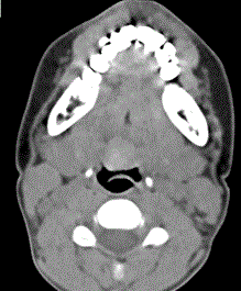
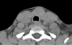
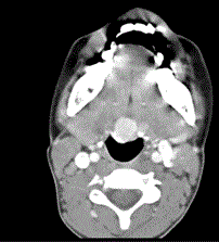
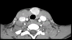
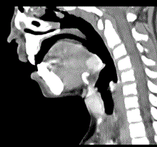
A 13 year old girl presented with asymptomatic lump in the anterior neck since one year. Contrast enhanced CT was revealed than normal bilobed thyroid gland was not present at its normal position. Instead two, soft tissue attenuation, well defined, rounded, intensely enhancing nodules were detected, one at the lingual base in midline just anterior to epiglottis in foramen cecum region and the second in pretracheal region just below the level of pharynx. In pre contrast images the nodule in the lung base was slightly hyper dense while the one in the pretracheal location was low attenuating.
Discussion
Clinical presentation
The majority of patients with ectopic thyroid are asymptomatic. In symptomatic patients the symptoms and signs are usually related to size and location of the ectopic gland as well as associated endocrine dysfunction [2]. The ETT may be the only functioning thyroid tissue or may coexist as a separate structure with a normally located thyroid gland [1].
Diagnostic workup
High resolution ultrasound is favored in the initial assessment, especially in patients presenting with neck masses. CT scan and MRI are especially useful when an ectopic thyroid gland is not identified by ultrasound.In cases of cervical lymphadenopathy or intra tracheal invasion by the tumor, CT and MRI may assist as the suspicion for malignancy is increased [4].
Scintigraphy is the most important diagnostic tool to detect ectopic thyroid tissue and shows the absence or presence of thyroid in its normal location. However, the possibility of false positive results must be taken into account [4] (Figure 1). Fine needle aspiration cytology (FNAC) is the only modality to differentiate between a benign and malignant lesion [4]. Other investigations may be required depending on the location of ETT. Echocardiography and coronary angiography may be necessary in intra-cardiac thyroid ectopy, while barium swallow is important in some patients presenting with dysphagia [2] (Figure 2). Secondary involvement and malignancy potential
All diseases capable of affecting the normal thyroid can affect the ectopic thyroid as well, like adenoma, hyperplasia, inflammation and malignancy. The rate of malignant transformation in ectopic thyroid is no greater than in normally placed thyroid [6]. Primary thyroid carcinomas arising from ectopic thyroid tissue are uncommon and have been reported in cases of lingual thyroid, thyroglossal duct cyst, lateral aberrant thyroid tissue, mediastinal and stroma ovarii [4] (Figure 3).
Management
Management is controversial. No treatment is required for asymptomatic lingual ETT with euthyroid state but patient should be followed for development of any complications. However due to capability of malignant transformation, some authors consider complete surgical removal of ETT as an appropriate treatment [7].
Conclusion
ETT is a rare entity with majority of cases located at the base of tongue. Dual and triple ectopies are extremely rare. The majority of patients are asymptomatic; however, symptoms may arise following enlargement of the gland during periods of stress. Scintigraphy, ultrasound, CT scan, MRI, biopsy and thyroid function tests are the main diagnostic tools.
References
|
Figures at a glance
 |
 |
 |
 |
 |
| Figure 1a | Figure 1b | Figure 2a | Figure 2b | Figure 3 |
Post your comment
Relevant Topics
- Abdominal Radiology
- AI in Radiology
- Breast Imaging
- Cardiovascular Radiology
- Chest Radiology
- Clinical Radiology
- CT Imaging
- Diagnostic Radiology
- Emergency Radiology
- Fluoroscopy Radiology
- General Radiology
- Genitourinary Radiology
- Interventional Radiology Techniques
- Mammography
- Minimal Invasive surgery
- Musculoskeletal Radiology
- Neuroradiology
- Neuroradiology Advances
- Oral and Maxillofacial Radiology
- Radiography
- Radiology Imaging
- Surgical Radiology
- Tele Radiology
- Therapeutic Radiology
Recommended Journals
Article Tools
Article Usage
- Total views: 13750
- [From(publication date):
June-2015 - Aug 29, 2025] - Breakdown by view type
- HTML page views : 9179
- PDF downloads : 4571
