Research Article Open Access
Effects of Repetitive Static Magnetic Field Exposure on Serum Electrolytes and Histology of Certain Tissues of Swiss Albino Rats
| Omer SA1, Sulieman A1*, Ayad CE1, Osman HM2, Abdulrahman MA3 and Saeed AM4 | |
| 1Basic Science Department, College of Medical Radiologic Science, Sudan University of Science and Technology P.O.Box 1908, Khartoum, Sudan | |
| 2Pathology and Diagnoses Department, Central veterinary research laboratory, Khartoum, Sudan | |
| 3Sudan Atomic Energy Commission, Statistic Unit, Khartoum, Sudan | |
| 4Physiology Department, Faculty of Medicine, University of Khartoum, Sudan | |
| Corresponding Author : | Abdelmoneim Sulieman College of Medical Radio- logic Science, Sudan University of Science and Technology P.O. Box 1908, Khartoum, Sudan Tel: +249-910874885 Fax: +249 183 785215 E-mail: abdelmoneim_a@yahoo.com |
| Received March 29, 2012; Accepted April 28, 2012; Published April 30, 2012 | |
| Citation: Omer SA, Sulieman A, Ayad CE, Osman HM, Abdulrahman MA, et al. (2012) Effects of Repetitive Static Magnetic Field Exposure on Serum Electrolytes and Histology of Certain Tissues of Swiss Albino Rats. OMICS J Radiology. 1:104. doi: 10.4172/2167-7964.1000104 | |
| Copyright: © 2012 Omer SA, et al. This is an open-access article distributed under the terms of the Creative Commons Attribution License, which permits unrestricted use, distribution, and reproduction in any medium, provided the original author and source are credited. | |
Visit for more related articles at Journal of Radiology
Abstract
Background: Due to the recent developments in electronic technology, daily exposure to strong static magnetic fields (SMF) is increasing. In particular is the increasing use of magnetic resonance imaging (MRI) for medical diagnoses. The intensity of SMF used at MRI due to development of MRI systems is increasing. Such strong-SMF exposure may have potential health hazards. Objectives: This experimental study aims to evaluate the effects of repetitive exposure to SMF on serum Na+, K+, and Ca++ concentrations. Methods: Fifty-three Albino rats were included in this study classified to 4 experimental groups that involved 4 different protocols of exposure to SMF. Blood samples were obtained from retro orbital venous sinus after exposing the rats to SMF (1.5 T) for 1 hour on day 1 (group 1), day 3 (group 2) and day 7 (group 3), then after 4 weeks from day 7 (group 4). The level of Na+, K+, and Ca++ were measured. The results were compared with blood samples taken pre- exposure, referred to as a control group results. The brain, spleen, liver, kidney, lung, pancreas, intestine, and muscle were dissected out and kept in formalin for histological study. Results: There was an increase in serum K+ concentration and a decrease in serum Na+ concentration after exposure in all groups. Serum Ca++ level fluctuated with a decrease in the groups 1 and 4 and an increase in the group 2. Various histological changes were observed in all tissues. Conclusions: The obtained results indicated that MRI techniques are potentially hazardous and affect electrolytes.
| Introduction |
| Exposure to electromagnetic fields occurs everywhere. Wherever there are electric wires, electric motors and electronic equipment, electromagnetic fields are created. Over the past two decades, there has been increasing interest in the biological effects and possible health outcomes of weak, low-frequency electric and magnetic fields. Epidemiological studies on the effect of magnetic fields on reproduction, cardiac functions and neurobehavioral reactions as well as carcinogenic effects on experimental animals have been presented [1]. Clinical magnetic resonance imaging (MRI) was introduced in the early 1980s and has become a widely accepted and heavily utilized medical imaging technology. This technique requires that the patients under study to be exposed to an intense magnetic field of strength not previously encountered on a wide scale by human [2]. With the growing number of operating Magnetic Resonance Systems in clinical practice, safety aspects gain increasing importance. Therefore, it is necessary to evaluate the possible risks and effects on human health [3]. |
| In this study, a repeated exposure protocol to static magnetic field will be used and the effect of this on serum electrolytes (sodium, potassium, calcium) concentration and some tissues (brain, liver, spleen, kidney, lung, pancreas, intestine, muscle) histology in rats is studied. |
| Materials and Methods |
| This experimental study was conducted at National Research Centre (NRC), in Khartoum, Sudan. Fifty-three Swiss Rodentia Albino rats were used in the experiments. The study was done to investigate the effects of static magnetic field exposure on serum sodium, potassium, and calcium and some tissues histology in Albino rats. MRI, which poses fewer hazards to organs than X-ray, was used for follow-up examination in pneumonia, pleural effusion and consolidation on days 1, 3 and 7, then after 4 weeks from day 7. A similar procedure using chest X-ray was described in the literature [4]. The MRI machine used was Philips interna (1.5) T super conductive system in Khartoum Advanced Diagnostic Center. Fifty-three healthy Swiss Rodentia Albino rats weighting (120 g) and aged 4 weeks were obtained from NRC. The rats were grouped to a control group and four experimental groups. The control group consisted of 10 normal rats matched for age and weight with the experimental groups of rats. Forty-three rats were included in the experiments to be subjected to 4 different protocols of exposure to SMF. Group one consisted of forty three Swiss Albino rats exposed to static magnetic field (1.5 T) for 1 hour in day one. Blood samples were collected from retro orbital venous sinus before and after the exposure and stored for Na+, K+, and Ca++ levels assessment. Ten rats were slaughtered and immediately; the brain, spleen, liver, kidney, lung, pancreas, intestine, and muscle were dissected out and kept in formalin for histological study. The blood collected before exposure in this group will be considered as a self-control. This procedure of blood and tissues collection was repeated in each experimental group. The 33 Swiss Albino rats (after slaughtering 10 rats in experiment 1) were exposed to the static magnetic field for another 1 hour in day 3 (group 2). Blood and tissues samples were collected. The remaining 18 Swiss Albino rats were exposed to the static magnetic field for another 1 hour in day 7 (group 3). Blood and tissues samples were collected. The last 10 Swiss Albino rats were exposed to the static magnetic field for another 1 hour after 4 weeks from day seven (group 4). Blood and tissues samples were collected. Staining was done by using Hematoxylin and Eosin (H & E stain). After preparing slides, they were studied and diagnosed under microscope with magnification 10 and 40 by 2 pathologists to confirm the slides readings and results. |
| Biochemical analysis |
| Blood samples were obtained from retro orbital venous sinus in lithium heparin tubes. Sera were obtained by centrifugation and were collected in plain tubes stored at -20°C for analysis. Serum Na+ and K+ were measured using Roche 9180 Electrolyte analyzer. Serum Ca++ was measured spectrophotometerically using Cromatest Calcium- Methylthymol Blue (Bio system calcium kit). |
| The data was analyzed using the Statistical Package for the Social Sciences (SPSS). Dependent T test was used for analysis and comparison between subjects on the same groups and independent T test was used for analysis of subjects on different groups. |
| Results |
| This study investigated the effects of static magnetic field exposure on serum Na+, K+, Ca++ and tissues histology in rats. Table 1 showed Serum Na+, K+, and Ca++ levels in the control group of the rats. In group 1 experimental rats, a significant decrease in serum sodium and calcium was obtained (P= 0.05, 0.00 respectively) when compared with controls, and a significant increase in potassium was recorded (P=0.00) as shown in Table 2. The same changes in sodium and potassium were noticed in group 2 but calcium significantly increased after exposure (Table 3). A decrease in serum sodium and an increase in serum potassium were observed in group 3. On the other hand, no significant change in calcium levels was observed in this group (Table 4). In group 4 a further decrease in sodium and an increase in potassium in serum were noticed. Calcium in this group was significantly decreased (Table 5). |
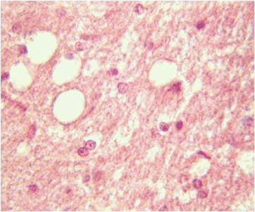
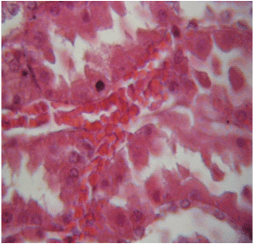
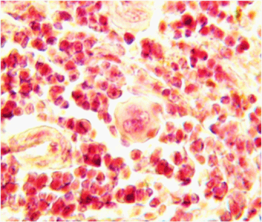
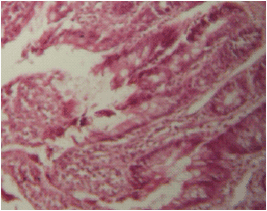
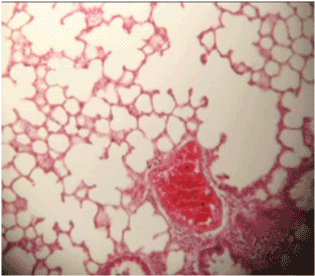
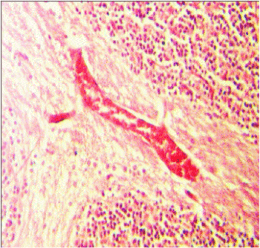
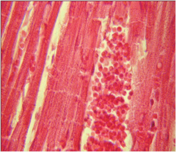
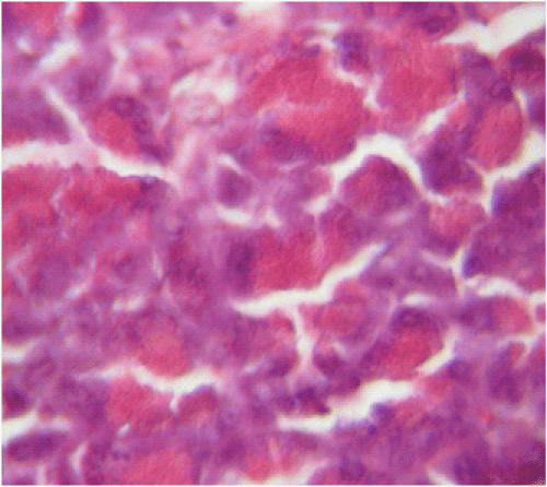
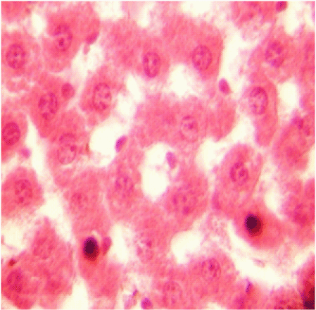
| Histological changes with various degrees were observed in all the organs studied after exposures to SMF in all the groups. The main changes were congestion, hemorrhage, necrosis and degeneration in different organs (Figures 1-9). The detected congestion of the different organs in the experimental rats was: the liver in 23.3%, the spleen in 11.6%, the kidney in 32.5%, the lungs in 32.5%, the brain in 62.8%, the intestine in 11.6% and the muscles in 46.5% of the experimental rats. Hemorrhagic changes were observed mainly in the spleen (55.8%), in the liver (20.9%), the kidney (23.2%) the lungs (20.9%) and in muscle (11.6%). Necrosis was seen in the kidney (86.0%) and the liver (51.2%). Degeneration was prominent in the kidney (86.0%) and muscles (83.7%) and to a lesser extent in the spleen (32.6%), the brain (20.9%), the pancreas (11.6%) and in the liver (9.3%). In addition to this, the kidney and the brain showed vacuolation while epithelial sloughing was confirmed in the intestinal mucosa. |
| Discussion and Conclusions |
| The study was designed to assess the effects of SMF of range used in MRI machine (1.5 T) on serum Na+, K+, Ca++ concentrations and some tissues histology in rats. The major findings were increase in serum K+ concentration, decrease in serum Na+ concentration and fluctuation in serum Ca++ concentration. The data are consistent with the findings of Gerasimova and Nakhil nitskaia [5], who reported an increase in K+ concentration during an hour exposure and a decrease in Na+ concentration during a three hour exposure to 4500 oersted CMF in rats. The results were also consistent with, Schober et al. [6] who reported similar findings. They studied the influence of weak magnetic fields on female white mice electrolytes balance and found that a one-day exposure to a 10 Hz rectangular field significantly lowered the Na+ and increased K+ levels. Similarly Banaszkiewicz et al. [7] found that exposure to 10 mT magnetic field combined with infrared laser radiation for 10 days on a 10 minutes/ once a day basis induced hyperkalaemia and hyponatremia in rats. Serafin et al. [8] reported similar findings on human. They observed a significant increase in the concentration of K+ in patients who are at risk of coronary heart disease when exposed to pulsed magnetic fields (maximum intensity of 0.07 mT) for 24 days, 8 minutes twice a day. |
| One of the suggested mechanism by which SMF can induce hyperkalaemia and hyponatraemia is a decrease in Na+-K+-ATP-ase activity under the influence of SMF [9]. Na+-K+-ATP-ase transports three Na+ ions outside and simultaneously two K+ ions inside across the cell membrane. In this way, each Na+-K+ pump transfers 9000 Na+ ions outside and 6000 K+ ions inside the cell in one minute [10]. However when the activity of the Na+-K+ pump decrease this spontaneously induce hyperkalaemia and hyponatraemia. |
| Various histological changes were observed studied tissues in the all groups of the rats. Similar observations showing severe hemorrhage in cardiac muscle, kidney, and liver in addition to center lobular necrosis and congestion in the liver, and congestion of the blood vessels in bone marrow in Albino rats repeatedly exposed to 1.5 T SMF were reported by Caroline [11]. Different histological, hematological changes were observed in previous studies in the literature with low magnetic fields in rats [12,13] and high magnetic fields in humans [14]. |
| This might increase health and environmental concern on the deleterious effects of repeated SMF on human beings. Further studies are needed to confirm these findings in animals and to explain the mechanism of these changes and to see the effect on humans. |
| Acknowledgements |
| This work was supported by The German Academic Exchange Service (DAAD). |
References |
|
Tables and Figures at a glance
| Table 1 | Table 2 | Table 3 | Table 4 | Table 5 |
Figures at a glance
 |
 |
 |
 |
 |
| Figure 1 | Figure 2 | Figure 3 | Figure 4 | Figure 5 |
 |
 |
 |
 |
|
| Figure 6 | Figure 7 | Figure 8 | Figure 9 |
Relevant Topics
- Abdominal Radiology
- AI in Radiology
- Breast Imaging
- Cardiovascular Radiology
- Chest Radiology
- Clinical Radiology
- CT Imaging
- Diagnostic Radiology
- Emergency Radiology
- Fluoroscopy Radiology
- General Radiology
- Genitourinary Radiology
- Interventional Radiology Techniques
- Mammography
- Minimal Invasive surgery
- Musculoskeletal Radiology
- Neuroradiology
- Neuroradiology Advances
- Oral and Maxillofacial Radiology
- Radiography
- Radiology Imaging
- Surgical Radiology
- Tele Radiology
- Therapeutic Radiology
Recommended Journals
Article Tools
Article Usage
- Total views: 7099
- [From(publication date):
April-2012 - Aug 23, 2025] - Breakdown by view type
- HTML page views : 2411
- PDF downloads : 4688
