Research Article Open Access
Hybrid Iterative Reconstruction Algorithm Improves Image Quality and Helps to Decrease Radiation Dose in 256-Slides Craniocervical CTA
| Bhoj Raj Sharma* | |
| Norman Bethune College of Medicine, Changchun, Jilin 130041, China | |
| Corresponding Author : | Bhoj Raj Sharma Norman Bethune College of Medicine Changchun, Jilin 130041, China Tel: +8615526653212 E-mail: bhojrajsharma2@gmail.com |
| Received October 29, 2014; Accepted November 13, 2014; Published November 19, 2014 | |
| Citation: Sharma BR (2014) Hybrid Iterative Reconstruction Algorithm Improves Image Quality and Helps to Decrease Radiation Dose in 256-Slides Craniocervical CTA. OMICS J Radiol 3:171. doi: 10.4172/2167-7964.1000171 | |
| Copyright: © 2014 Sharma BR. This is an open-access article distributed under the terms of the Creative Commons Attribution License, which permits unrestricted use, distribution, and reproduction in any medium, provided the original author and source are credited. | |
Visit for more related articles at Journal of Radiology
Abstract
Purpose: The purpose of this study is to prospectively compare the image quality and radiation dose in two different 256- slice MDCT using Filter Back Projection and iDose4 (Hybrid iterative reconstruction) algorithm craniocervical CT angiography.
Materials and methods: Thirty patients were randomly assigned into two groups (Group A and B), each comprising 15 patients. All patients underwent craniocervical CTA examination. Group A (10 men, 5 women; mean age 57.13 ± 11.27; age range 41-73 years) and Group B (9 men, 6 women; mean age 58.80 ± 12.55; age range 41-76 years) received different amount of X-ray tube potential and tube current and underwent on two different version of reconstruction algorithm. Then two blinded radiologist analyzed image quality of craniocervical CTA independently. They performed the subjective image quality and objective image quality assessment. The Effective Radiation Dose (ED) was calculated using CT dose volume Index (CTDIvol.), Dose-Length Product (DLP) and conversion coefficient for chest (conversion factor k=0.014 mSv mGy-1cm-1). We used chi-square test, and Maan-Whitney U test for non-parametric independent t test for subjective evaluation of image quality and to assess inter-observer reproducibility of the image quality by the subjective method between two observers, we used the interclass correlation test.
Result: The quantitative image quality of iterative reconstruction algorithm was significant compared to FBP. The mean image noise in group B was 0.51 times lesser than group A (P-value <0.001).The mean image scores in both the groups by radiologist 1 and radiologist 2 was (3.65 ± 0.48 vs. 3.33 ± 0.63 respectively, p=0.002) and (3.73 ± 0.45 vs. 3.40 ± 0.69 respectively, p=0.002). The radiation dose was found to be reduced by 55% in group B compared to group A (P<0.0001) in craniocervical CTA.
Conclusion: Hybrid Iterative Reconstruction (HIR) Algorithm significantly improves the image quality in craniocervical CT, as well as helps to lower the radiation dose while maintaining the image quality.
| Keywords |
| Hybrid Iterative Reconstruction (HIR); Craniocervical computed tomography angiography (CTA); Image quality; Radiation dose |
| Introduction |
| Since Computed Tomography (CT) became commercially available, the number of CT procedures has increased rapidly. As we are aware that with the development of CT it is widely used and we have huge concerns regarding the risk of malignancies induced by the application of medical ionizing radiation [1]. Although automatic tube current modulation has lead more balanced image quality [2], problem still persists, particularly in modern dose –optimized protocols. Several approaches have been taken to reduce CT radiation doses, the most important of which is appropriate use by the development of evidencebased recommendations on when the benefits of CT image acquisition outweigh the risks as well as costs. Different iterative reconstruction algorithm has been shown to improve image quality in various CT examinations [3-6]. The old method were low tube voltage, low tube current have been tested in several studies to decrease radiation dose but clinical implementation is limited because of increased noise and artifacts which can affect the accuracy of diagnosis [7-10]. Assessment of image quality can be done by subjective as well as objective parameters. |
| 4th generation iterative reconstruction technique also called as [hybrid iterative reconstruction algorithm (iDose4)-[dose right tools] that provides significant improvements in image quality combined with dose reduction capabilities. 4th generation iterative reconstruction technique is better in preventing photon starvation artifacts (streaks, bias) prior to image creation and in maintaining image texture. iDose4 performs iterative processing in both the projection and image domains. The reconstruction algorithm starts first with projection data where it identifies and corrects the nosiest CT measurement those with poor signal to Nosie ratio, or very low photon counts. In the present study, we tested the hypothesis that iDose4 [Hybrid Iterative Reconstruction (HIR)) could yield images with diagnostic quality for the head and neck vessels when low tube voltage and tube current as well as reduces the radiation dose. |
| Materials and Methods |
| The present study was approved by our local hospital Institutional Review Board (IRB). The written informed consents were obtained from all the patients prior to CTA procedures. The study population consisted of 30 consecutive patients (men 19, women 11) underwent CTA for various clinical conditions. The inclusion criteria was All patient who presented to the emergency department with suspected atherosclerotic disease of the carotid and vertebro-basilar vascular system with stroke-like symptoms, including dizziness, transient ischemic attacks. (b) Patients with previous intra and extra-cranial surgery including angioplasty, bypass and stent were included in the study. (c) Patients without severe heart failure. (d) Patients with venous access through the right antecubital vein to lessen the venous reflex of contrast medium into the cervical veins [11]. |
| Pregnant women, patients with documented allergies to iodinebased contrast medium, respiratory failure, history of asthma, suspected dissection of the cervical artery and poor renal function (serum creatinine concentration >100 mmol/L or glomerular filtration rate <50 mmol/L) and patients with left antecubital vein were not included in this study. These patients were randomly divided into two groups of 15 patients each and underwent craniocervical CTA. Group A (10 men, 5 women ;) received the conventional scan with X-ray tube voltage of 120kVp and with current-rotation time product of 250 mAs; while Group B (9 men, 6 women;) received the X-ray tube voltage of 100kVp and current-rotation time product of 225mAs. |
| Image acquisition |
| All patients in this study underwent craniocervical CTA with a 256-slice MDCT scanner (Brilliance iCT, Philips Healthcare, Cleveland, Ohio, USA). X-ray tube voltage was 120 kV and 100 kV and an effective tube current-rotation time product of 250 mAs and 225mAs respectively. The contrast agent used was Xenetix 350 (Iobitridol 350 mg of iodine/mL; Guerbet Asia Pacific, Shanghai, China) and the volume for CTA was 50 ml for both groups. The contrast agent (Xenetix 350), volume for cervical CT angiography was 50 ml for both the groups at a rate of 5.0ml/s. It was followed by 0.9% normal saline chaser bolus by using a flow rate of 4 mL/s using automated dual-syringe injector (Empower CTA dual-syringe injector) with the pressure limit set at 300 PSI. The scanning delay was predetermined by using a contrast agent bolus tracking method (Bolus Pro, Philips Healthcare, Cleveland, OH, USA) for assessment of the optimal time delay for CT scanning, to optimize contrast material enhancement in carotid arteries; the Region Of Interest (ROI) indicator was placed on a reference image obtained from the aorta [12]. A summary of the acquisition protocol is given in Table 1. Image analysis was performed on a digital image workstation (Extended Brilliance Workspace [EBW] Version V4.5.2.40007, Philips Healthcare, Cleveland, Ohio, USA). Xres Standard filter (XCB, Philips Healthcare, Cleveland, Ohio, USA) was used for the purpose of image reconstruction with axial, coronal and MIP images in Group A, while in Group B Intelli Space Portal (ISP) version 5.0.1.10050, Philips Healthcare, Cleveland Ohio, USA was used. Raw data reconstruction was acquired by using 0.9 mm slices. For the qualitatively analysis, source axial and coronal MIP with the slice thickness of 3.0 mm was reconstructed for the study of the subclavian vein and the aorto-carotid and vertebro-basilar arteries. |
| Quantitative image assessment |
| The CT attenuation (Cv) value i.e. Hounsfield Unit was measured at circular ROI, placed in the center of the vessels at the following sites: ascending aorta (around the crania of trachea), bilateral common carotid arteries (around the ventricular bands), bilateral internal carotid arteries (around the glottis), bilateral middle cerebral arteries. The additional ROI (25 cm2) was placed at the Pectoralis major muscle, sternocleidomastoid muscle. The CT attenuation value of the additional ROIs (CA) was used to calculate the Contrast to Noise Ratio (CNR). All vessel ROIs were made of equal size as possible according to vessel size but should avoid vessel wall, calcifications or metallic artifacts to prevent partial volume effects. Image noise (N) was defined as the mean standard deviation of the attenuations of the vessels computed form aforementioned four positions. CNR was determined by the equation: CNR = (CV-CA)/N |
| Qualitative image assessment |
| Image quality was rated on axial, curved planar reconstructions and volume renderings by two experienced radiologists and was blinded to all scanning and processing conditions. We evaluated vessel images of significant segments (D.1.5 mm) for graininess, vessel sharpness, streak artifact and the overall image quality with a 4-point scale. Image graininess was graded as following: 4, excellent with small and homogeneous graininess; 3, good; 2, acceptable; 1, unacceptable with excess grain. Vessel sharpness was graded as following: 4, sharpest; 3, good; 2, suboptimal; 1, blurry. Streak artifact was graded as following: 4, none or minimal artifact; 3, artifacts occupying parts of the image, but not interfering with diagnostic decision making; 2, artifacts occupying the entire image, but diagnosis still possible; 1, unable to evaluate, severe artifact makes diagnosis impossible. The overall image quality was graded as following: 4, excellent; 3, good; 2, acceptable; 1, undiagnosable. The image with 1 point was considered as unacceptable. In case of inter-observer disagreement, the final decisions were reached by consensus [13]. |
| Radiation dose analysis |
| The Dose-Length Product (DLP) and Computed Tomography Dose Index (CTDI) displayed on the CT system were used to calculate the radiation dose. The estimated Effective Dose (ED) in mSv per patient was calculated by product of DLP and conversion coefficient for chest (conversion factor k=0.014 mSv mGy Cm) [14]. |
| Statistical analysis |
| All statistical analyses were done by using the Statistical Package for the Social Sciences (SPSS) for Windows 32 bit edition, version 21.0.0.0 (IBM Corporation, 2012). We transferred all variables and data into SPSS software from Excel. The quantitative or continuous variables were defined as mean ± standard Deviation. The categorical variables were defined as frequencies or percentages. We considered associations significant at P values<0.05. We applied independent t-test to compare between the means of continuous variables. While doing comparison between the categorical variables, we used chi-square test, and Maan- Whitney U test for non-parametric independent t test for subjective evaluation of image quality. To assess inter-observer reproducibility of the image quality by the subjective method between two observers, we used the interclass correlation test. A Cronbach α> 0.9 will indicate a strong correlation between them, and value less than 0.4, >0.4-<0.7, >0.7-<0.9 will indicate week, good, very good correlation respectively. |
| Results |
| In the two divided protocol group A, included (10 Males, 5 Females; Mean age 57.13 ± 11.27 years) patients while in group B, included (9 Males, 6 Females; Mean age 58.80 ± 12.55 years) patients. There was no significant variability between two groups of 100 kV and 120 kV protocols in age (58.80 ± 12.55 vs. 57.13 ± 11.27, P=0.747), sex (male/ female distribution; 10/5 vs. 9/6, P=0.705). The BMI (23.99 ± 0.86 vs. . 23.82 ± 0.82, P=0.576) (Table 1) for quantitative image assessment, the mean image noise, the CT attenuation values for ascending aorta, bilateral common carotid arteries, bilateral internal carotid arteries, and bilateral middle cerebral arteries, Pectoralis major muscle and sternocleidomastoid muscle were different according to different protocols of CTCA. The mean image noise of 100 kV protocol was 0.51 times lesser than 120 kV protocol (5.96 ± 1.24 vs. 11.59 ± 1.41 respectively; P-value <0.001). We found the attenuation value in CCA with 100 kV is 0.67 times lesser than in 120 kV protocol (232.62 ± 13.90 vs. 344.35 ± 47.45 respectively; P-value<0.0001). In reduced dose group, the attenuation value in ICA is 0.7 times lesser than in 120 kV protocol (238.58 ± 23.04 vs. 338.97 ± 35.62 respectively; P-value <0.0001). The attenuation value in MCA with 100 kV is 0.67 times lesser than in 120 kV protocol (216.16 ± 37.56 vs. 321.97 ± 24.56) respectively; P-value <0.0001). The mean attenuation value with 100 kV is 1.45 times lesser than in 120 kV protocol (230.52 ± 26.83 vs. 333.96 ± 35.68 respectively; P-value <0.0001) (Table 2). The mean SNR was 1.2 times higher in 100 kV than in 120 kV (39.97 ± 7.33 vs. 29.08 ± 3.04 respectively; P<0.001). The mean CNR was 1.2 times higher than 120 kV (29.11 ± 4.65 vs. 23.98 ± 1.96 respectively; P<0.0001) as shown in (Table 2). |
| The subjective image quality between two protocols (120 kV protocol and 100 kV protocol) by 2 radiologists. In subjective image quality assessment parameters image graininess and artifact was statistically significant in radiologist I while only image graininess in radiologist II. The different scores in both the groups by radiologist 1 and 2 are. Image graininess (3.93 ± 0.26 vs. 3.60 ± 0.51 respectively, p=0.034), for vessel sharpness (3.67 ± 0.49 vs. 3.53 ± 0.64 respectively, p=0.622), for streak artifact (3.47 ± 0.52 vs. 2.93 ± 0.59 respectively, p=0.041), for overall image quality (3.53 ± 0.52 vs. 3.26 ± 0.59 respectively, p=0.285). For radiologist 2, the different scores in group A (120 kV) and B (100 kV) was for image graininess (3.93 ± 0.26 vs. 3.60 ± 0.50 respectively, p=0.034), for vessel sharpness (3.80 ± 0.41 vs. 3.40 ± 0.74 respectively, p=0.098), for streak artifact (3.60 ± 0.51 vs. 3.13 ± 0.74 respectively, p=0.073), for overall image quality (3.60 ± 0.50 vs. 3.47 ± 0.74 respectively, p=0.773). The mean image scores in both the groups by radiologist 1 and radiologist 2 was (3.65 ± 0.48 vs. 3.33 ± 0.63 respectively, p=0.002) and (3.73 ± 0.45 vs. 3.40 ± 0.69 respectively, p=0.002). The inter-observer reproducibility was slight 0.47 for all four parameters. The subjective image quality score obtained by 2 radiologists in two protocols were summarized in (Table 3). |
| The radiation dose estimated in two different protocols was summarized in (Table 4). There was no any significant difference between the scan length of 100 kV protocol and the standard protocol of 120 kV (33.73 ± 1.56 vs. 33.31 ± 1.27 respectively; P=0.433). The reduction of tube voltage in 100 kV protocol reduced all three parameters of CTDI vol. (9.07 ± 0.08 vs. 16.53 ± 0.0; P<0.0001), DLP (445.44 ± 32.47 vs. 810.60 ± 58.73; P<0.0001), and ED (6.24 ± 0.45 vs. 11.35 ± 0.83; P<0.0001) in comparison to standard protocol of using 120 kV during CCTA respectively. In comparison between them, there was marked reduction of ED by 55% in 100 kV protocols (Table 4). |
| Discussion |
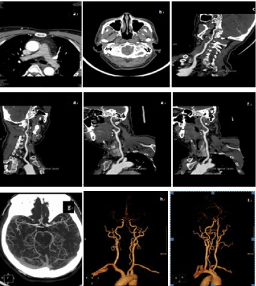
| Iterative reconstruction resulted in generally lower mean attenuation than FBP, both in the vessels and in the reference structures. In many cases the differences were significant but supposedly not clinically relevant in craniocervical CTA. A “hybrid iterative reconstruction algorithm (iDose)” has recently been developed, which consists of the following two de-noising components: (i) an iterative maximum likelihood-type sinogram restoration method based on Poisson noise distribution; and (ii) a local structure model fitting on image data that iteratively decreases the uncorrelated noise. When performed, iDose allows users to adjust the image noise level by inputting a parameter called “iDose level”. The larger the iDose level is, the larger the noise reduction is, allowing users to prospectively decrease the dose at the time of the scan (expecting that iDose will cancel the associated increased noise level during the reconstruction process) [15]. Thus resulting in image appearance or look that is close representation of conventional FBP reconstruction [16]. In our study the noise was reduced (5.96 ± 1.24 vs. 11.58 ± 1.41) in iterative reconstruction with subsequent improvement of both SNR (29.08 ± 3.04 vs. 39.97 ± 7.33) and CNR (23.97 ± 1.96 vs. 29.12 ± 4.65). This improvement indicates that the use of iterative reconstruction enables dose reduction with preservation of diagnostic value. The question may arise that instead of using different kVp and mAs we could have used same value to see how the iDose algorithm works and improves the image quality but we intend to show that iDose algorithm is far better even in lower parameter compared to filter back projection. However there are very limited studies which have tried to find out the effectiveness of the iDose in craniocervical CTA but there are studies in coronary CTA. Daisuke Utsunomiya et al suggest that using iDose yields higher CNR and better image quality than FBP. He also stated that there is not so significant change in image quality using level 3 iDose compare with iDose 7 [17] (Figure 1). |
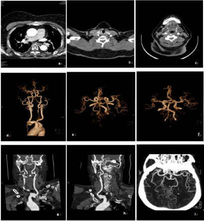
| This study found that the radiation estimated dose can be reduced while maintaining the image quality using the new algorithm. The ED in Group B was significantly lower than in Group A (6.24 ± 0.45 vs 11.35 ± 0.83), we found that a 55% reduction in the radiation dose and lower CT attenuation of vessels can be achieved by using 100 kVp and 225 mAs, Askell Love et al. also stated that lower attenuation and radiation dose can be achieved using lower kVp and mAs [18]. When combined with HIR algorithm, the improved imaging of head and neck arteries were sufficient for clinical diagnosis. Some studies in coronary CTA shows that iterative reconstruction facilitates dose reduction ranging 32-64%, depending on patient BMI. By using low tube voltage technique, a better vascular enhancement can be achieved with less iodine amount than only using low tube voltage technique as we can do it by combining low contrast and high sapital resolution. Normally in filter back projection high sapital resolution reconstructions amplify image noise levels to clinically unacceptable levels and make them suboptimal for low contrast assessments but with iDose, the noise in sharp reconstruction can be maintained at sufficiently low level to permit soft tissue and detailed high contrast assessment [19]. Excessive image noise reduction tends to result in image blurring and resolution degradation and reconstructed images appear “artificial”. Leipsic et al. [20,21] who used a different iterative reconstruction technique suggested that highly iterative reconstructions produce images that are significantly different in appearance from images acquired with FBP; their noise texture appears different and their borders manifest a significantly higher degree of smoothness. Similar observations were made by Silva et al. [22] who noted that noise-freeness may manifest as an artifactual over-smoothing of the image. Unlike image-based adaptive filtering techniques, iDose use a combination of processing in the projection and image domain providing clinical advantages such as the ability to effectively remove photon starvation related artifacts e.g. streaks [15]. Even though there is inter-observer reproducibility is not so significant but the image graininess and streak artifact is reduced significantly. The vessel plaques can ne visualized more clearly than in the filtered back projection (Figure 2). |
| The strategy of this study was to reduce the estimated radiation dose during the craniocervical CTA and have better image quality by using iDose4 in image reconstruction compared to FBP CT. Thus we conclude that HIR algorithm not only improves the image quality but also helps to reduce the radiation dose. |
| In conclusion, we can say that new HIR algorithm (iDose4) definitely reduces the radiation dose bye 55% however; further study with large sample size is required to say, it also improves the image quality compared to the filter back projection because of weak interobserver reproducibility. |
References |
|
Tables and Figures at a glance
| Table 1 | Table 2 | Table 3 | Table 4 |
Figures at a glance
 |
 |
| Figure 1 | Figure 2 |
Relevant Topics
- Abdominal Radiology
- AI in Radiology
- Breast Imaging
- Cardiovascular Radiology
- Chest Radiology
- Clinical Radiology
- CT Imaging
- Diagnostic Radiology
- Emergency Radiology
- Fluoroscopy Radiology
- General Radiology
- Genitourinary Radiology
- Interventional Radiology Techniques
- Mammography
- Minimal Invasive surgery
- Musculoskeletal Radiology
- Neuroradiology
- Neuroradiology Advances
- Oral and Maxillofacial Radiology
- Radiography
- Radiology Imaging
- Surgical Radiology
- Tele Radiology
- Therapeutic Radiology
Recommended Journals
Article Tools
Article Usage
- Total views: 13833
- [From(publication date):
December-2014 - Aug 20, 2025] - Breakdown by view type
- HTML page views : 9223
- PDF downloads : 4610
