Make the best use of Scientific Research and information from our 700+ peer reviewed, Open Access Journals that operates with the help of 50,000+ Editorial Board Members and esteemed reviewers and 1000+ Scientific associations in Medical, Clinical, Pharmaceutical, Engineering, Technology and Management Fields.
Meet Inspiring Speakers and Experts at our 3000+ Global Conferenceseries Events with over 600+ Conferences, 1200+ Symposiums and 1200+ Workshops on Medical, Pharma, Engineering, Science, Technology and Business
Case Report Open Access
Iatrogenic Injury to the Superior Vena Cava and Brachiocephalic Vein
| Jiri Herman1*, Petr Bachleda1, Marie Cerna2, Vojtech Prasil2and Petr Santavy3 | ||
| 1IInd Department of Surgery, Palacky University, Cardiovascular Centre, Olomouc, Czech Republic | ||
| 2Department of Radiology, Palacky University, Cardiovascular Centre, Olomouc, Czech Republic | ||
| 3Department of Cardiosurgery, Palacky University, Cardiovascular Centre, Olomouc, Czech Republic | ||
| Corresponding Author : | Doc MD Jiri Herman PhD, IInd Department of Surgery Cardiovascular Centre, Palacky University I.P.Pavlova 6, 775 20, Olomouc, Czech Republic Tel: +420 736 488 998 E-mail: jiriherman@seznam.cz |
|
| Received August 26, 2014; Accepted September 23, 2014; Published September 30, 2014 | ||
| Citation: Herman J, Bachleda P, Cerna M, Prasil V, Santavy P (2014) Iatrogenic Injury to the Superior Vena Cava and Brachiocephalic Vein. J Infect Dis Ther 2:169. doi:10.4172/2332-0877.1000169 | ||
| Copyright: © 2014 Jiri Herman, et al. This is an open-access article distributed under the terms of the Creative Commons Attribution License, which permits unrestricted use, distribution, and reproduction in any medium, provided the original author and source are credited. | ||
Related article at Pubmed Pubmed  Scholar Google Scholar Google |
||
Visit for more related articles at Journal of Infectious Diseases & Therapy
Abstract
We report two serious complications related to cannulation and angioplasty of the central venous system. In patient 1, superior vena cava rupture occurred in an attempt to dilate a stenosis; in patient 2, perforation of the brachiocephalic vein occurred during the placement of a dialysis catheter. These rare complications are mostly fatal. Our two female patients were treated surgically and both survived despite major blood loss. Early detection of venous system perforation and immediate treatment are of crucial importance.
| Abstract |
| We report two serious complications related to cannulation and angioplasty of the central venous system. In patient 1, superior vena cava rupture occurred in an attempt to dilate a stenosis; in patient 2, perforation of the brachiocephalic vein occurred during the placement of a dialysis catheter. These rare complications are mostly fatal. Our two female patients were treated surgically and both survived despite major blood loss. Early detection of venous system perforation and immediate treatment are of crucial importance. |
| Keyword |
| Vena cava rupture; Vena cava perforation; Hemopericardium; Angioplasty complication Introduction |
| The use of a central venous catheter was first described by Aubaniac in an injured soldier in 1952 [1]. At present, central venous catheter has an essential role in the care of patients with various conditions, particularly in intensive care units. The main indications include the need for intravenous administration of drugs, particularly of vasoactive ones and of those irritating the vein wall, parenteral nutrition, large volume replacement, hemodialysis, and hemodynamic monitoring. It is most commonly inserted via the subclavian vein or the internal jugular vein. The choice between the two veins is more or less empirical, depending on the custom of the center. Exceptionally, the cannula is inserted via the femoral vein; however, there is a higher risk of infection and thrombosis [2], even though some papers have questioned this statement [3]. |
| Although a routine procedure, central venous cannulation is associated with a wide range of complications some of which may even be life-threatening. |
| We report a successfully treated perforation of the superior vena cava (SVC) and brachiocephalic vein associated with hemorrhagic shock. |
| Case Reports |
| Case 1 |
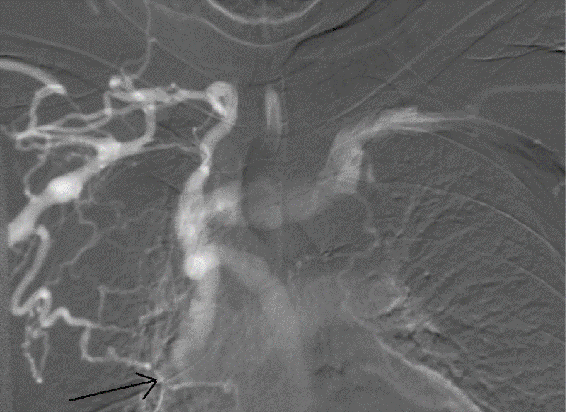
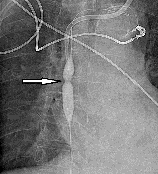
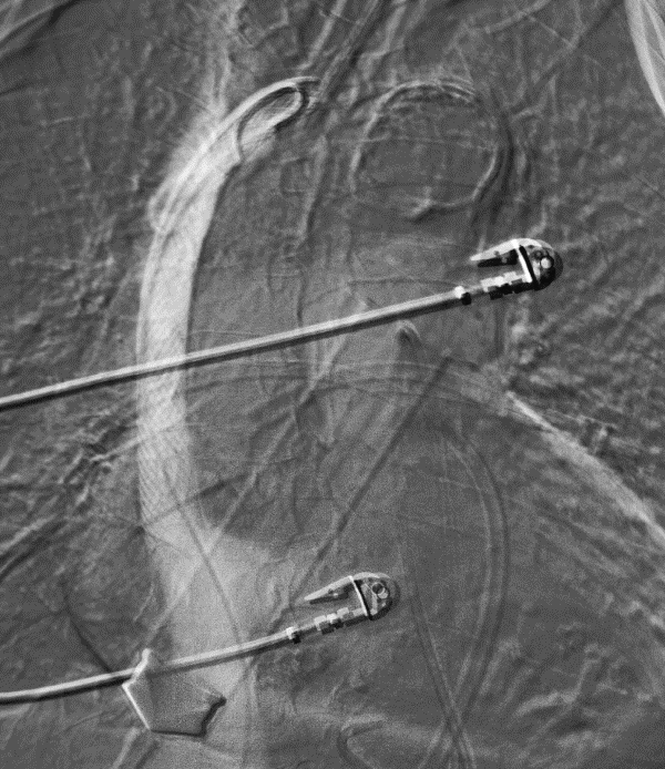
| We report a case of a 68-year-old female patient with chronic renal insufficiency, a ten-year history of peritoneal dialysis, and occluded arteriovenous fistula (AVF). Creation of a new AVF would only be possible by using an artificial vascular graft, which had been refused by the patient; therefore, a long-term dialysis catheter has been used for the last three years. In a local hospital, it had repeatedly been inserted bilaterally via the subclavian veins, bilaterally via the jugular veins as well as via the femoral vein. The last procedure had failed; therefore, the patient was referred to our center for dialysis catheter insertion. Phlebography was performed that revealed occlusion of the arm's superficial venous system as well as occlusion of the axillary and subclavian veins. The SVC was filled by means of collateral circulation and had a tight stenosis prior to its entry into the right atrium (Figure 1A). A free deep venous system was found on the left; therefore, a dialysis catheter (Kflow® Epic®. Kimal plc. Arundel Road, Uxbridge, Middlesex, England) was inserted under ultrasound guidance via the left subclavian vein and, subsequently, an interventional radiologist performed dilatation of the tight SVC stenosis via the right femoral vein (Figure 1B). At this point, rupture of the SVC and hemopericardium occurred. The interventional radiologist inserted a stent to the site of perforation which closed the rupture only partially (Figure 1C). Simultaneously, a pericardial drain was inserted and 1850 ml of blood were evacuated. The patient was indicated for surgical management. A suture of the SVC was performed via a sternotomy. A dialysis catheter was inserted via the right femoral vein. On the fourth postoperative day, the patient was transferred in a good condition to a local hospital near where she lived. She was discharged from the hospital in a good condition five days later. The patient has been on regular dialysis performed with a catheter inserted via the femoral vein. |
| Case 2 |
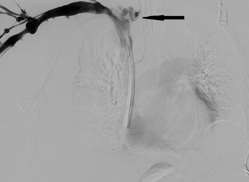
| We report a case of a 72-year-old multimorbid female patient (chronic renal insufficiency, uremic syndrome, Wegener's granulomatosis, hypertension, hyperlipoproteinemia, cholecystolithiasis, a history of stroke, atrial fibrillation) newly indicated for dialysis. There had been failed attempts to place a central venous catheter in a local hospital; therefore, the patient was referred to our center for dialysis catheter insertion. Initially, phlebography was performed that revealed a fragile superficial venous system of both upper extremities and a free deep venous system. Thus, puncture of the subclavian vein was performed below the right collar bone; the guidewire was introduced into the SVC under skiascopic control, followed by the dilator, sheath and the dialysis catheter that would not run smoothly in the sheath and higher pressure had to be applied for insertion. Suddenly, the patient started to be restless, reporting pain between the shoulder blades. An x-ray showed a pneumothorax that was managed by performing a tube thoracostomy. The patient became hemodynamically unstable, requiring blood transfusions, and a central vein injury was suspected. Consequently, phlebography was performed via a cannula inserted in the right cubital vein that failed to confirm bleeding from the venous system. The patient became stable and the pain started to subside; however, when being moved from the operating table, she suddenly became hypotensive and tachycardiac, and 1700 ml of blood were evacuated with a chest drain. She was intubated and phlebography via the right cubital vein was performed again that showed perforation of the brachiocephalic vein just below the junction of the jugular and subclavian veins (Figure 2). The initial phlebography failed to prove the bleeding, which was likely due to compression by a hematoma. The patient was indicated for revision surgery. Partial sternotomy with cranial extension was used to seek the right brachiocephalic vein; on its dorsomedial aspect, a perforation that was bleeding was found. A suture was performed. No further hemorrhage occurred. Once stabilized, the patient was transferred to the Department of Anesthesiology and Intensive Care Medicine; on the 14th postoperative day, she was transferred to a hospital near where she lived and discharged from it a week later. The patient has been in a good condition. Dialysis is performed with a catheter inserted via the right subclavian vein. |
| Discussion |
| Perforation of the SVC is a rare, but serious complication. To date, only a few cases have been reported. Perforation occurs during the management of SVC syndrome rather than with central venous catheter placement [4]. |
| Iatrogenic injury to the central venous system is a serious complication. Pericardial hemorrhage with subsequent heart tamponade and circulatory shock can be life threatening. Immediate resuscitation, pericardiocentesis or chest drainage, and, in particular, urgent surgical management of the site of the injury are of crucial importance. Nevertheless, the prognosis is usually not good [5]. Most cases include elderly multimorbid patients in whom hemorrhagic shock or hemopericardium represent a fatal complication. The quality of SVC tissue also tends to be poor in these patients. |
| However, perforation of a central vein is not always associated with a fatal outcome. Mansour treated a perforation of the SVC with emergency stent implantation and the patient survived [6]. Dilatation of a malignant stenosis of the SVC was performed during which a vein wall rupture occurred followed by mediastinal as well as pericardial hemorrhage and the development of shock. It was crucial for the survival of the patient that the lesion was treated with an immediate reinflation of the venoplasty balloon. |
| Both our patients survived as well, even though an invasive surgical procedure had been chosen in both of them and stent placement had failed in one of them. The advantage is that the procedures are carried out in a hybrid operating room equipped with a Philips angiogram. |
| The time factor is decisive for survival. Azizzadeh reported an iatrogenic SVC injury [7]. A 54-year-old female patient underwent a left subclavian vein tunneled hemodialysis catheter placement. No complications occurred during insertion; however, a post-operative x-ray revealed right-sided hemothorax and perforation of the SVC by the catheter. The perforation was successfully repaired using a 10-mm Viabahn stent graft delivered through a femoral approach. In this case, there was a blood loss of 1 liter, the patient received blood transfusions, but no hemorrhagic shock developed. An even later presentation of perforation was described in a case report by Siani [8]. Three months following angioplasty of the SVC with stent placement, there was development of hemorrhagic shock with a fatal outcome. The autopsy showed SVC perforation due to stent rupture and bare-metal stent penetration into the vena cava wall. The pericardial sac was filled with coagulated blood. No mediastinal bleeding was found. |
| An interesting case was reported by Kuzniec [9]. Even though the catheter was inserted via the right internal jugular vein, perforation of a central vein occurred. Videothoracoscopic-guided management of the site of perforation was used. In this case, the injury to the vein wall was likely to be minimal. |
| The diagnosis may be challenging, particularly when there is not a rapid development of hemorrhagic shock. Even when phlebography is negative, it does not imply the absence of perforation, as illustrated by our case report. Electrocardiography cannot distinguish between intra- and extravasal positions of a central venous catheter [10]. Bedside echocardiography is the simplest and fastest method for diagnosing hemopericardium. |
| These events are very rare. In the last five years, 725 central venous cannulations, including long-term dialysis catheters, and 325 dilatations of the central venous system have been performed in our cardiovascular center. Perforation and bleeding occurred only in two patients in this group. |
| Despite the rare nature of these complications, extreme caution must be taken during the placement of central venous catheters and angioplasty of the central venous system. These procedures should be carried out by an experienced operator and, in the case of pain, hypotension, or dyspnea, vein wall injury should be kept in mind and venography performed. CT is time consuming and requires transfer of the patient from the operating room; therefore, it should only be reserved for completely stable patients. Pericardial hemorrhage is demonstrated by echocardiography. It is necessary to rule out an allergic reaction to drugs or contrast agents as well as a vasovagal episode, which should not be difficult. When perforation is suspected, the access to the vein is preserved and venography is carried out. This can also be performed via a peripheral vein. It is more important to use the route of access to the central system to insert a venoplasty balloon for temporary tamponade of the rupture site. In the case of hemopericardium, pericardiocentesis is performed. Rapid and effective hemorrhage control is of importance. This can be done by endovascular technique, which may not be successful, as in our patient. We consider surgical treatment to be more suitable. Total or partial sternotomy provides a quick overview of the central venous system and allows for a definitive management of the vein wall injury. |
References
- Aubaniac R (1952) [Subclavian intravenous injection; advantages and technic]. Presse Med 60: 1456.
- Parienti JJ,du Cheyron D, Timsit JF, Traoré O, Kalfon P, et al. (2012) Meta-analysis of subclavian insertion and nontunneled central venous catheter-associated infection risk reduction in critically ill adults. Crit Care Med 40: 1627-1634.
- Marik PE,Flemmer M, Harrison W (2012) The risk of catheter-related bloodstream infection with femoral venous catheters as compared to subclavian and internal jugular venous catheters: a systematic review of the literature and meta-analysis. Crit Care Med 40: 2479-2485.
- Khalid I,Omari M, Khalid TJ, Castillo E, Khandelwal A, et al. (2010) Pericardial tamponade after superior vena cava stent: are nitinol stents safe? Asian CardiovascThorac Ann 18: 294-296.
- Schummer W,Schummer C, Fritz H (2001) [Perforation of the superior vena cava due to unrecognized stenosis. Case report of a lethal complication of central venous catheterization]. Anaesthesist 50: 772-777.
- Mansour M, Altenburg A, Haage P (2009) Successful emergency stent implantation for superior vena cava perforation during malignant stenosis venoplasty. CardiovascInterventRadiol 32: 1312-1316.
- Azizzadeh A, Pham MT, Estrera AL, Coogan SM, Safi HJ (2007) Endovascular repair of an iatrogenic superior vena caval injury: a case report. J VascSurg 46: 569-571.
- Siani A,Marcucci G, Accrocca F, Antonelli R, Mounayergi F, et al. (2012) Endovascular central venous stenosis treatment ended with superior vena cava perforation, pericardial tamponade, and exitus. Ann VascSurg 26: 733.
- Kuzniec S, Natal SR, Werebe Ede C, Wolosker N (2010) Videothoracoscopic-guided management of a central vein perforation during hemodialysis catheter placement. J VascSurg 52: 1354-1356.
- Schummer W, Schummer C, Paxian M, Stock U, Richter K, et al.Extravasal position of central venous catheters despote unsuspicious ECG-guidance. AnästhesiolIntensivmedNotfallmedSchmerzther. 2005; 40: 91-96.
Figures at a glance
 |
 |
 |
 |
|||
| Figure 1a | Figure 1b | Figure 1c | Figure 2 |
Post your comment
Relevant Topics
- Advanced Therapies
- Chicken Pox
- Ciprofloxacin
- Colon Infection
- Conjunctivitis
- Herpes Virus
- HIV and AIDS Research
- Human Papilloma Virus
- Infection
- Infection in Blood
- Infections Prevention
- Infectious Diseases in Children
- Influenza
- Liver Diseases
- Respiratory Tract Infections
- T Cell Lymphomatic Virus
- Treatment for Infectious Diseases
- Viral Encephalitis
- Yeast Infection
Recommended Journals
Article Tools
Article Usage
- Total views: 16753
- [From(publication date):
December-2014 - Aug 29, 2025] - Breakdown by view type
- HTML page views : 11949
- PDF downloads : 4804
