Make the best use of Scientific Research and information from our 700+ peer reviewed, Open Access Journals that operates with the help of 50,000+ Editorial Board Members and esteemed reviewers and 1000+ Scientific associations in Medical, Clinical, Pharmaceutical, Engineering, Technology and Management Fields.
Meet Inspiring Speakers and Experts at our 3000+ Global Conferenceseries Events with over 600+ Conferences, 1200+ Symposiums and 1200+ Workshops on Medical, Pharma, Engineering, Science, Technology and Business
Case Report Open Access
Idiopathic Late Subacute Epidural Hematoma: An Uncommon Cause of Pure Lumbar Radiculopathy with MR of Spinal Epidural Lesions Differentials; Revisited
| Mohamed Ragab Nouh*, Mostafa Abd El-Monem El-Shimy, Aymen Ahmed Sakr and Alaa Talaat | |
| Department of Radiology, Alexandria University, Egypt | |
| Corresponding Author : | Mohamed Ragab Nouh Department of Radiology Alexandria University Alexandria-21526, Egypt Tel: +966537476313 E-mail: mragab73@yahoo.com |
| Received June 08, 2015; Accepted June 26, 2015; Published June 30, 2015 | |
| Citation: Nouh MR, El-Shimy MAEM, Sakr AA, Talaat A (2015) Idiopathic Late Subacute Epidural Hematoma: An Uncommon Cause of Pure Lumbar Radiculopathy with MR of Spinal Epidural Lesions Differentials; Revisited. OMICS J Radiol 4:192 doi: 10.4172/2167-7964.1000192 | |
| Copyright: © 2015 Nouh MR, et al. This is an open-access article distributed under the terms of the Creative Commons Attribution License, which permits unrestricted use, distribution, and reproduction in any medium, provided the original author and source are credited. | |
Visit for more related articles at Journal of Radiology
Abstract
Spontaneous spinal epidural hematoma is an increasingly recognized entity in clinical practice. MR is a valuable tool in diagnosis yet its chronological signal changes and different pattern of enhancement may impose a diagnostic difficulty. Other worrisome differential diagnostics with different management, prognoses represent the daily radiologist challenge. We present a case of pure lumbar radiculopathy caused by accidentally discovered spontaneous late sub-acute epidural hematoma and discuss the diagnostic differentials of lumbar spinal epidural mass lesions.
|
Abstract
Spontaneous spinal epidural hematoma is an increasingly recognized entity in clinical practice. MR is a valuable tool in diagnosis yet its chronological signal changes and different pattern of enhancement may impose a diagnostic difficulty. Other worrisome differential diagnostics with different management, prognoses represent the daily radiologist challenge. We present a case of pure lumbar radiculopathy caused by accidentally discovered spontaneous late sub-acute epidural hematoma and discuss the diagnostic differentials of lumbar spinal epidural mass lesions.
Introduction
The incidence of spinal epidural hematoma (SEH) is estimated to be 1 per million patients. SEHs approximately constitute 0.3%-0.9% of spinal epidural space-occupying lesions [1]. SEHs are classified etiologically into: (a) secondary; if it is associated with an underlying cause as trauma, coagulopathy and spinal procedures, etc. (b) spontaneous; if it is associated with a minor traumatic event with no identifiable underlying etiologic factor and (c) idiopathic when non-of the above events are present [2].
The increased recognition of idiopathic spinal epidural hematoma (ISEH) acknowledges the advance of MR imaging [1,2]. On the basis of its morphological findings, specific signal intensity changes and enhancement pattern, SEH can be easily recognized on MR imaging [3]. However, epidural hematomas could be confused with other epidural inflammatory or neoplastic mass lesions especially following intravenous gadolinium administration. These may deceit the inexperienced and inadvertent radiologist and/or treating physician, delay patient diagnosis and increases patient's morbidity [4]. The authors present an absurd case of idiopathic late subacute epidural hematoma. It occurred in the lumbar region, which isn't a typical location and manifested only as a pure right L3 lumbar radiculopathy in a patient with a previous history of lumbar surgery. Moreover, it spontaneously resolved on a 3-months follow-up examination in a conservative trial. Case Report
A 46 year old male patient referred to our MR unit, by his spinal surgeon, for imaging of his lumbosacral spines. The patient was having a sudden onset of severe back pain and right sciatica since 7 days. His past medical history was unremarkable except for previous back surgery 2 years earlier for LV4/5 disc protrusion. He denied any recent trauma. The patient reported that he is frequently taking acetyl salycilates and paracetamol prescriptions for headaches. No other relevant medical history was given. The spinal surgeon referred the patient for suspected recurrent disc pathology. Revision of previous post-operative MRI showed limited LV4-5 laminectomies with no other detectable abnormality.
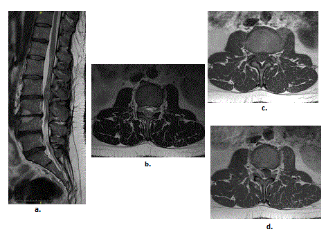
On examination he was afebrile; the pulse was 70 beats/m regular, BP 120/80 mmHg. Neurological examination of the lower limbs revealed normal sensation and preserved motor power of his lower limbs. There was no history of sphincteric disturbances or erectile dysfunction. The plain radiographs of the lumbar spine as well the routine laboratory and diagnostic studies, including coagulation parameters, were unremarkable. The patient underwent MRI evaluation of his dorso-lumbar spines that revealed a right posterolateral mass measuring 2.5 × 1.2 × 0.6 cm in greater dimensions in the epidural space opposite LV2-3 disc space level, displacing the dura and compressing the cauda equine roots anteriorly and to the left side (Figure 1). The mass has a small foraminal extension dorsal to the corresponding right L3 nerve root. It exhibited T1W iso-intense signal to the adjacent cauda equine roots and high-signal on corresponding T2W images. No signal changes were observed for the corresponding cauda equine roots. The neighboring bony structures were intact. The lesion showed significant peripheral enhancement following intravenous gadolinium administration. This enhancement pattern aroused the suspicion for an epidural abscess as the primary suspect. Extra-medullary nerve-sheath tumor, lymphoma or metastases were the most likely diagnostic differentials. However, good general condition, afebrile state, normal blood profile investigations, absent motor neurologic deficit and history of habitual NSAID intake favored the diagnosis of acute spontaneous epidural hematoma. Accordingly, a conservative trial was done. The patient had a repeat MRI scan after 3 months, which showed complete resolution of the previously seen epidural hematoma (Figure 2). Discussion
Idiopathic SEH is defined as a spontaneous spinal epidural hemorrhage in the absence of any associated risk factor as trauma, coagulopathy, anticoagulant therapy, vascular malformation, neoplasia, or systemic disease [1]. Spontaneous SEH are commonly located in the cervico-dorsal region with higher prevalence in males than females [1,5].
Spontaneous SEHs are most commonly located in the posterior or posterolateral epidural space due to intimate fibrous adherence of the dura to the posterior longitudinal ligament on the ventral surface of the spinal canal hindering hematoma formation anteriorly [4]. Their most common sources are venous bleeding from the epidural veins interconnected to the Batson venous plexus [1]. Arterial sources from the poorly vascularized ligamentum flavum have been postulated [6]. Patients with SSEHs often present with acute onset of intense, knife-like back pain radiating along the distribution of the corresponding dermatome, with rapidly developing sensory impairment, progressive distal motor weakness and even sphincteric disturbances in more severe cases [7]. These symptoms may mimic other more common conditions such as a prolapsed intervertebral disc, vertebral degenerative diseases, spinal fractures, epidural abscess formation as well as primary and secondary tumors. Hence, imaging is pivotal in the differential diagnosis of these cases [3,4]. MRI is the method of choice for screening different spinal neuroaxis pathologies, so far [3,4]. Epidural space-occupying lesion detected by MRI of the spine may pose a diagnostic challenge to the inexperienced radiologists. Currently, imaging diagnosis of epidural hematoma is eased by its characteristic epidural morphology and specific signal intensities on MR imaging (Table 1), at different phases of hematoma formation [3,7,8]. However, these variable signal intensities along with the diversity of the pattern of enhancements either peripherally or centrally, are worrisome or keep other diagnostic differentials in suspicion. The MR differential diagnosis of spinal epidural hematomas will comprehend various pathologic spectra including the atypical degenerative spinal diseases as sequestrated or migrated discs and synovial cysts/ganglia, tumors, and abscesses. Most of these pathologies share the same morphology of elliptical lesion located between the low signaled dura and spine bony elements with or without creeping through inter-vertebral foramen. Beside associated vertebral infiltration, pathologic fractures and soft-tissue masses, most epidural spinal neoplastic processes, including metastasis will have T1W ISO-to hypointense signal and T2W hyperintense signals; compared to the subjacent thecal contents; with variable patterns of gadolinium enhancement. Variations will include high-signal on T1W images on epidural lipomatous lesions and melanomas [9] as well as low signal intensity on T2-weighted images of epidural lymphomas due to their dense cellularity [10]. In the rare entity of epidural extra-medullary hematopoiesis, the diffuse signal intensities of the vertebral bone marrow, presence of paravertebral soft-tissue component and homogenous pattern of enhancement in a patient with chronic anaemic state will provide clues in the differential diagnosis [11]. Epidural abscesses are usually located anteriorly in the spinal epidural space. The presence of adjacent disc and/or endplate involvement will favor the diagnosis along with marked peripheral gadolinium enhancement. The diagnosis of epidural abscess can be clued by the clinical background of fever, laboratory findings of leucocytosis and elevated acute phase reactants [12]. Degenerative spinal entities that may overlap with SEH on MR will include both the facet synovial cysts and extruded disc fragments. Synovial cysts of the facet may develop in the spectrum of degenerative spinal disease, especially at the most mobile segment level i.e L4-5. They usually reside at the medial aspect of the facet joint encroaching on the epidural space and protrude into the spinal canal and there may be associated degenerative spondylolithesis [13]. Extruded disc fragments are usually located in the anterior epidural space, parallel the adjacent parent disc with a peripheral enhancement after gadolinium administration, due to reactive inflammation or vascular granulation tissue [13]. SSEH are self-limited process that can resolve spontaneously [1,8,7]. Although, surgical evacuation is the standard treatment for spinal epidural hematoma (SEH), conservative management is increasingly recognized in literature as in our case. Hence, in patients with small hematomas and minor symptoms repeated imaging follow-up may obviate the need for unjustified surgical procedures, especially in patients with given co-morbidities [8,14]. Summary
Spontaneous epidural hematomas should be kept in mind in patients with a recent history of evolving sciatica with no associated underling etiology. Also, it can be easily differentiated from other epidural lesions using MRI. Moreover, small hematomas can spontaneously resolve on a conservative watch trial using follow-up MR imaging.
References
|
Tables and Figures at a glance
| Table 1 |
Figures at a glance
 |
 |
| Figure 1 | Figure 2 |
Post your comment
Relevant Topics
- Abdominal Radiology
- AI in Radiology
- Breast Imaging
- Cardiovascular Radiology
- Chest Radiology
- Clinical Radiology
- CT Imaging
- Diagnostic Radiology
- Emergency Radiology
- Fluoroscopy Radiology
- General Radiology
- Genitourinary Radiology
- Interventional Radiology Techniques
- Mammography
- Minimal Invasive surgery
- Musculoskeletal Radiology
- Neuroradiology
- Neuroradiology Advances
- Oral and Maxillofacial Radiology
- Radiography
- Radiology Imaging
- Surgical Radiology
- Tele Radiology
- Therapeutic Radiology
Recommended Journals
Article Tools
Article Usage
- Total views: 14696
- [From(publication date):
June-2015 - Aug 30, 2025] - Breakdown by view type
- HTML page views : 10094
- PDF downloads : 4602
