ISSN:2167-7964
Journal of Radiology
Make the best use of Scientific Research and information from our 700+ peer reviewed, Open Access Journals that operates with the help of 50,000+ Editorial Board Members and esteemed reviewers and 1000+ Scientific associations in Medical, Clinical, Pharmaceutical, Engineering, Technology and Management Fields.
Meet Inspiring Speakers and Experts at our 3000+ Global Conferenceseries Events with over 600+ Conferences, 1200+ Symposiums and 1200+ Workshops on Medical, Pharma, Engineering, Science, Technology and Business
Image Open Access
Image Description of Lipomatous Hypertrophy of the Inter-Atrial Septum
| Aribal Serkan1*, Ozturk E2 and Yalc�?±nkaya E3 | |
| 1Aksaz Military Hospital, Radiology Service Marmaris, Mugla, Turkey | |
| 2GATA Haydarpasa Teaching Hospital, Department of Radiology Uskudar, Istanbul, Turkey | |
| 3Aksaz Military Hospital, Cardiology Service Marmaris, Turkey | |
| Corresponding Author : | Ar�?±bal Serkan Aksaz Military Hospital Radiology Service Marmaris Mugla, Turkey E-mail: serkanaribal@gmail.com |
| Received: January 06, 2016; Accepted: January 11, 2016; Published: January 13, 2016 | |
| Citation: Serkan A, Ozturk E, Yalc�?±nkaya E (2016) Image Description of Lipomatous Hypertrophy of the Inter-Atrial Septum. OMICS J Radiol 5:213. doi:10.4172/2167-7964.1000213 | |
| Copyright: © 2016 Serkan A, et al. This is an open-access article distributed under the terms of the Creative Commons Attribution License, which permits unrestricted use, distribution, and reproduction in any medium, provided the original author and source are credited. | |
Visit for more related articles at Journal of Radiology
| Keywords |
| Dyspnoea; Hypertrophy; Lipomatous |
| Image |
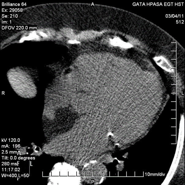 |
| A 58-year-old female patient was suffering from exortional dyspnea was admitted to our hospital’s cardiology service. She was underwent the coronary computed tomography angiography (CTA) for further evaluation. The CTA revealed variable non-critic stenosis through coronary arteries. Also there is an expansion and increase in thickness at inter-atrial septum because of a hypo dense mass-like area. The density of this area is -37 HU which is compatible with fatty tissue. The final diagnosis is lipomatous hypertrophy of the inter-atrial septum (LHIS). |
| The prevalence of LHIS is estimated to be between 1-8%. It’s asymptomatic and detected incidentally in most of the cases. In some cases, there could be superior vena cava obstruction and cardiac rhythm disorders. In these complicated cases the only treatment choice is surgical. However, it is benign in many individuals and often does not warrant any treatment. |
Relevant Topics
- Abdominal Radiology
- AI in Radiology
- Breast Imaging
- Cardiovascular Radiology
- Chest Radiology
- Clinical Radiology
- CT Imaging
- Diagnostic Radiology
- Emergency Radiology
- Fluoroscopy Radiology
- General Radiology
- Genitourinary Radiology
- Interventional Radiology Techniques
- Mammography
- Minimal Invasive surgery
- Musculoskeletal Radiology
- Neuroradiology
- Neuroradiology Advances
- Oral and Maxillofacial Radiology
- Radiography
- Radiology Imaging
- Surgical Radiology
- Tele Radiology
- Therapeutic Radiology
Recommended Journals
Article Tools
Article Usage
- Total views: 10134
- [From(publication date):
February-2016 - Aug 28, 2025] - Breakdown by view type
- HTML page views : 9216
- PDF downloads : 918