Research Article Open Access
Image Quality and Radiation Dose Comparison between Filtered Back Projection and Adaptive Statistical Iterative Reconstruction in Non-contrast Head CT Studies
| Seth Alper*, Prasoon Mohan, Ian Chaves, Richard Kane and Joseph Calandra | |
| St. Francis Hospital, Evanston, Illinois, USA | |
| Corresponding Author : | Seth Alper St. Francis Hospital, Evanston, Illinois, USA E-mail: seth-alper@comcast.net |
| Received June 26, 2013; Accepted October 01, 2013; Published October 07, 2013 | |
| Citation: Alper S, Mohan P, Chaves I, Kane R, Calandra J (2013) Image Quality and Radiation Dose Comparison between Filtered Back Projection and Adaptive Statistical Iterative Reconstruction in Non-contrast Head CT Studies. OMICS J Radiology 2:147. doi: 10.4172/2167-7964.1000147 | |
| Copyright: © 2013 Alper S, et al. This is an open-access article distributed under the terms of the Creative Commons Attribution License, which permits unrestricted use, distribution, and reproduction in any medium, provided the original author and source are credited. | |
Visit for more related articles at Journal of Radiology
Abstract
Purpose: The goal of the study was to compare the total radiation dose and subjective image quality between conventional Filtered Back Projection (FBP) and Adaptive Statistical Iterative Reconstruction (ASIR) in non-contrast head CT. Method: Forty seven patients underwent non contrast head CT with image reconstruction using 100% FBP. The same set of patients underwent subsequent non contrast head CT within 6 months using combination of ASIR and FBP reconstruction. Institutional Review Board approval was obtained. Objective assessment of total radiation dose was obtained by measuring the dose length product (DLP) and effective radiation dose. Subjective image quality was assessed by 2 attending radiologists who were blinded to the scanning technique. All images were graded for sharpness, image noise and overall image quality on a 5 point Likert scale. Both subjective and objective measurements from the two sets of images were compared. Paired Student’s t-Test was used for statistical analysis. Results: There was a significant reduction in DLP with the application ASIR from 1781.7 (310) to 1098.9 (SD 281.4) mGy-cm (p<0.01), resulting in mean total dose reduction of 38.4%. The total effective dose also showed similar reduction from 3.9 (SD 0.68) to 2.4 (SD 0.62) mSv (P<0.01). Subjective score for image noise was 4.44 (SD 0.43) and 4.40 (SD 0.48), before and after application of ASIR, respectively (p=0.59). Overall image quality scores were 4.87 (SD 0.33) and 4.85 (0.36) before and after application of ASIR, respectively (p=0.12). Subjective scores for image sharpness decreased after application of ASIR from 4.94 (SD 0.24) to 4.77 (SD 0.47) (p=0.04) Conclusion: Compared to conventional FBP reconstruction, combination of ASIR with FBP reconstruction results in significant reduction in total radiation dose without affecting the overall image quality in non-contrast head CT.
| Keywords |
| Image quality; Filtered Back Projection (FBP); Adaptive Statistical Iterative Reconstruction (ASIR); Effective radiation dose |
| Introduction |
| Dependency on radiological studies as primary means for diagnosis of disease continues to rise. Specifically, CT scan utilization has been shown to have markedly increased over the last decade, and has tripled from 1996 to 2010 [1]. While advances in diagnostic imaging has led to shorter delays in diagnosis and treatment, increasing media coverage on the harmful effects of radiation dose has led to rising concerns for the general public. Data from epidemiological studies following atomic bomb survivors, as well as from prospective studies following patients receiving radiation therapy as treatment for various conditions has shown a link between cumulative radiation dose and an increased risk for development of cancer [2]. Therefore, an underlying conflict exists between the reliance on medical imaging and preventing exposure to the potentially harmful ionizing radiation that is inherent with its increasing utilization. |
| Ionizing radiation from medical imaging now accounts for nearly half of the radiation exposure experienced by the population in the United States [3]. Computed Tomography (CT) scans are commonly ordered and account for approximately 66% of the total radiation dose received by patients from medical imaging [4,5]. Several technical factors are important in determining the radiation dose from a CT scan, such as x-ray beam energy, tube current and scan time. The dose-length-product (DLP) is a measure of the total radiation dose, which takes into account the weighted CT Dose Index (CTDI) and the scan time [6,7]. The CTDI is a measure of radiation exposure per slice. The DLP represents the product of the CTDI and scan length, and therefore, represents the radiation exposure for the entire image series. The effective dose (E) is a concept originally designed to describe occupational exposure for workers, and is used as a descriptor that reflects risk, or an estimated radiation detriment averaged over age and gender. It is often used in dose comparisons between various diagnostic exams, and is estimated from the DLP multiplied by a coefficient k, which is dependent on the region of the body being scanned [8]. |
| The total CT radiation dose can be reduced by decreasing the tube current or the tube voltage [9,10]. However, low dose CT is associated with in increased image noise [11]. Several mathematical algorithms have been used to minimize noise from medial images to improve diagnostic quality. Filtered Back Projection (FBP) has been the most popular of these mathematical models. FBP uses mathematical filters to reduce image noise as the image is being back projected on itself. FBP is considered relatively mathematically simple and requires relatively low computational power, which results in shorter processing time. This method gained popularity in the early years of CT as the computational processing power of the older machines was limited. However, when the tube current is further lowered to decrease the dose, FBP images become unreliable, as they have unacceptable noise level. |
| Iterative reconstruction is a more complicated algorithm used in two- and three-dimensional image processing that involves correction of certain data points based on mathematical models of various projections that an image is acquired in. While having an improved insensitivity to image noise, long computational times compared to FBP was an initial limitation to iterative reconstruction methods that were historically used in PET and early CT image processing. In Adaptive Statistical Iterative Reconstruction (ASIR), only one mathematical model is used to correct for image noise during processing of raw data, thereby allowing for a less computationally expensive method of reducing image noise compared to conventional iterative reconstruction. |
| With ASIR, statistical models and an iterative reconstruction approach allow for reduction in noise by limiting pixel variance that is statistically unlikely to represent true anatomy [12]. This is accomplished by iteratively comparing an acquired image to a statistically modeled projection. Several studies comparing ASIR techniques with standard FBP have showed improved Signal to Noise Ratios (SNR), increased subjective image quality and lower total DLP delivered to the patient with both chest and abdominal CT protocols [13-17]. Additionally, newer studies looking at using ASIR with head CT have shown potential in limiting effective dose, while lowering SNR and producing acceptable subjective image quality [18,19] (Table 1). |
| The primary benefit of ASIR over FBP is reduced image noise with decreased radiation dose. This is achieved by allowing for lower tube current settings (and thus lower radiation dose) when acquiring the raw CT data, and counteracting the increased noise inherent with these images. |
| An optimal combination using FBP data initially followed by ASIR algorithms has been shown to produce diagnostically acceptable images at low tube currents, with lower noise levels compared to images processed with FBP alone. In our study, a combination of the two algorithms was chosen because an image that has been processed using 100% adaptive statistical iterative reconstruction tends to have a homogenous attenuation that is not diagnostically desirable, thus producing a diminishing returns effect as the proportion of ASIR reconstruction approaches 100%. Based on previous studies, a 20%- 60% proportion of ASIR (and thus 40-80% relative proportion of filtered back projection) has been shown to be most effective, by both producing an image with a quality similar to that produced by FBP alone, and allowing for significant dose reduction (Figure 1a). |
| The aim of our study was to compare subjective image quality and total radiation dose delivered with head CT at our institution before and after application of ASIR. |
| Methods |
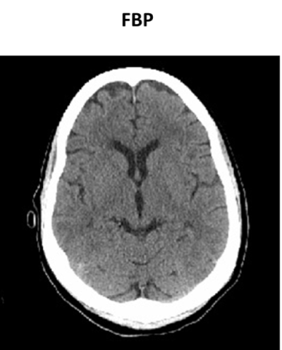
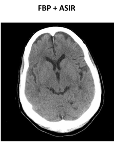
| The institutional review board approved this study with waiver of informed consent. Data was collected retrospectively. Forty seven adult patients who had undergone two non-contrast head CT examinations at our institution within a six month period of time were selected. The initial set of images was reconstructed using 100% filtered back projection. The second set of images was reconstructed using a combination of 30% ASIR and 70% FBP. This combination produces images similar in quality to FBP alone, while allowing for significant dose reduction. CT settings included 2.5 mm sections through posterior fossa (140 keV), and 5 mm sections through the vertex of the skull (120 keV). Dose modulation software was used for variable milliamperage. Image acquisition was obtained using a LightSpeed VCT 64 slice MDCT by GE, which was equipped with both ASIR and FBP algorithms. |
| Objective assessment of total radiation dose was obtained by measuring the dose length product and the total Effective Radiation Dose (ERD). Subjective image quality was assessed by two attending radiologists, with at least 10 years of experience in clinical practice. The radiologists were blinded to the reconstruction technique. Images were graded for sharpness, noise and overall image quality on a 5 point Likertrating scale from 1 (worst) to 5 (best). The Likert scale was defined as 1: poor, unacceptable for diagnostic purposes; 2: adequate but poorer than average quality; 3: average quality of a diagnostic acceptable image; 4: above average quality; 5: best quality. Images were analyzed on the same PACS workstation. The image analyzers did not review previous imaging studies for the selected patients, and were not aware of pre-existing intracranial pathology. Both subjective and objective measurements from the two sets of images before and after application of ASIR were compared. Paired Student’s t-Test was used for statistical analysis. |
| Radiation dose measurements were provided by an included image in PACS generated by the manufacturer’s software, displaying both the CTDI and DLP for each exam. The effective dose was converted from the DLP into mSV by multiplying by the conversion factor 0.0024 mSV×mGy-1×cm-1 [7,8]. |
| Results |
| There was a significant reduction in DLP with the application ASIR, which decreased from 1781.7 (SD 310.0) to 1098.9 (SD 281.4) mGy-cm (p<0.01), resulting in mean reduction of 38.4% (Figure 1a). |
| The total effective dose also showed similar reduction from 3.9 (SD 0.68) to 2.4 (SD 0.62) mSv (P<0.01) (Figure 1b). Given that the effective dose was calculated by multiplying the DLP by a conversion factor, this also resulted in a 38% decrease in the effective dose with the combination of ASIR and FBP. |
| Subjective score for image noise was 4.44 (SD 0.43) and 4.40 (SD 0.48) before and after application of ASIR respectively (p=0.59). Therefore, our observers found no significant difference in noise level between the two image sets. Overall image quality scores were 4.87 (SD 0.33) and 4.85 (0.36) before and after application of ASIR, respectively (p=0.12). There was no observable change in image quality with the addition of ASIR. The observers did notice a small but significant decrease in image sharpness with the addition of ASIR. Subjective scores for image sharpness decreased after application of ASIR from 4.94 (SD 0.24) to 4.77 (SD 0.47) (p=0.04) (Table 2). |
| Discussion |
| Compared to 100% FBP reconstruction, combination of ASIR with FBP reconstruction results in significant reduction in total radiation dose, without affecting the overall image quality in non-contrast head CT. Applying the ASIR algorithm allows for approximately 38% reduction in radiation dose to the patient. This is achieved by allowing for lower dose tube settings ASIR is applied. In our study, this was primarily achieved by lowering the tube current. This is similar to the results shown by other authors, such as Hara et al. [12] and Cornfeld et al. [17] who looked at dose reduction using ASIR applied to abdominal CT and aortic dissection protocols, respectively. Other methods of achieving a lower radiation dose have been shown by authors, such as Marin et al. [13] who used a lower kVp protocol to lower dose with ASIR reconstruction with abdominal CT [17]. In fact, in the lower kVp group that Marin used, the resulting milliamperage was actually higher. The technical method of actually achieving lower dose protocols using ASIR has been shown to be variable, with the optimal approach varying with both patient and imaging protocol parameters. |
| Our results are congruent with previous studies cited in this paper where radiation dose reduction was measured for ASIR algorithms applied to other types of CT examinations. For example, Marin et al. [13] showed effective dose reductions as great as 70% when applying ASIR to abdominal CT exams, while simultaneously achieving a statistically significant improvement in signal-to-noise ratio. Sagara et al. [20] also applied ASIR to abdominal CT imaging, demonstrating decreased radiation doses ranging from 23-66% and simultaneously lowering noise levels. Interestingly, they also showed a reduction in subjective image sharpness with some patient subsets, similar to our study. |
| Leipsic et al. [14] applied ASIR to coronary CT angiography and chest CT, demonstrating improvements in study quality and interpretability with various proportions of ASIR applied, while allowing for statistically significant decreased effective radiation dose. More recently, the application of ASIR has been described to lower both noise and radiation dose in head CT in studies by Rapalino et al. [18] and Kilic et al. [19] showed similar results, noting that the reduction in image noise for head CT was less than that observed with abdomen and chest CT, however, our study did not show significant difference in image noise levels, as other studies have shown. While the primary interest in this study was achieving lower radiation dose while maintaining acceptable quality, we hypothesize based on the findings of other studies that by modifying certain aspects of the image protocol, this can be simultaneously achieved. For example, the ratio of ASIR to FBP that was used in our algorithm was a user defined proportion of 30% to 70%, respectively. This ratio was chosen based on the fact that image quality was similar to images obtained using FBP alone, and this allowed for significant dose reduction. When the proportion of ASIR is increased toward 100%, noise levels continue to decrease toward a point at which there is a “noise-less” appearance to the image, which produces over-smoothing artifact, and is not diagnostically desirable. Making smaller adjustments to the ASIR proportion could however be investigated in future studies. A method of applying a proportion of ASIR based on patient-depended factors (such as BMI) in real time is of particular interest. |
| The increased computational demand that ASIR requires has been described in several of the previous studies cited in this paper, which can theoretically lead to increased post-processing times. However, at our institution, there is no significant time delay for image processing using the combination protocol. Images are available for transfer to the PACS system within seconds after acquisition and processing, and thus, the use of ASIR does not adversely affect radiology reporting times. |
| There is no significant change in the subjective assessment of image quality or noise after addition of ASIR to FBP, however; there was a small but statistically significant reduction in subjective image sharpness. Currently, the significance of small differences in image sharpness on diagnostic or interpretative accuracy is not known. Differences in image sharpness are also dependent on physical features of the patient, such as BMI or overall size of the patient. Variations in cranial thickness, shape and amount of subcutaneous tissue surrounding the calvarium are factors that may contribute to subjective image sharpness. Additionally, sharpness itself as a measure of diagnostic quality may also be more crucial in vascular imaging, where quantifying the degree of stenosis or demonstrating vascular dissection is necessary for diagnosis. This represents a variable for future studies. |
| Ultimately, there was agreement between the two interpreting radiologists that there was no significant difference in overall image quality. |
| Several limitations related to this study can be mentioned. First, a relatively low patient population was used for this study. This was in part due to time limitations in selecting patients who had undergone repeat CT head examinations within a 6 month period. This may have led to selection bias, as well. Second, this analysis was done retrospectively, which does limit tight control of certain variables and characteristics of the cohort selected. Third, only noncontrast head CT examinations were used for the study, and our results cannot be directly applied to contrast-infused exams or other types of commonly ordered CT exams of other anatomical regions. Specifically, confirming these results by comparing ASIR and non- ASIR reconstruction algorithms with cardiac and abdominal CT examinations at our institution are areas of interest which could be tested in the future. Finally, only two radiologist observers were used, which could account for some inter-observer variability when scoring for the subjective measurements of image quality and noise. |
| Future research could investigate further the relationship between image sharpness and dose reduction with ASIR. Expanding the number of radiologist observers is a goal for further work, which could perhaps minimize error from observer variability. Examining the impact of ASIR reconstruction on subjective interpretation of specific pathologies in neuroradiology would also be of interest, for example, carotid dissection, brain tumor or stroke. |
| In summary, our results confirm that applying a combination of ASIR with conventional FBP algorithms for non-contrast head CT image processing demonstrated no statistically difference in image quality or image noise levels, while significantly reducing the dose of radiation delivered to the patient. There was a small but statistically significant reduction in image sharpness, which is a target of future research. |
References |
|
Tables and Figures at a glance
| Table 1 | Table 2 |
Figures at a glance
 |
 |
| Figure 1a | Figure 1b |
Relevant Topics
- Abdominal Radiology
- AI in Radiology
- Breast Imaging
- Cardiovascular Radiology
- Chest Radiology
- Clinical Radiology
- CT Imaging
- Diagnostic Radiology
- Emergency Radiology
- Fluoroscopy Radiology
- General Radiology
- Genitourinary Radiology
- Interventional Radiology Techniques
- Mammography
- Minimal Invasive surgery
- Musculoskeletal Radiology
- Neuroradiology
- Neuroradiology Advances
- Oral and Maxillofacial Radiology
- Radiography
- Radiology Imaging
- Surgical Radiology
- Tele Radiology
- Therapeutic Radiology
Recommended Journals
Article Tools
Article Usage
- Total views: 14223
- [From(publication date):
October-2013 - Aug 16, 2025] - Breakdown by view type
- HTML page views : 9565
- PDF downloads : 4658
