Make the best use of Scientific Research and information from our 700+ peer reviewed, Open Access Journals that operates with the help of 50,000+ Editorial Board Members and esteemed reviewers and 1000+ Scientific associations in Medical, Clinical, Pharmaceutical, Engineering, Technology and Management Fields.
Meet Inspiring Speakers and Experts at our 3000+ Global Conferenceseries Events with over 600+ Conferences, 1200+ Symposiums and 1200+ Workshops on Medical, Pharma, Engineering, Science, Technology and Business
Case Report Open Access
Paediatric Renal Tumors with Subcapsular Fluid Sign: Is it Specific for Rhabdoid Tumors
| Seema Kembhavi1*, Sneha Deshpande1 and Sajid Qureshi2 | |
| 1Department of Radiodiagnosis, Tata Memorial Center, Mumbai, India | |
| 2Department of Surgery, Tata Memorial Center, Mumbai, India | |
| Corresponding Author : | Seema Kembhavi Associate Professor and Consulting Radiologist Tata Memorial Centre, Parel, Mumbai, India Tel: +912224177160 E-mail: seema.kembhavi@gmail.com |
| Received April 05, 2013; Accepted May 09, 2013; Published May 13, 2013 | |
| Citation: Kembhavi S, Deshpande S, Qureshi S (2013) Paediatric Renal Tumors with Subcapsular Fluid Sign: Is it Specific for Rhabdoid Tumors. OMICS J Radiology 2:128. doi: 10.4172/2167-7964.1000128 | |
| Copyright: © 2013 Kembhavi S, et al. This is an open-access article distributed under the terms of the Creative Commons Attribution License, which permits unrestricted use, distribution, and reproduction in any medium, provided theoriginal author and source are credited. | |
Visit for more related articles at Journal of Radiology
Abstract
Paediatric renal tumors are commonly encountered entities in clinical oncology, Wilms’ being the most common
histology. Other histological types include Rhabdoid tumor, Clear Cell Sarcoma, PNET, etc. Though CT/MRI scan is
perfomed for staging, it cannot differentiate between the histological types. However, there are a few imaging features
which indicate certain pathologies. Rhabdoid tumors of kidney are associated with subcapsular haemorrhage which
leads to ‘Subcapsular fluid sign’. We present our case to acquaint the radiologists with this sign and recommend
exercising caution while interpreting it.
| Abbreviations |
| RTK: Rhabdoid Tumor of Kidney; CCSK: Clear Cell Sarcoma of the Kidney; PNET: Primitive Neuroectodermal Tumors; RCC: Renal Cell Carcinoma |
| Introduction |
| Paediatric renal tumors are commonly encountered entities in clinical oncology, with the incidence of the individual tumors being largely age dependent. Though CT is considered to be the imaging modality of choice, its accuracy is limited by the fact that histologically varied tumors present with overlapping radiological features [1]. Rhabdoid tumor is an aggressive renal malignancy which exclusively affects the pediatric population. Imaging features of this rare tumor have been studied and few finding have been described to be suggestive of this pathology. ‘Subcapsular fluid sign’ is one of these features which though not pathognomonic, are closely associated with rhabdoid tumors of kidney. |
| Case Report |
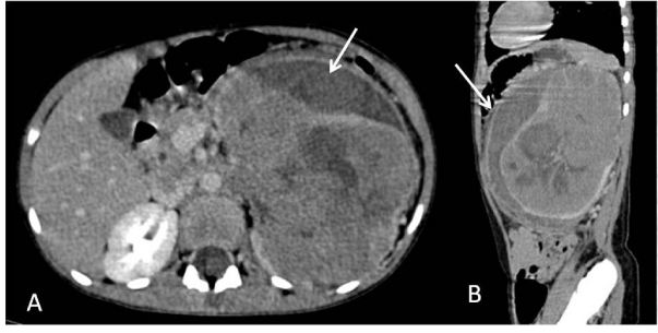
| Parents of a three year old girl came to our hospital after sonographic detection of an abdominal lump. Ultrasound was done for fullness of the abdomen; otherwise, there were no associated symptoms like haematuria. The patient underwent routine blood examinations and a contrast enhanced CT scan of the chest and abdomen. CT revealed a single large heterogeneously enhancing mass arising from the left kidney. The mass measured about 9×12×13 cm and involved almost the entire parenchyma, including the renal sinus. There were no separate tumor nodules identifiable. It did not have any calcifications or fat within. A lentiform subcapsular hypodense fluid collection was noted under the Gerota`s fascia (Figures 1A and 1B). There was no infiltration of the adjacent organs. A tumor thrombus was noted involving left renal vein with extension into IVC up to the infrahepatic level. The right kidney was normal. No evidence of lymphadenopathy or pulmonary or hepatic metastases was seen. The visualized bones were also unremarkable. An overall impression of a non-metastatic malignant renal tumor was formed, which most often is a Wilms’ tumor in this age group. However due to the presence of sub-capsular fluid sign, a probability of this tumor being a Rhabdoid Tumor of Kidney (RTK) was raised. CT guided biopsy revealed a classical triphasic Wilms’ tumor and not a RTK. |
| Discussion |
| ‘Subcapsular fluid sign’ implies visualization of a cresentic peripheral collection under the Gerota’s fascia. Sisler and Siegel first described this sign in 1989 as a characteristic feature of RTK [2]. They found that these tumors present as centrally placed soft tissue masses surrounded by subcapsular fluid collection. They attributed this to be due to the aggressive nature of the tumor. Subcapsular fluid collection is contemplated to be due to tumor necrosis (in which case it appears hypodense) or hemorrhage (appears hyperdense). |
| Chung et al. studied the imaging characteristics of RTKs and found this sign to be present in about 44% of rhabdoid tumors (8 out of 18 patients) [3] while Han et al. described this sign to be present in 57% of their RTKs [4]. Agrons et al. evaluated the specificity of imaging features in diagnosing RTK [5]. In their study of 21 patients, 71% of the tumors showed subcapsular fluid sign. However, this sign was also present in 12% of non-RTK tumors of the comparative age group, which comprised of Wilms’ tumor, Clear Cell Sarcoma, Mesoblastic Nephroma, renal cell carcinoma and undifferentiated sarcoma. In children less than one year of age, the other common cause of this sign was Mesoblastic Nephroma while in older children it was Wilms’ tumor. |
| Wilms’ tumor is the most common renal tumor in children less than 5 years of age, accounting for 96.2% in children while RTK, though exclusively a paediatric malignancy, is a rare neoplasm representing about 1.3% of renal tumors [6]. Thus because of the sheer exceedingly greater incidence of Wilms’ tumor as compared to rhabdoid tumors, this sign may be more frequently encountered with Wilms’ tumor in clinical practice than with RTK. |
| RTK is an aggressive renal malignancy with a different management protocol. This tumor needs more than usual chemotherapeutic agents and a different chemotherapeutic regimen: Ifosfamide, Carboplatin, etoposide and cyclophophamide are added to the drugs used in Wilms’ tumor (Vincristine, Dactinomycin, Doxorubicin). Also, it has a propensity for brain metastasis or even a synchronus primary in the brain and hence a screening CT scan of brain is essential for staging this tumor. Similarly it has a propensity for bone metastases. As regards the prognosis, Wilms’ tumor has an excellent prognosis with an overall 5 year survival of 92%, while rhabdoid tumor has the worst prognosis amongst the pediatric renal neoplasms, with an 18-month survival rate of only 20% [3,7]. However, imaging cannot reliably differentiate between Wilms’ tumor and RTK. Even the other pediatric renal tumors like Nephroblastoma, Mesoblastic Nephroma, Clear Cell Sarcoma of the Kidney (CSSK), Primitive Neuroectodermal Tumors (PNET), Renal Cell Carcinoma (RCC), Intra-renal Neuroblastoma, Renal Medullary Tumor, Metanephric Adenoma, Lymphoma, etc. present with non-specific imaging features. Miniati et al. [1] studied the accuracy of imaging in preoperative diagnosis of pediatric renal tumors in 92 patients. They found the diagnostic accuracy of CT to be only 82% [7]. Thus is the importance of obtaining tissue diagnosis before instituting a definitive management in pediatric renal masses. |
| Thus, pediatric renal tumors present with varied histology but only subtle clinically and radiologically appreciable differences. Therapeutic and prognostic variability however mandates accurate diagnosis. Imaging is of limited value in accurately differentiating these tumors. ‘Subcapsular fluid sign’ which is considered to be an imaging characteristic of rhabdoid malignancies is however also appreciated in other pediatric renal malignancies. Hence we present this case to acquaint the radiologists with the fact that though it definitely aids to narrow the list of differentials, the ‘subcapsular fluid sign’ sign in radiology needs to be interpreted with caution. |
| Conclusion |
| “Thus, pediatric renal tumors present with varied histology but only subtle clinically and radiologically appreciable differences. Therapeutic and prognostic variability however mandates accurate diagnosis. Imaging is of limited value in accurately differentiating these tumors. ‘Subcapsular fluid sign’ which is considered to be an imaging characteristic of rhabdoid malignancies is however also appreciated in other pediatric renal malignancies. Hence we present this case to acquaint the radiologists with the fact that though it definitely aids to narrow the list of differentials, the ‘subcapsular fluid sign’ sign in radiology needs to be interpreted with caution.” |
References |
|
Figures at a glance
 |
| Figure 1 |
Post your comment
Relevant Topics
- Abdominal Radiology
- AI in Radiology
- Breast Imaging
- Cardiovascular Radiology
- Chest Radiology
- Clinical Radiology
- CT Imaging
- Diagnostic Radiology
- Emergency Radiology
- Fluoroscopy Radiology
- General Radiology
- Genitourinary Radiology
- Interventional Radiology Techniques
- Mammography
- Minimal Invasive surgery
- Musculoskeletal Radiology
- Neuroradiology
- Neuroradiology Advances
- Oral and Maxillofacial Radiology
- Radiography
- Radiology Imaging
- Surgical Radiology
- Tele Radiology
- Therapeutic Radiology
Recommended Journals
Article Tools
Article Usage
- Total views: 15422
- [From(publication date):
July-2013 - Aug 29, 2025] - Breakdown by view type
- HTML page views : 10801
- PDF downloads : 4621
