Research Article Open Access
Patient Radiation Dose Assessment in Pelvic X-ray Examination in Ghana
| Eric K Ofori1*, William K Antwi1, Diane N Scutt2 and Matt Ward3 | |
| 1Department of Radiography, School of Allied Health Sciences, College of Health Sciences, University of Ghana, Korle-Bu-Accra, Ghana | |
| 2School of Health Sciences, University of Liverpool, United Kingdom | |
| 3Integrated Radiological Services Limited, Liverpool, United Kingdom | |
| Corresponding Author : | Eric K Ofori Department of Radiography School of Allied Health Sciences College of Health Sciences University of Ghana, Korle-Bu-Accra, Ghana Tel: 233-244-655-156 E-mail: erikof2001@yahoo.co.uk |
| Received July 27, 2013; Accepted October 22, 2013; Published October 27, 2013 | |
| Citation: Ofori EK, Antwi WK, Scutt DN, Ward M (2013) Patient Radiation Dose Assessment in Pelvic X-ray Examination in Ghana. OMICS J Radiology 2:151. doi: 10.4172/2167-7964.1000151 | |
| Copyright: © 2013 Ofori EK, et al. This is an open-access article distributed under the terms of the Creative Commons Attribution License, which permits unrestricted use, distribution, and reproduction in any medium, provided the original author and source are credited. | |
Visit for more related articles at Journal of Radiology
Abstract
Protecting the gonads of children and adults is of particular importance during diagnostic imaging of the pelvis since evidence suggests that X-rays could cause direct damage to the gonad which could result in mutation. Gonad shielding during diagnostic X-ray procedures is an effective way of reducing dose to patients’ reproductive organs and reduces the risk of genetic effects in future generations. Given the potential harmful effects associated with exposure to ionizing radiation, it is important not just to provide gonad shielding, but also to measure patient doses, and reduce them where possible. The aim of this study was to provide patient dose estimates for pelvic examination being undertaken at selected diagnostic centers in Ghana as a baseline data for pelvic dose optimization in Ghana. Dose measurements were calculated on 323 patients (137 (42%) male, 186 (58%) female, ages, 38.56 yr ± 9.0; range 20–68). The Entrance Surface Dose (ESD) was determined by an indirect method, using the patient’s anatomical data and expo¬sure parameters utilized for the specific examination. The Quality Assurance Dose Database software (QADDs) developed by Integrated Radiological Services Ltd. in Liverpool, UK was used to generate the ESD values. There were variations in the technique factors used in all the centers as compared to the recommendations in the European Commission (EC) quality criteria. Eighty percent of the hospitals recorded lower ESD values below IAEA recommended diagnostic reference levels (10 mGy) and 40% of the hospitals exceeded the UK national reference value (4 mGy). The varia¬tions in the data recorded demonstrate the importance of creating awareness by the radiographic staff on quality assurance and standardization of protocols to ensure satisfactory standards and optimized radiation dose to patients and staff.
| Keywords |
| Gonadal dose; Patient dose assessment; Optimization |
| Introduction |
| Protecting the gonads of children and adults is of particular importance during diagnostic imaging of the pelvis since evidence suggests that X-rays could cause direct damage to the gonad which could result in mutation [1]. Gonad shielding during diagnostic X-ray procedures is an effective way of reducing dose to patients’ reproductive organs and reduces the risk of genetic effects in future generations [2]. Given the potential harmful effects associated with exposure to ionizing radiation, it is important not just to provide gonad shielding, but also to measure patient doses, and reduce them where possible. |
| The most reliable dosimetry quantities commonly used in diagnostic radiology to give an indication of the typical dose that is being delivered to an average adult patient are the patient Entrance Surface (skin) Dose (ESD) including backscatter for simple X-ray projections, and the Dose Area Product (DAP) for complex examinations [3,4]. The ESD, in particular, is recommended as the most appropriate dosimetry quantity for simple X-ray projections since it meets the three basic conditions set out by the International Atomic Energy Agency (simple to measure, permits direct measurement on patient during the examination, and is representative of the dose received by the patient). It is also recommended by the Commission of the European Communities (CEC) in the document on quality criteria for the most common radiographic images. In addition, the measurement of ESD permits easy comparison with published diagnostic guidance or reference levels [4-7]. |
| Patient radiation protection in pelvis X-ray examination has not been given much attention in Ghana. Therefore this study was set out to provide an estimate of patient dose in pelvic examination being undertaken at selected diagnostic centers in Ghana as a baseline data for pelvic dose optimization in Ghana. The estimated mean ESD values were compared with the International Atomic Energy Agency [6], the European Commission (EC) guidance on diagnostic reference levels for medical exposures [8], and the 2005 United Kingdom reviewed reference levels [9]. This comparison was felt to be appropriate because at the time of the study, there were no accepted local or national diagnostic level values in Ghana for comparison. |
| Materials and Methods |
| Subjects |
| Patient Radiation dose assessment was conducted on 323 patients over 18 years, who underwent pelvic examinations during the study period. Inclusion criteria were patients over 18 years, who underwent pelvic examinations in ten selected diagnostic centers in Ghana from January to April, 2011. The pelvic examination was selected for this study because during this examination, critical organs (testes, ovaries) that contribute to effective dose are irradiated. Data was collected on 323 patients who underwent Antero–Posterior (AP) pelvis examination in 10 selected hospitals. Ten radiographers and ten radiographic technicians participated in the study and completed the data collection sheets after each examination. The data sheets required for the study were placed near the console of the X-ray room and were completed by the radiographers when a patient entered and required pelvic examination. The examination rooms were chosen for practical and logistical reasons, and were representative of the regional and district hospitals in Ghana. A tape measure of a least count of 0.1 cm was used to measure the Focus-Film-Distances (FFDs). All FFD measurements were from the centre of the tube to the film or the table top. |
| 0X-ray equipment |
| Table 1 shows the characteristics of the X-ray machines in the 10 hospitals used for the study, all of which were constant potential generator (80 kVp) with 2.5 mm Al filtration. Two manufacturers’ cassettes were in use during the study, namely Agfa and Kodak with two different screen-film combination speeds; 200 and 400. Since the study was aimed to provide patient dose estimates based on the patient’s anatomical data and exposure parameters utilized for the specific examination, the performance of these cassettes was not assessed. The ESD was determined from the radiographic examinations, which were of adequate image quality. For purposes of ethical consideration the study sites were coded H-1 to H-10. |
| Patient physical measurements |
| The anatomical thickness (cm), weight (kg), height (m), and gender of the patients, as well as radiographic exposure factors (kV and mAs) used for each patient’s examination, were recorded. Patients who met the inclusion criteria (aged 18 years and above) in the selected hospitals were weighed and a specially designed caliper of least count of 1 mm was used to measure the anatomical thickness for the body part under examination, which in turn was used to estimate the focus skin distances for the examination. The anatomical thicknesses of the patient were measured at the level of anterior–superior iliac spines. A tape measure of least count 0.1 cm was used to measure the Focus Film Distances (FFD). All FFD measurements were from the center of the tube to the film or the table top. The datasheets were placed near the console of the X-ray room and were completed by the radiographic staff for each patient requiring pelvis examination. |
| Patient radiation dose assessment |
| The ESD for patients was assessed by an indirect method, using data of radiation output of the X-ray tubes and exposure factors (peak kilovoltage (kVp) and milliampere seconds (mAs)). In order to determine the ESD from the generator output, air kerma at different kVp settings was first measured using a calibrated solid state detector Unfors Xi Platinum (SN: 127871). It was placed at one-meter focusdetector distance on top of the table at different kVp settings. The Focus-patient Surface Distance (FSD) and radiographic exposure factors (kVp and mAs) used for selected examinations were recorded on a self-designed Excel sheet. Data sheets were collected on a weekly basis and the exposure factors recorded were crosschecked against actual practice with the radiographers who recorded them, in order to validate the figures. The information on the data sheets were prepared in Excel formats which were transferred into a QA Dose Database Software (QADDS) developed by Integrated Radiological Services (IRS) Ltd (Liverpool, UK) for the computation of the ESD. In order to perform calculations of ESD, several items of information such as kV selected, mAs seconds delivered and the FSD were entered into a front page data sheet of the program. Having entered the required information on to the front page the program performs a calculation of ESD using the formula: |
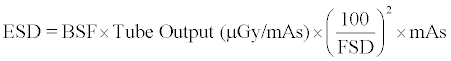 (1) (1) |
| Where BSF is the backscatter factor = (1 + backscatter fraction), and FSD is the focus-to-skin distance used. An Excel output data file was then generated from the QADDS which were converted into an SPPS version 17 file to facilitate descriptive and inferential analysis. |
| Results |
| Descriptive statistics of examinations and patient data |
| Dose measurements were calculated on 323 patients of which 137 (42.4%) were males and 186 (57.6%) were females. Descriptive statistics on patients’ age, weight, body mass index, and the body part thickness are shown in Table 2. The age range for all patients in the hospitals was 20.0–68.0 years, with mean and standard deviation values as 38.6 yrs and 9.0, respectively. |
| The EC criteria for ESD calculations assume a 20.0 cm AP trunk thickness and average weight of 70.0 kg for a standard adult patient. However, in this sample of Ghanaian patients, the range of AP trunk thicknesses for pelvis was 20.0–40.0 cm. This influenced focus-toskin distance used to calculate ESD because, if the Focus-to-Film Distance (FFD) is constant, then a range of patient size will present a corresponding range of FSD values, which have a direct impact on ESD. |
| Analysis of entrance surface dose (ESD) |
| The mean and the Standard Deviation (SD) of the Entrance Surface Doses (ESD) estimated for the individual examinations for all the ten rooms in addition to the Range Factor (RF)-defined as the ratio of maximum to minimum dose for the same type of examination- were calculated and are presented in Table 3. |
| The range factor, which highlights the spread/variation in the ESD values for the same type of examination either within or between the rooms, as well as the minimum, maximum, and range factor of ESD values for the same type of examination in the same room (intra-room variation) are also shown in Table 3. In this way, the factor by which the dose of radiation can vary for the same examination in the same room is indicated by the quotient of highest and lowest dose for an examination. |
| Summary of radiographic exposure data |
| The mean and the range of the exposure factors (the tube voltage (kVp), the tube current (mAs), and the Focus Film Distance (FFD) in centimeters for the individual hospitals are presented in Table 4, alongside the European Commission’s recommended exposure factor values [8] for good radiographic techniques. |
| Discussion |
| A total of 323 dose measurements on antero–posterior pelvis examinations were recorded during the study. The proportion of males (46.7%) to females (53.3%) in this study reflects the demography in Ghana where females constitute about 55% of the population [10]. In the EC quality criteria and the IPEM Report 91 [11], it is recommended that the ESD measurements be made on statistically significant sample of patients (minimum 10) whose weights are near the standard adult patient of average weight 70.0 kg ± 10. This study complied with this recom mendation and therefore the estimate of ESDs for the various examinations could be considered sufficiently representative value for the specific rooms. |
| This study has provided some initial base line data on the size of the average adult patient in Ghana and the corresponding dose for pelvis examinations. The mean weight recorded for all patients was 80.72 ± 13.0 kg. This is different from the mean weight recorded in the IAEA study in 2004 [6] on patients undergoing radiographic examinations in some European and Asian countries. In the IAEA study, an average weight of 70.0 ± 10 kg was considered appropriate for the European participating countries, while 65 ± 10 kg was used for the Asian countries. The average weight of the only African country that participated in the study, Morocco, was not stated, but a compromise was made to enable a comparison of the measured doses with reference doses. It is therefore relevant to compare the estimated dose recorded from this study with the reference values based on the average weight in the Ghanaian context. The fact is that, the weight of the Ghanaian average man differs considerably from the European and Asian male suggests that more applicable data are needed for the Ghanaian situation. |
| The radiographic technique parameters recorded show that there were variations in the technique factors when compared with the recommendations in the EC quality criteria [8]. Vary ing radiographic voltages and reduced focus film distances were used. All these factors have adverse influence on the outcome of the dose to patient. The above problem was not isolated to Ghana, but is common in other developing countries [12-14]. These problems probably could partly be associated with the inadequate training of imaging staff, variation in patient physique, different types of equipment, and variety of techniques used in different hospitals. Also, the different methods of documentation of data on radiation dose could also lead to apparent dose variations [14,15]. |
| This study also revealed that there were inconsistencies in the use of the focus film distances as recommended in the EC quality criteria. The EC criteria recommend an average FFD of 115 cm and a range of 100– 150 cm. Most diagnostic centers used FFD values below the average values (115 cm) but equal to the minimum recommended value (100 cm). Since ESD is inversely proportional to the square of the FFD, for the same kV and mAs the dose reaching the patient is expected to be high. Although the general trend across all centers is the use of lower FFDs and this, in part, might explain higher ESDs, it can be seen that the results do not show this as a universal trend (some centers with low FFDs present mean ESDs around 2 mGy, some much, much higher). It is worth noting that changing FFD could be a good change, but will still not solve all discrepancies found in the study. It is therefore essential that policies on quality control and assurance monitoring programs be enforced in the hospitals to protect the patient against unnecessary exposures through repeat examinations [16]. |
| Generally, ESD values for the same type of examination in the same room will vary due to the differences in patient size and in the radiographic technique used by different radiographers. Variations in the ESD values between different X-ray rooms will additionally be due to differences in radiographic equipment, film type, processing, chemistry, and processing conditions. The mean ESD values for the individual examinations varied considerably across all hospitals and within hospitals. A particular hospital, H-7, recorded consistently higher ESDs than the other departments [17]. On closer investigation, it was revealed that the Automatic Exposure Control (AEC) device was consistently being incorrectly used or was frequently overridden by the radiographer for no apparent reason. Automatic exposure devices are intended to take some of the human error out of exposure factor selection, but overriding them has a detrimental effect on patient dose. This particular issue (of not using AEC where they were available) was not confined to this hospital; some hospitals with AECs had disconnected them but the reasons for this were not clear and warrant further investigation. It is likely that staff had not been trained in the use of automatic exposure devices, lacked confidence, and therefore defaulted to the original methods of manual selection of exposure factors. The lower doses recorded in the other hospitals in comparison with H-7 was because lower mAs were used. |
| The above results suggest that hospitals with lower ESD values than the reference dose values provided by UK, EC, and IAEA are acceptable in terms of dose to the patient. However, optimization has to be interpreted in the light of the number of the acceptable radiographs produced with adequate image quality. The use of lower ESD values in pelvic examinations is encouraging but should not be at the expense of image quality. There should be always a balance between the patient dose and the quality of the radiographic images produced [18]. |
| There was a considerable variation in the range factor for ESD for the same type of examination in the same room. The range factor highlights the spread/variation in the ESD values for the same type of examination either within or between the rooms. This implies that in the same X-ray room there were variations in radiographic technique between radiographers (usually differences in exposure time) which could be, in part, related to differences in patient size, but could not be accounted for by this parameter alone. In terms of inter-room variations, the mean ESDs showed variations in dose between rooms. The ratio of the maximum dose in one room to the minimum dose in another was 69.8. These variations in the ESD for the same type of examination between the rooms may be due in part to the different technical characteristics of radiographic equipment, but are mainly due to the techniques employed, and particularly technique inconsistency. These inter- and intra-hospitals’ dose variations for the same type of examination confirm the variability in the operational conditions within and among the hospitals. Diagnostic References dose levels are yet to be established in Ghana and other West African Countries. |
| Conclusions |
| Patient radiation doses in pelvic X-ray examinations in ten selected diagnostic centres in Ghana were assessed along with the radiographic technique factors and compared with the IAEA, EC and UK recommended DRLs. The study showed inconsistent practice techniques and radiation dose delivered to patients for the same kind of radiographic examinations which suggest lack of optimization of radiographic practice in Ghana. Eighty percent of the hospitals recorded lower ESD values below EC/IAEA recommended diagnostic reference levels (10 mGy), and 40% of the hospitals exceeded the UK national reference value (4 mGy). There is therefore urgent need for standardization of practice and implementation of appropriate measures that will protect the patient against unnecessary radiation exposure. Also these variations, which are assumed to be present in most of the X-ray departments operating in the country, point to the need for the introduction of a national protocol and QA system, and frequent dose audits. However in Ghana due to the high illiteracy rate and lack of awareness the effects of radiation are little known nor taken seriously among the population. Although patient concerns about radiation exposure have been somewhat muted in Ghana coverage of these issues in the media and on the Internet may produce an increasing public awareness about radiation doses. There is therefore the need for more aggressive quality assurance measures in the performance of general radiographic examinations. Once there is awareness and appropriate quality assurance programmes are implemented, it will no doubt contribute to the provision of high-quality care in the country. This is the key to improving patient safety, quality service delivery, and the performance of the imaging facilities. |
| Acknowledgement |
| The authors acknowledge the support and cooperation received from the staff of the participating hospitals, and are grateful to Professors Edwin Wiredu and Cyril Schandorf for their technical support. |
| Funding |
| This project was funded by the authors from research and book allowance given to academic staff of the University of Ghana. |
References |
|
Tables and Figures at a glance
| Table 1 | Table 2 | Table 3 | Table 4 |
Relevant Topics
- Abdominal Radiology
- AI in Radiology
- Breast Imaging
- Cardiovascular Radiology
- Chest Radiology
- Clinical Radiology
- CT Imaging
- Diagnostic Radiology
- Emergency Radiology
- Fluoroscopy Radiology
- General Radiology
- Genitourinary Radiology
- Interventional Radiology Techniques
- Mammography
- Minimal Invasive surgery
- Musculoskeletal Radiology
- Neuroradiology
- Neuroradiology Advances
- Oral and Maxillofacial Radiology
- Radiography
- Radiology Imaging
- Surgical Radiology
- Tele Radiology
- Therapeutic Radiology
Recommended Journals
Article Tools
Article Usage
- Total views: 16500
- [From(publication date):
November-2013 - Aug 30, 2025] - Breakdown by view type
- HTML page views : 11619
- PDF downloads : 4881