Case Report Open Access
Pseudomeningocele after Surgical Fenestration of A Posterior Fossa Arachnoid Cyst
| Alcy R Torres* and Edgar Andrade | |
| Department of Pediatric Neurology, Boston Medical Center, Boston, USA | |
| Corresponding Author : | Alcy R Torres Department of Pediatric Neurology 725 Albany Street, Boston, MA 02118, USA Tel: +617-414-4501 Fax: +617-414-4502 E-mail: alcy.torres@bmc.org |
| Received May 13, 2015; Accepted June 11, 2015; Published June 15, 2015 | |
| Citation: Torres AR, Andrade E (2015) Pseudomeningocele after Surgical Fenestration of A Posterior Fossa Arachnoid Cyst. OMICS J Radiol 4:189. doi: 10.4172/2167-7964.1000189 | |
| Copyright: © 2015 Torres AR, et al. This is an open-access article distributed under the terms of the Creative Commons Attribution License, which permits unrestricted use, distribution, and reproduction in any medium, provided the original author and source are credited. | |
Visit for more related articles at Journal of Radiology
Abstract
We report a 7-year-old boy with a history of headaches, staring spells and speech delay. His neurological examination showed difficulties with tandem gait but no other cerebellar signs. Head CT showed an incidental mass suggestive of large arachnoid cyst of the posterior fossa. Brain MRI showed a cystic lesion at the inferomedial aspect of the left cerebellar hemisphere that follows CSF on all sequences and measured 5.3 cm transverse × 4.1 cm AP × 3.4 cm craniocaudal also consistent with an arachnoid cyst. The posterior wall of the cyst was then resected to create an opening end of the cyst. The symptoms improved but the procedure was complicated days later with the development of pseudomeningocele, a rare complication in children.
|
Abstract
We report a 7-year-old boy with a history of headaches, staring spells and speech delay. His neurological examination showed difficulties with tandem gait but no other cerebellar signs. Head CT showed an incidental mass suggestive of large arachnoid cyst of the posterior fossa. Brain MRI showed a cystic lesion at the inferomedial aspect of the left cerebellar hemisphere that follows CSF on all sequences and measured 5.3 cm transverse × 4.1 cm AP × 3.4 cm craniocaudal also consistent with an arachnoid cyst. The posterior wall of the cyst was then resected to create an opening end of the cyst. The symptoms improved but the procedure was complicated days later with the development of pseudomeningocele, a rare complication in children.
Keywords
Arachnoid cyst; Pseudomeningocele; Children
Case Report
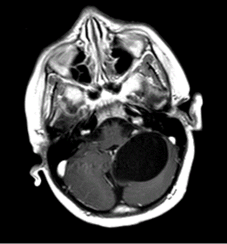
A 7-year-old boy with a history of speech delay, headaches, and staring spells. His neurological examination showed subtle ataxia (difficulties with tandem gait) but no other significant cerebellar signs. Head CT obtained because of the headaches in an outside ER showed an incidental mass. Brain MRI showed a cystic lesion at the inferomedial aspect of the left cerebellar hemisphere that follows CSF on all sequences and measures 5.3 cm transverse × 4.1 cm AP × 3.4 cm craniocaudal (Figure 1).
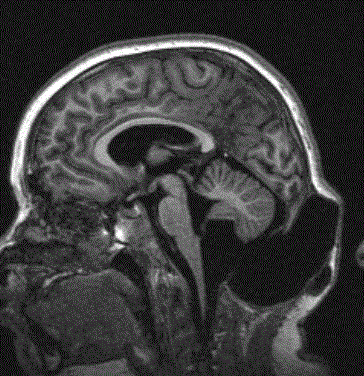
The posterior wall of the cyst was then resected to create an opening end of the cyst. The symptoms improved but the procedure was complicated days later with the development of an asymptomatic but visible pseudomeningocele (Figure 2). Denouement and Discussion
Arachnoid cyst and post-operative pseudomeningocele
In 1879, English anatomist Cunningham published an autopsy report on a young patient with a left hemispheric cyst in a young patient with acromegaly who died from diabetes insipidus and consistent with now we call arachnoid cysts. These cysts are benign collections that occur in the cerebrospinal axis in relation to the arachnoid membrane and that do not communicate with the ventricular system. They usually contain clear, colorless fluid but with different chemical composition compared to the cerebrospinal fluid [1,2].
Most are developmental anomalies. A small number of arachnoid cysts are acquired, such as those occurring in association with neoplasms or those resulting from adhesions occurring in association with leptomeningitis, hemorrhage, or surgery. They constitute approximately 1% of intracranial masses; 50-60% occur in the middle cranial fossa. Cysts in the middle cranial fossa are found more frequently in males than in females; they occur predominantly on the left side [3]. Usually, arachnoid cysts are asymptomatic; this is true even of cysts that are quite large. The most commonly associated clinical features are headache, calvarial bulging, and seizures; focal neurologic signs occur less frequently. Controversy surrounds the treatment of arachnoid cysts. Some clinicians advocate treating only patients with symptomatic cysts, whereas others believe that even asymptomatic cysts should be decompressed to avoid future complications. The most effective surgical treatment appears to be excision of the outer cyst membrane and cystoperitoneal shunting [4]. DNA from affected families has revealed 4 molecular variations in the G protein–signaling modulator 2 gene, GPSM2. Subsequent brain imaging of these individuals revealed frontal polymicrogyria, abnormal corpus callosum, and gray matter heterotopia, consistent with a Chudley-McCullough syndrome diagnosis, but no ventriculomegaly. The gene product, GPSM2 is required for orienting the mitotic spindle during cell division in multiple tissues, suggesting that the brain malformations are due to defects in asymmetric cell divisions during development [5]. MRI is the diagnostic procedure of choice because of its ability to demonstrate the exact location, extent, and relationship of the arachnoid cyst to adjacent brain or spinal cord. Every effort must be made to reliably detect arachnoid cysts, because most arachnoid cysts are an incidental finding and most patients are asymptomatic. Arachnoid cysts must be differentiated from the more serious cystic intracranial and intraspinal tumors. In cases involving larger arachnoid cysts, consideration should be given to the use of serial scans, because such cysts may enlarge over time; patients with such cysts may become candidates for surgery [3]. The most important differentiation to make is between arachnoid cysts and epidermoid cysts; MRI diffusion-weighted images (DWIs) make differentiating the 2 masses easier. Some arachnoid cysts contain proteinaceous fluid or blood; in such cases, signal loss on DWIs may not be marked, which may pose diagnostic problems. Also, tissue contrast with fluid-attenuated inversion recovery (FLAIR) imaging is similar to that with a T2-weighted image, but FLAIR shows no signal arising from the CSF. Thus, unlike with epidermoid cysts, arachnoid cysts containing CSF demonstrate a suppressed (low) signal on FLAIR [3]. A large cisterna magna (mega cisterna magna) occasionally may be confused with an arachnoid cyst. Mega cisterna magna may represent a normal variant (intact cerebellum and vermis), but it may be associated with Dandy-Walker syndrome, either full blown or a variant in which the vermis is either completely or partially absent. Mega cisterna magna and arachnoid cysts show CSF characteristics on T1-weighted, T2-weighted, DWI, and FLAIR sequences. However, whereas an arachnoid cyst may demonstrate mass effect with an en bloc displacement of the cerebellum and vermis, normal-variant mega cisterna magna demonstrates no mass effect, and the cerebellum and vermis remain intact [4]. References
|
Figures at a glance
 |
 |
| Figure 1 | Figure 2 |
Relevant Topics
- Abdominal Radiology
- AI in Radiology
- Breast Imaging
- Cardiovascular Radiology
- Chest Radiology
- Clinical Radiology
- CT Imaging
- Diagnostic Radiology
- Emergency Radiology
- Fluoroscopy Radiology
- General Radiology
- Genitourinary Radiology
- Interventional Radiology Techniques
- Mammography
- Minimal Invasive surgery
- Musculoskeletal Radiology
- Neuroradiology
- Neuroradiology Advances
- Oral and Maxillofacial Radiology
- Radiography
- Radiology Imaging
- Surgical Radiology
- Tele Radiology
- Therapeutic Radiology
Recommended Journals
Article Tools
Article Usage
- Total views: 15204
- [From(publication date):
June-2015 - Aug 30, 2025] - Breakdown by view type
- HTML page views : 10513
- PDF downloads : 4691
