Pulmonary Hydatid Disease in an Adolescent: A Case Report
Received: 30-Aug-2017 / Accepted Date: 06-Oct-2017 / Published Date: 10-Oct-2017
Abstract
Pulmonary echinococcosis is a parasitic disease caused by larval forms of Echinococcus granulosus sensu lato. A 10-years-old patient from the rural central region of the province of Tungurahua, Ecuador with previous history of recurrent respiratory and intestinal infections was admitted in the Hospital. The patient was having symptoms of cough, hemoptysis, weight loss, sore throat, headache and malaise at the time of admission. Preliminary physical examination has shown that the O2 saturation was greater than 95%. The image studies revealed a chest level radioopaque lesion in the right upper lobe of the lung. The blood tests revealed leukocytosis with neutrophilia, eosinophilia and PCR negative. Clinical examination, laboratory findings and imaging reports concluded atypical pneumonia. A simple lung tomography was performed as the patient’s condition didn’t improve with the treatment for pneumonia. Tomography studies suggested a picture of active tuberculosis but the patient showed negative to tuberculin skin test (TST). So, the pulmonology department performed lung biopsy and the subsequent histopathological tests were found to be positive for pulmonary echinococcosis. Then, the patient was given with specific antiparasitic therapy as treatment. It is highly recommended to consider parasitic zoonosis pulmonary location during the differential diagnosis in infants with hemoptysis.
Keywords: Pulmonary echinococcosis; Hemoptysis; Echinococcus granulosus
66966Introduction
Respiratory infections are one of the leading causes of infant morbidity and mortality; and in this context, cough is an important initial symptom. Hemoptysis is also an important symptom in respiratory syndrome associated with tuberculosis. However, a test for parasitosis in the respiratory system is a must for the patients living in areas having high probability of endozootic and endemic disease breakouts [1]. Parasites mainly use three mechanisms for the symptoms to become evident viz., hypersensitivity mechanisms, in situ invasion of the pleura or lung parenchyma and post infection by other organisms [1].
Hydatid disease is a zoonotic disease caused by hydatid cysts of the genus Echinococcus, where the dog is the definitive host and man becomes an accidental intermediate host. There are four species granulosus sensu lato that can infect Human beings who are closely associated with livestock and poor socio-economic conditions [2]. In 90% of cases, liver and lungs are the major infected organs by these parasites [3]. Hydatid cyst may go unnoticed for years depending on the organ they infect, before causing symptoms. Symptoms such as cough, expectoration, dyspnea generally start to appear when the cyst reaches 5 to 6 cm in diameter [2,4]. The diagnosis must be made with regard to the epidemiological history, clinical manifestations, imaging tests and serological tests [4].
Echinococcosis is a zoonotic disease of health interest throughout the country. Due to the prolonged hospitalizations, health services, sequential clinical, serological tests to establish the definitive cause of the problem it causes financial burden to common man [5].
Clinical Case
A 10-year-old patient from the highlands of Ecuador region with a history of high and recurrent respiratory infections from the last three months was admitted in the hospital. Family history had a death of paternal grandfather with lung disease. The patient was known to be associated closely with Livestock such as sheep and pigs. The patient admitted with symptoms of cough, heavy hemoptysis, sore throat, headache, fatigue, weight loss and hyporexia.
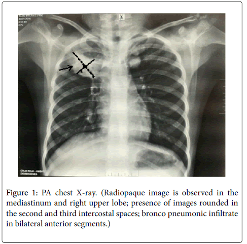
During physical examination, oxygen saturation (over 95%) was found to be normal but, bilateral submandibular and retroauricular lymph nodes are less than one centimetre. However, the pulmonary auscultation during air inhalation was found to be normal. Blood tests showed leukocytosis with neutrophilia, eosinophilia and negative for PCR & procalcitonin. A radiopaque lesion in the right upper lobe was found in the chest radiograph (Figure 1) suggesting that the patient might have atypical pneumonia or tuberculosis. Initially patient was treated with double antibiotic regimen.
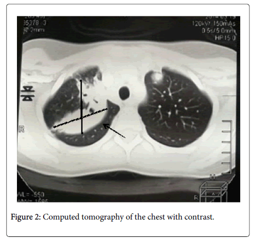
The patient continued hemoptysis despite treatment and hence a contrasted chest CT is performed with air bronchograms. It reported a consolidation in the upper lobe of the right lung and cavitation inside. In the upper lobe of the left lung, two hypodense nodules (28 mm and 12 mm) were observed. Another node was observed at paracardiac location with low density along with a hyperdense adjacent node. These findings suggested active tuberculosis (Figure 2).
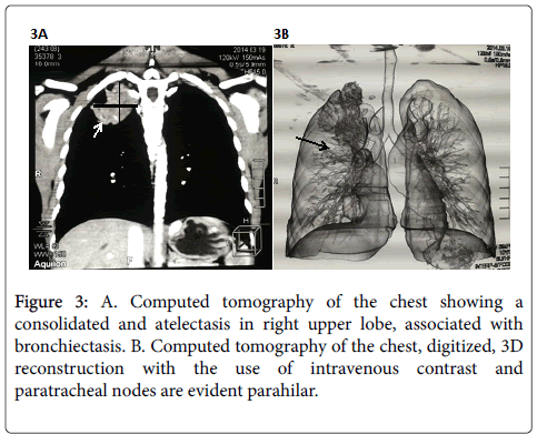
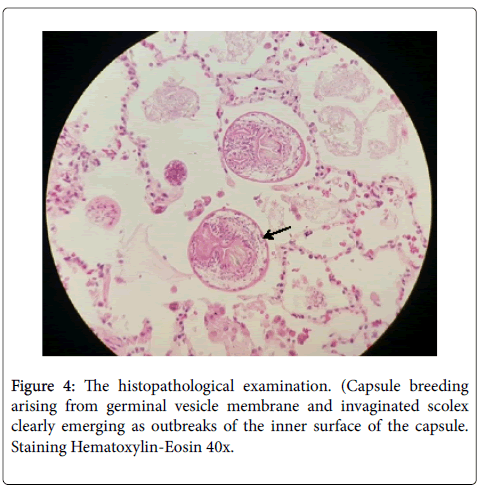
Interferon gamma release assays (IGRA), TST, sputum AFB staining seriate and cultures with Lowenstein-Jensen sputum were done to complement the diagnosis of Tuberculosis. But all the tests gave negative results. Then, bronchoscopy was performed along with a chest CT and lung biopsy. The bronchoscopy result was inconclusive, chest CT gave a clear evidence of atelectasis of the right upper lobe level associated with bronchiectasis and three oval images of cystic characteristics. It also showed evidence of walls prone to calcification that are located in sections 5 right (1.4 cm) and 1-2 left (2.1 cm and 2.7 cm) (Figures 3A and 3B). A small segment of lung tissue was obtained by using thoracoscopy a lung biopsy and it was found with numerous hydatid cysts and scolex of Echinococcus granulosus sensu lato.
All these observations concluded pulmonary hydatidosis. Oral antiparasitic therapy was started with 200 mg of albendazole twice daily for one month; and a satisfactory response was found in the patient (Figure 4).
Discussion
Cystic echinococcosis (CE) is an endemic zoonosis in several countries, with highest incidence in South America. It mainly affects agricultural and pastoral regions in Argentina, Chile, Turkey and Saudi Arabia [2,3,6-8,21].
Ungulates get parasitized permanently with E. granulosus sensu lato eggs by ingestion. After ingestion, these eggs get hatched into larvae and enter the bloodstream and reaches different organs. These larvae develop posterior vesicles and transforms into hydatid cyst that can infect a definitive host. The cycle is completed when an animal ingests viscera infected with hydatid cysts having protoscolex [2]. The dog and other canines are the definitive hosts and are considered as a risk factor in our case [3].
The lung is one of the most affected sites [4] and the lower lobe is most commonly involved. In this case, the cyst was found in the right upper lobe [6,9,10]. Clinical manifestations depend on the integrity of the cyst and the patient remains asymptomatic for a long duration. The most important characteristic feature is the presence of nux associated with repetitive hemoptysis after disease outbreak. This is because of dissection of contents into a bronchus, which can mimic the symptoms of tuberculosis, giving a wrong diagnosis as happened with our patient. This type of situation has occurred in several reported cases [7,8].
Regarding diagnosis in the hemogram, eosinophilia is rare but can occur in 20% of patients and our patient was observed with eosinophilia 5.1% [2,5]. Chest radiography is the primary choice of examination for pulmonary hydatid disease. However, in some special cases or to confirm the disease magnetic resonance, tomography and X-rays will be used. Serological tests like enzyme immunoassay ELISA or Western Blot with high sensitivity and specificity can be used to detect the presence of specific circulating antibodies (IgE) against parasite antigens [4]. In the absence of ELISA and Western Blot, the presumption of disease must be based on diagnostic imaging studies that need to be confirmed by a biopsy.
Thoracotomy surgery is the treatment of choice in pulmonary echinococcosis and albendazole can be used as an adjuvant theraphy in cases where the size of the cyst is around 5 cm in diameter [4]. However, there are reports of anaphylactic shock in 1 to 7.5% by spontaneous rupture of the cyst or during pericystectomy with capitonaje [2].
Conclusions
Echinococcus granulosus sensu lato infestation is cosmopolitan. It is mainly observed in Latin American countries like Argentina, Bolivia, Brazil, Peru, Uruguay and Ecuador where people are associated with Livestock. Hydatid disease is a major public health concern in endozootic areas; and domestic dogs are important in the epidemiological chain.The slow growth of the cyst allows the patient to remain asymptomatic for a long period of time. Most of the times the disease was diagnosed incidentally during imaging studies for other reasons. For its long latent period and asymptomatic infestation, it has been under reported in kids. In our epidemiological framework it is recommended to consider diagnostic possibility of Hydatid disease, besides pneumonia and tuberculosis in a patient with cough and hemoptysis, from endemic and endozootics areas.
Conflict of Interest
The authors declare no conflict of interest.
References
- Valle M (2005) PatologÃa respiratoria importada: parasitosis. An Pediatr 62: 18-26.
- Rodulfo J, Carrión M, Freitas M, Real J, Merchán M (2013) Hidatidosis pulmonar. Neumol Pediatr 8: 5-9.
- Berberian G, Rosanova T, Inda L, Sarkis C, Questa H, et al. (2017) Hidatidosis en niños: experiencia en un hospital de alta complejidad fuera del área endémica. Arch Argent Pediatr 115: 274-286.
- Pinto P (2017) Diagnóstico, tratamiento y seguimiento de la hidatidosis. Rev Chil Cir 69: 94-98.
- Ministerio de Salud Pública Argentina (2012) Enfermedades Infecciosas Hidatidosis. GuÃa para el equipo de salud Nro. 1: 1-50.
- Jans J, Bórquez P, Marambio A, Manolis P, Hollsteing A, et al. (2012) Resultados del tratamiento de la hidatidosis pulmonar complicada y no complicada. Rev Chil Cir 64: 346-351
- Ünver E, Kaymak M, Caylan Ö, Kaptanoğlu M (2017) Ruptured Pulmonary Cyst Echinococcosis Mimicking Tuberculosis in Childhood: A Case Report. Turkiye Parazitol Derg 41: 126-129.
- Hafiz B, Al-qurani A, Al-Malki A, Kheel E, Esheba G (2016) Pulmonary Hydatid Cyst in a 13 Years Old Child: A Case Report . J Clin Case Rep 6: 1-3.
- Akgul Ozmen C, Onat S (2017) Computed Tomography (CT) Findings of Pulmonary Hydatid Cysts in Children and the Factors Related to Cyst Rupture. Med Sci Monit 29: 3679-3686.
- Calle C, Rosales F, MacÃas E (2014) Hidatidosis Pulmonar. Rev Fac Cien Med (Quito) 39: 101-104.
Citation: Paredes P, Toapanta I, Aguayo A, Salazar M, Bravo A (2017) Pulmonary Hydatid Disease in an Adolescent: A Case Report. Arch Parasitol 1:113.
Copyright: © 2017 Paredes-Lascano P, et al. This is an open-access article distributed under the terms of the Creative Commons Attribution License, which permits unrestricted use, distribution, and reproduction in any medium, provided the original author and source are credited.
Select your language of interest to view the total content in your interested language
Share This Article
Open Access Journals
Article Usage
- Total views: 6216
- [From(publication date): 0-2017 - Jul 15, 2025]
- Breakdown by view type
- HTML page views: 5092
- PDF downloads: 1124
