Case Report Open Access
Right Ventricular Rupture and Active Contrast Extravasation on MDCT in a Trauma Patient: A Case Report
| Bradford D Moore, Charles H Cockrell, Siddhi Shah and Yang Tang* | |
| Department of Radiology, Virginia Commonwealth University Health System, Richmond, Virginia, 23298, USA | |
| Corresponding Author : | Yang Tang Department of Radiology Virginia Commonwealth University Health System Richmond, Virginia, 23298, US Tel: 804-828-8691 Fax: 804-628-1132 E-mail: ytang2@vcu.edu |
| Received February 18, 2013; Accepted November 18, 2013; Published November 22, 2013 | |
| Citation: Moore BD, Cockrell CH, Shah S, Tang Y (2013) Right Ventricular Rupture and Active Contrast Extravasation on MDCT in a Trauma Patient: A Case Report. OMICS J Radiology 3:153. doi: 10.4172/2167-7964.1000153 | |
| Copyright: © 2013 Moore BD, et al. This is an open-access article distributed under the terms of the Creative Commons Attribution License, which permits unrestricted use, distribution, and reproduction in any medium, provided the original author and source are credited. | |
Visit for more related articles at Journal of Radiology
Abstract
Cardiac chamber rupture is very rare in traumapatients who reach the emergency department alive. Here, we report a case of cardiac injury after Motor Vehicle Crash (MVC), in which sternal fracture fragment was thought to penetrate the pericardium as well as the right ventricular free wall and result in eventual exsanguinations and death. The diagnosis of this case was confounded by patient’s vague symptoms and prior history of coronary disease. This case highlights the utility of using Multi Detector Computed Tomography (MDCT) in accurately diagnosing these rare but potentially fatal injuries.
| Keywords |
| Multidetector computed tomography-MDCT; Motor vehicle collision-MVC; Blunt cardiac injury |
| Case Report |
| 83-year-old man with past medical history of coronary artery disease status post bypass surgery and recent percutaneous coronary intervention presented to our level I trauma center after being involved in a head-on, rollover MVC. Upon arrival, he complained of chest pain but was hemodynamically stable with blood pressure of 102/64 mmHg, heart rate of 112/min, respiratory rate of 26/min and Glasgow coma scale of 15. Shortly afterwards, he became slightly hypotensive with systolic blood pressure of 90 mmHg for a brief period but responded to transfusion of packed blood cells and intravenous fluid. On physical examination, he had tenderness over the sternum and mild abdominal pain. He was also found to have an elevated troponin at 3.5 and hemoglobin of 9.6 gram/dl. A bedside transesophageal echocardiogram demonstrated a small amount of pericardial fluid without evidence of cardiac tamponade. His Electrocardiogram (EKG) revealed 3 mm ST elevation in the inferior leads and 2 mm ST depression in the lateral leads, which was initially concerning for acute myocardial infarction. |
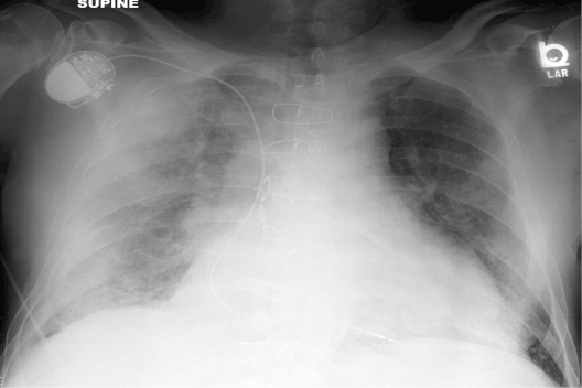
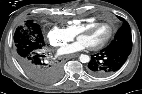
| His chest radiography showed multiple rib fractures and diffuse opacity of right lung concerning for pulmonary contusion and hemothorax (Figure 1). Subsequent MDCT of chest revealed ruptured right ventricular free wall and pericardium with active contrast extravasation into the anterior pericardial space and anterior mediastinum (Figures 2 and 3). This was presumably caused by a penetrating injury from the overlying sternal fracture. |
| The patient was immediately taken to the operating room for repair of cardiac injury. However, shortly after introduction of general anesthesia, the patient developed profound hypotension. Upon opening the sternum, the surgeons found an approximately 5×3 cm perforation in the right ventricular free wall, which was too large to repair. Despite efforts of resuscitation, blood quickly exsanguinated through the perforation and the patient expired. |
| Discussion |
| Cardiac injury is believed to be second most common cause of traumatic death in the United States after central nervous system injury [1]. These injuries are either due to blunt force causing compression of the heart between the sternum and the spine, or due to penetrating trauma, which is typically from gunshot wound or stabbing [2]. Our case represents somewhat a hybrid of these two typical scenarios with blunt force causing sternal fracture which then penetrated the pericardium and right ventricle. |
| Mild blunt injury may only result in myocardial concussion or contusion. However, severe blunt injury or penetrating injury can cause cardiac chamber rupture, hemopericardium, pericardial tamponade, valvular injury, cardiac herniation etc, and is among the most lethal emergencies with prehospital mortality as high as 94% and subsequent mortality rate of initial survivors of 50% according to one study [3]. Historically, these injuries are not diagnosed through imaging modalities and are usually identified during surgery or autopsy [4]. |
| As demonstrated in this case, a subset of patients with these catastrophic injuries may be initially stable, and the diagnosis can be masked by other concomitant injuries to lungs or chest wall. Symptoms such as chest pain are nonspecific and often arise from a noncardiac source. Chest radiographs may show abnormal cardiomediastinal contour, which is also frequently artifactual due to portable technique and patient’s positioning. Echocardiography can detect abnormalities associated with cardiac injury, such as increased myocardial echogenicity and dyskenesis, ventricular septal defect and pericardial fluid with tamponade [5]. However, it has not been proved to be useful as a primary screening modality for cardiac injury. In addition, it is known to provide false negative results for hemopericardium when there is concurrent violation of the pericardial sac [6]. In our case, the echocardiogram failed to directly demonstrate the right ventricular rupture, and only showed a small amount of pericardial fluid. |
| This case also highlights the difficulty in distinguishing acute coronary syndrome from traumatic cardiac injury, as it is certainly possible that this patient might have had an acute coronary ischemic event, which would impair the driving ability and result in a MVC. Cardiac enzymes such as Creatinine Kinase (CK) and troponins are well known diagnostic markers for myocardial ischemia. However, it has been reported that CK levels are commonly elevated in trauma patients suffering from either cardiac or noncardiac damage [7]. Troponin level may also be elevated due to myocardial injury and their level is proportional to the extent of myocardial damage [8]. Likewise, although routinely used to diagnose myocardial infarction, EKG changes are commonly seen in patients with traumatic myocardial contusion and it has been reported that patients with abnormal ECG findings had more significant complications [9]. Therefore, enzyme markers and EKG changes are not particularly useful in differentiating myocardial injury from coronary ischemia in trauma patients with known coronary history, As a matter of fact; it is possible that this patient might indeed have suffered from secondary myocardial ischemia from traumatic injury to the coronary arteries, which resulted in dramatic troponin elevation and EKG changes. |
| With the excellent speed, spatial resolution and capability of multiplanar reformation, MDCT has quickly become the modality of choice for suspected cardiac injury. At most trauma centers, patients presenting with significant trauma typically undergo a routine trauma series of MDCT exams spanning the head to pelvis. MDCT is a proven diagnostic tool in the setting of thoracic trauma with high sensitivity for identifying pericardial and myocardial lacerations as well as concomitant aortic, lung or chest wall injuries [10]. Specifically, in patients with penetrating thoracic trauma who are hemodynamically stable, CT has been shown to be both sensitive and specific for diagnosing hemopericardium [11]. In addition, with the advent of modern dual energy technique or coupling with EKG gating, MDCT is now able to detect subtle injuries to the aortic root, coronary arteries or valves. |
| Conclusions |
| In summary, we report MDCT findings of right ventricular rupture in a hemodynamically stable blunt trauma patient, whose injuries were initially masked by vague symptoms and past history of coronary artery disease. It is important for the trauma surgeons and radiologists to be familiar with this condition since early detection of these lethal injuries using MDCT is essential for improving survival in a subset of patients whose injuries may be amenable to surgical repair. |
References |
|
Figures at a glance
 |
 |
 |
| Figure 1 | Figure 2 | Figure 3 |
Relevant Topics
- Abdominal Radiology
- AI in Radiology
- Breast Imaging
- Cardiovascular Radiology
- Chest Radiology
- Clinical Radiology
- CT Imaging
- Diagnostic Radiology
- Emergency Radiology
- Fluoroscopy Radiology
- General Radiology
- Genitourinary Radiology
- Interventional Radiology Techniques
- Mammography
- Minimal Invasive surgery
- Musculoskeletal Radiology
- Neuroradiology
- Neuroradiology Advances
- Oral and Maxillofacial Radiology
- Radiography
- Radiology Imaging
- Surgical Radiology
- Tele Radiology
- Therapeutic Radiology
Recommended Journals
Article Tools
Article Usage
- Total views: 14207
- [From(publication date):
January-2014 - Aug 18, 2025] - Breakdown by view type
- HTML page views : 9557
- PDF downloads : 4650
