ISSN: 2165-7025
Journal of Novel Physiotherapies
Make the best use of Scientific Research and information from our 700+ peer reviewed, Open Access Journals that operates with the help of 50,000+ Editorial Board Members and esteemed reviewers and 1000+ Scientific associations in Medical, Clinical, Pharmaceutical, Engineering, Technology and Management Fields.
Meet Inspiring Speakers and Experts at our 3000+ Global Conferenceseries Events with over 600+ Conferences, 1200+ Symposiums and 1200+ Workshops on Medical, Pharma, Engineering, Science, Technology and Business

Three-Dimensional Motion Analysis: Relevant Concepts in Physiotherapy Movement Dysfunction Management

Derek J Rutherford1,2* and Cheryl Hubley-Kozey1,2
1School of Physiotherapy, Dalhousie University, Halifax, Nova Scotia, Canada
2School of Biomedical Engineering, Dalhousie University, Halifax, Nova Scotia, Canada
Corresponding Author : Derek J Rutherford
School of Physiotherapy, Dalhousie University
5869 University Avenue, Halifax, Nova Scotia, B3H 3J5, Canada
Tel: 1 (902) 494 4248
Fax: 1 (902) 494 1941
E-mail: djr@dal.ca
Received February 24, 2014; Accepted May 23, 2014; Published May 25, 2014
Citation: Rutherford DJ, Kozey CH (2014) Three-Dimensional Motion Analysis: Relevant Concepts in Physiotherapy Movement Dysfunction Management. J Nov Physiother 4:214. doi: 10.4172/2165-7025.1000214
Copyright: © 2014 Rutherford JD, et al. This is an open-access article distributed under the terms of the Creative Commons Attribution License, which permits unrestricted use, distribution, and reproduction in any medium, provided the original author and source are credited.

Visit for more related articles at Journal of Novel Physiotherapies

Abstract

The explorations of typical and dysfunctional movements are recognized physiotherapy assessment and management strategies. With the development of state-of-the-art three-dimensional (3-D) motion capture systems, traditional two-dimensional and observational movement quantification are being used less frequently in research initiatives. This technology is slowly infiltrating physiotherapy practice as systems become more accessible. Knowledge of 3-D acquisition methodology will provide insight into advancing movement dysfunction theories in physiotherapy. The objective of this commentary is to introduce the clinician to 3-D motion capture while providing a basic theoretical framework for understanding this methodology. The current use of 3-D systems and the limitations of this study methodology are reviewed. Familiarization with the use of this technology and understanding the methods in capturing 3-D motion will provide the physiotherapist with the latest knowledge in movement dysfunction. These concepts are an exceptional addition to the clinical reasoning strategies currently employed in the profession of physiotherapy.

Keywords

Physiotherapy; Human movement analysis; Joint kinematics; Three-dimensional analysis

Introduction

Physiotherapy is moving beyond the technical application that once dominated the profession [1]. Investigating pathological and non-pathological movement qualities, facilitating function and augmenting healthy lifestyles and quality of life are recognized strategies in physiotherapy management. A great deal of study has been conducted to improve the analysis of human motion [2,3] as many challenges face the clinician attempting to assess and quantify musculoskeletal kinematics. With the evolution in state of the art three-dimensional (3-D) motion capture systems, two-dimensional and observational movement analysis techniques are becoming less frequently utilized in research initiatives. Over almost two decades, 3-D systems have been shown to be highly accurate and able to capture simultaneous multi-segmental movement characteristics of human motion [4-7]. The methodology employs skin surface marker/receiver coordinates that can be converted to values that correspond to joint motion nomenclature typically employed in a clinical setting [2,8,9]. These data can provide understanding of normal human kinematics and insight into improving assessment and management of movement dysfunction.

Although these methods provide a realistic representation of joint motion, limitations exist. Studies have shown that soft tissue artifact [10-12], kinematic crosstalk [11,13,14] and accuracy in anatomical land marking [13] affects the accuracy of capturing joint ranges of motion during human movement. Despite methodological and analytical limitations, 3-D study is currently state-of-the-art and provides detailed knowledge not available through conventional two-dimensional and observational analyses.

The objective of this commentary is to introduce the basic concepts of 3-D motion capture while providing a theoretical framework to assist in the interpretation of literature where this methodology is utilized.

Material and Methods

Three-Dimensional analyses

Three-dimensional biomechanical research is well established and advanced analysis systems have continued to evolve. These systems may have passive (VICON (b) and Qualysis (c)), active (Northern Digital Inc. (a)) or electromagnetic (Polhemus (d) and Ascension (e)) motion capture properties that are independently unique yet synonymously implemented in research to capture movement in 3-D. Recently, systems to track 3-D motions have targeted a clinical audience (f), (g), where previously this technology was primarily used for a research focus. If this is the future, clinicians should have a basic understanding of 3-D methodologies. To start, a brief mathematical description is warranted to provide the necessary background.

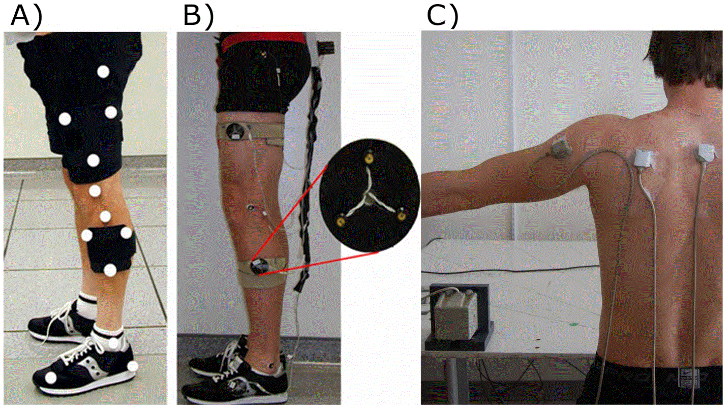
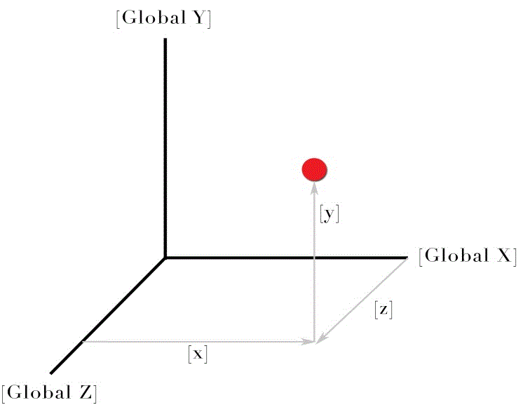
Three-dimensional motion capture utilizes, what is often referred to as an X, Y, Z Cartesian coordinate system and is considered a standard in reporting kinematic data as recommended by the International Society of Biomechanics (ISB) [2,9]. During camera-based analysis, this coordinate system can be derived from any set of three skin surface markers that are not all in a straight line (non-collinear) [2,15] (Figure 1). In the case of electro-magnetic motion capture [4,5], specially designed magnetic sensors are employed that are sensitive to movement in each of the three Cartesian axes. In practical terms, each marker/sensor can be described, relative to a global coordinate system (i.e. the room), by the corresponding units X, Y, and Z (Figure 2).

Using these principles, an organized system of coordinates is mathematically established that gives reference to the rigid segments monitored throughout movement [2,15]. These rigid segments relate to segment anatomy based on anatomical landmark identification. Rigid segments in the lower extremity typically analyzed during gait include the pelvis, thigh, shank and foot segments [16-19]. The thorax, scapula and humerus are often described as rigid bodies during shoulder movement analysis [20-22]. Three-dimensional (X, Y, Z) movement can be captured through the analysis of one rigid segment coordinate system with respect to another (i.e. tibia with respect to femur). As each Cartesian coordinate axis orientation within each rigid segment is orthogonal (i.e. perpendicular), three independent movements can now be derived [15]. These movements correspond to clinical osteokinematic nomenclature; flexion/extension, abduction/adduction and internal/external rotation. The scapular convention has included; medial/lateral rotation, anterior/posterior tilt and upward/downward rotation [5].

Research investigating the dynamics of human movement, with the exception of the electro-magnetic properties of the Polhemus 3-space Fastrak (d) and Ascension Flock of Birds (e) systems, utilize cameras with shutter speeds typically between 50-120 Hz for most clinical applications. These cameras are standardized to laboratory position and orientation [15,23]. The number of camera positions can be task specific. If the movement involves a walking task, cameras in two positions might be sufficient in ensuring that all the markers are captured [24]. In contrast, if the movement involves cutting and twisting, more positions will be required to guarantee that all of the markers are identified [12,25]. This concept of redundancy ensures the markers are captured continuously, regardless of subject orientation.

Although marker-less motion capture systems such as the Microsoft kinect™ are being developed in an effort to evaluate human movement in a natural setting [26], marker based systems are currently state-of-the-art. Skin surface markers can be passive or they can be active. The passive markers are retro-reflective and the specially designed cameras that emit infrared stroboscopic illumination are used to capture the position of the marker (VICON (b) and Qualysis (c)). These systems do not require the use of wires and connection hardware. Conversely, active markers contain light emitting diodes, pulsed in sequence allowing the motion capture system to detect their location (Northern Digital Inc. (a)). Figure 1 illustrates both marker systems. The size of the markers, as well as the calibration volume can affect the accuracy of the measurement [6,7,27,28].

Previous comparison studies have found mean absolute errors in spatial recognition to be less than five mm for the VICON (b), Qualysis (c) and Northern Digital Inc. (a) systems [6,7]. Generally, active markers produce a more accurate result, although require lead wires and power equipment that may encumber natural movement. For instance, the optoelectronic Northern Digital Inc. Optotrak (a) camera system utilizes an active LED marker system and was found to have a 1.0 mm mean average error during standardized measurement tasks [6]. In clinical nomenclature, knee flexion angle error has been found to be less than two degrees using optoelectronic camera equipment such as the Optotrak (a) system [29]. For camera-based systems, the volume of measurement area and skin marker size is inversely related to the accuracy of the measurement. Each of these elements of motion capture must be taken into account when extrapolating the results.

Camera based systems track skin surface markers during movement where the Polhemus 3-space Fastrak (d) system and the Ascension Flock of Birds (e) are motion capture devices that utilize changes in electro-magnetic fields to capture movement [4,5]. These systems appear to be more prevalent in studies involving the kinematics of the spine [30-32], scapula and shoulder [5,20,21,33]. Electro-magnetic motion capture relies on receiver sensors mounted to the skin and must be in close orthogonal approximation to the central transmitter. The absolute errors of such systems have been shown to be within 1-2 mm, however the accuracy of these measures were linearly dependent on the sensors distance from the central transmitter [34]. In addition, these systems are sensitive to magnetic distortion created from metal within the capture volume [34]. Karduna et al. [5] validated the use of electromagnetic motion capture for scapular kinematics and found low root mean square errors when capturing movement below 120 degrees of humeral-thoracic elevation. For instance, in the case of upward scapular rotation, errors were less than 10% of the total range of motion [5]. In the case of spinal and scapular kinematic investigation, the motion capture volume has been controlled within a range from the central receiver [35,36]. This requirement would not provide sufficient volume for capturing dynamic gross motor function, such as over ground walking. In these instances, camera-based systems have been employed.

Many motion 3-D motion capture systems are technologically advanced in comparison to traditional two-dimensional motion capture systems. The application of the 3-D systems in rehabilitation and medical research is vast and continuing to expand as access to this technology increases. For instance, the EMOVI (g) KneeKG™ and 3D Gait (f) platforms have emerged in recent years. The implementation of relevant literature in clinical practice will further expand the value of these research methods.
Where is this technology being used?

Attempting to quantify human movement through motion capture allows for an increased scope of understanding not available with qualitative assessments. Coutts [37] reviewed the literature in observational movement analysis and concluded that there is much skepticism surrounding the reliability and validity of clinically produced movement analysis strategies.

Three-dimensional techniques of movement analysis have fostered a greater understanding of upper and lower extremity movement dysfunction and have been found to be reliable [24,38,39]. For example, the shoulder complex has the largest amount of movement among all of the body joints [40]. Various studies support that shoulder kinematics are dysfunctional when various impairments are present [41-43]. With many impairments of the gleno-humeral joint, scapular mechanics are also compromised [36,44]. Recent 3-D studies suggest that under normal circumstances, scapular external rotation and posterior tilting occur at end of range along with upward rotation, helping to prevent humeral encroachment on the sub-acromial space [5,22]. Altered scapular patterns of movement have been investigated as a result of muscle fatigue [45], impingement [43], gleno-humeral restrictions [46] and kyphotic postures [42]. The use of 3-D motion analyses has produced a quantitative objective report on scapulo-thoracic and gleno-humeral movement characteristics not available with observational and two-dimensional analysis.

In comparison to the upper extremity, the evaluation of the lower extremity has encompassed 3-D methodologies pertaining primarily to gross motor function. Gait [47-53], ascending and descending stairs [50,54] and running [25,55] have been investigated in many populations including individuals with tibialis posterior fatigue, anterior cruciate ligament injuries, knee and hip osteoarthritis. Many studies acknowledge the limitations of kinematic data, in that it provides a measure of outcome only and does not address the causal factors related to the forces required to produce movement. In general, studies will often utilize 3-D motion capture methodology to assist in multi-dimensional outcome measurement. In the lower extremity, concurrent measures typically include kinetic and electromyographic recordings [18,51,56,57]. These traits are important for a complete investigation of muscle function and the forces involved in movement production and joint loading.

As 3-D technology continues to improve and become more accessible, research into the effectiveness of various interventions aimed at altering physical movement characteristics will emerge. Although 3-D motion capture remains state of the art, methodological and interpretive limitations exist.

Limitations

The International Society of Biomechanics has outlined recommendations aimed to improve the collection and reporting of kinematic data [2,9,58]. Despite current recommendations, human movement is multi-dimensional and intrinsic differences in study methodology and subject populations can affect results interpretation.

Currently, state-of-the-art motion capture systems rely on capturing rigid segment movement through skin surface marker motion in a particular volume of space. The accuracy of identifying boney landmarks used for marker placement and virtual point digitization can affect the derivation of coordinate systems used to calculate the resultant joint angles [13]. As a result of poorly placed markers, the mathematically derived joint axes will not correspond to the individual anatomical axes. For instance, a frontal knee axis that is orientated obliquely in the transverse plane will result in flexion and extension being partially represented by movements about the other axes. This coupling of movements is termed kinematic crosstalk and severely affects the outcome of the motion capture [11,13,14].

Aberrant marker movement can also limit the interpretability of the results. Soft tissue artefact has been shown to affect the representation of a rigid segment during motion capture [10,12]. For instance, the thigh segment contains relatively large amounts of associated soft tissue and produces the greatest amount of soft tissue artefact in the lower extremity [10]. This suggests that small joint rotations should be regarded with caution as soft tissue artefact may have magnitudes that are comparable to the relevant joint rotations [10,12].

In deriving the final representation of the motion, methods used to reduce, filter and present the data can be varied throughout the literature. In addition, the motion description (i.e. Osteokinematics) between two rigid segments is important in relating the results to clinical practice [59]. For instance, in the lower extremity, the standard convention records the distal segment moving about the proximal segment [8,9]. While sagittal plane motions can easily be interpreted, frontal and transverse plane motions can be more difficult (i.e. understanding whether the femur, tibia or both bones are moving during the movement of knee joint internal/external rotation).

Although limitations are evident, this methodology has been accepted as a standard in biomechanical research and is slowing being adopted in clinical practice. As with most methodology, literature must be analyzed with respect to the question being asked. Understanding that limitations do exist is a positive step in extrapolating clinically feasible and applicable results.

Conclusion

This paper introduced the basic concepts of 3-D motion capture while providing a brief theoretical framework for accurate critical appraisal. Familiarization with the use of this technology and understanding the methods in capturing 3-D motion will provide the physiotherapist with access to the latest knowledge in movement dysfunction. Where knowledge exchange is fundamental to the growth of scientific inquiry, collaborative associations in scrutinizing human movement are essential. The scientific community has accepted this technology and continues to provide the clinician with the medium to enhance understanding and develop mutual links in the investigation of human movement and dysfunction.

(a) Northern Digital Inc, Waterloo, ON, Canada

(b) Oxford Metrics, Oxford, UK

(c) Qualisys Medical, Gothenburg, Sweden

(d) Polhemus Inc., Colchester, VT, USA

(e) Ascension Technology Inc., Burlington, VT, USA

(f) The Running Injury Clinic, http://3dgaitanalysis.com

(g) EMOVI Inc. http://www.emovi.ca

References


--

Figures at a glance

Figure Figure
Figure 1 Figure 2
Post your comment

Share This Article

Article Usage

  • Total views: 17320
  • [From(publication date):
    July-2014 - Sep 01, 2024]
  • Breakdown by view type
  • HTML page views : 12756
  • PDF downloads : 4564
Top