Research Article Open Access
Value of 18F-FDG-PET/CT Initial Staging in No Metastasic Breast Cancer with Poor Prognostic Factors
| Alberto Martinez Lorca1*, Alejandro Gallego2, Cristina Escabias1 and Pilar Zamora2 | |
| 1Nuclear Medicine Department, Hospital Universitario La Paz, Nuclear Medicine, Spain | |
| 2Medical Oncology Department, Universitary Hospital La Paz, Madrid, Spain | |
| Corresponding Author : | Alberto Martinez Lorca Department of Nuclear Medicine Hospital Universitario La Paz Madrid-28046, Spain Tel: +0034679732928 E-mail: albertoml85@yahoo.es |
| Received: July 3, 2015 Accepted: August 06, 2015 Published: August 10, 2015 | |
| Citation: Lorca AM, Gallego A, Escabias C, Zamora P (2015) Value of 18F-FDG-PET/CT Initial Staging in No Metastasic Breast Cancer with Poor Prognostic Factors. OMICS J Radiol 4:201. doi:10.4172/2167-7964.1000201 | |
| Copyright: © 2015 Lorca AM, et al. This is an open-access article distributed under the terms of the Creative Commons Attribution License, which permits unrestricted use, distribution, and reproduction in any medium, provided the original author and source are credited. | |
Visit for more related articles at Journal of Radiology
Abstract
Purpose: This study analysed retrospectevely the impact of 18F-FDG-PET/CT initial staging, in women with poor prognosis factors breast cancer, in our usual practice.
Methods: From January 2009 to December 2012, initial breast cancer underwent PET/CT in 298 women with one of these poor prognosis factors: Her-2+ or triple negative phenotype, Ki 67 ≥ 14%, tumoral size and/or positive axilary lymph nodes. Only 254 patients diagnosed with mammography, ultrasonography, MRI, and bone scintigraphy accomplished inclusion criteria.
Results: 18F-FDG-PET/CT changed 13.4% to IV clinical stage, detected metastatic disease in 3.3% of patients with stage I, 13% with stage II and 17.4% with stage III. Statistical significance was found between the PET/CT metastatic disease findings and patients with stage IIB and III(A-C)(OR:3.04;IC95%:1.2-7.2;p=0.009). PET/CT also revealed N3 disease in 7.9%. According to the location, visceral affectation was found in 4.7% of the patients, metastatic lymph nodes in 3.2% and secondary bone disease in 8.7%. Her-2 negative and hormonal positive receptors group submitted a higher incidence of distant metastases, 15.5%. The more visceral and lymph node disease (N3 and distant-M1) was found in the triple negative group. Metastatic disease was observed in 28.6% of the patients above 70 years, a higher incidence than those patients with lower age (OR: 3.7; IC95%: 1.7 - 8; p = 0.01).
Conclusion: 18F-FDG-PET/CT showed to be useful in the metastatic disease detection from IIB stage patients in initial poor prognosis breast cancer patients. The incidence of distance affectation was higher in patients above 70 years and in the Her-2 negative and hormonal positive receptor group.
|
Abstract
Purpose: This study analysed retrospectevely the impact of 18F-FDG-PET/CT initial staging, in women with poor prognosis factors breast cancer, in our usual practice.
Methods: From January 2009 to December 2012, initial breast cancer underwent PET/CT in 298 women with one of these poor prognosis factors: Her-2+ or triple negative phenotype, Ki 67 ≥ 14%, tumoral size and/or positive axilary lymph nodes. Only 254 patients diagnosed with mammography, ultrasonography, MRI, and bone scintigraphy accomplished inclusion criteria. Results: 18F-FDG-PET/CT changed 13.4% to IV clinical stage, detected metastatic disease in 3.3% of patients with stage I, 13% with stage II and 17.4% with stage III. Statistical significance was found between the PET/CT metastatic disease findings and patients with stage IIB and III(A-C)(OR:3.04;IC95%:1.2-7.2;p=0.009). PET/CT also revealed N3 disease in 7.9%. According to the location, visceral affectation was found in 4.7% of the patients, metastatic lymph nodes in 3.2% and secondary bone disease in 8.7%. Her-2 negative and hormonal positive receptors group submitted a higher incidence of distant metastases, 15.5%. The more visceral and lymph node disease (N3 and distant-M1) was found in the triple negative group. Metastatic disease was observed in 28.6% of the patients above 70 years, a higher incidence than those patients with lower age (OR: 3.7; IC95%: 1.7 - 8; p = 0.01). Conclusion: 18F-FDG-PET/CT showed to be useful in the metastatic disease detection from IIB stage patients in initial poor prognosis breast cancer patients. The incidence of distance affectation was higher in patients above 70 years and in the Her-2 negative and hormonal positive receptor group. Keywords
18F-FDG-PET/CT; Breast cancer; Her-2; Hormonal receptors; ki67
Introduction
Breast cancer is the most common tumor and the first cause of women cancer mortality in our area [1]. The incidence has progressively increased in the last decades, at the same time diagnosis and therapeutic strategies has been improving. Nowadays, different imaging techniques are available in the loco-regional diagnosis, such a mammography, ultrasonography and breast MRI [2,3], with high sensibility and specificity. Although in the metastatic disease study we do not have a “gold standard” technique, in the last years some reports have documented the value of 18FDG-PET-CT in patients with locally advanced breast cancer and it has been used in clinical practice. Also it has shown a very high yield in terms of finding extra-axillary nodal involvement in patients who were not suspected of having it and of distant lesions. In these patients with poor prognostic factors, performing 18FDG-PET-CT might modify the treatment course by revealing occult distant metastases.
In using 18F-FDG the nature of the tumor and metastases can be evaluated at an earlier time than any other anatomic imaging study. When an unexpected focus of FDG uptake is detected during and FDG examination it is necessary to explore with other techniques or biopsy because of the high risk of malignancy. FDG uptake intensity correlates with breast cancer grade. There is also a correlation between the tumor proliferation index and the intensity of FDG uptake as well as HER2/neu expression. Triple negative breast tumors are currently a subject of major interest because of their aggressiveness, poor prognosis and lack of targeted therapy. For this reason, the aim of this study is to evaluate the utility of PET/CT in the initial breast cancer staging, and determine the correlation between findings in PET/CT and the clinipathologic and inmunohistochemical factors in a group of women with poor prognostic factors breast cancer. Study Design
Inclusion criteria
This retrospective study was carried out in the La Paz Universitary Hospital, Madrid, and the data of 254 newly diagnosis breast cancer patients without known metastatic disease were gathered and analyzed between January 2009 and December 2012. In those patients with at least one of this poor prognosis factors: Her-2 positive, triple negative tumor phenotype, tumoral size more than 2 cm, Ki 67% higher or equal value of 14% and/or positive axilar lymph node, underwent 18F-FDG-PET/CT as imaging technique of choice for the patients of our study.
Initially 298 patients were analyzed, but only were selected for the study those patients with ductal or lobulillar histology. Ten patients were excluded due to their uncommon histopathology [neuroendocrine, sarcomatoids, squamous o microcytic carcinoma]. Of the 288 remaining patients were ruled out six patients with a initially stage IV diagnosis, five of them had spine bone affectation, including one with a medullar compression, and the other one showed liver injury in the blood analytics. Also were excluded seven patients who had a previous cancer and one diagnosed of breast and ovarian hereditary syndrome cancer. Finally, 20 patients were left out from the study because the follow-up was not available in our hospital. Loco-regional and histology diagnosis
Local: For initial imaging, mammography and ultrasounds were performed in all patients and breast MRI was done in some selected cases when it was suspected of mulricentricity or the results of the other imaging modalities were inconclusive.
Histological: The histological diagnosis was done by CNB [core needle biopsy]. The molecular parameters analyzed were hormonal receptors [estrogenic and progestagen], human epidermic growth factor receptor 2 [Her-2] and profileration index Ki67. The Ki67 cutoff was 14%, according to the current criteria of Saint Gallen 2011 at the time of data collection [4]. Nowadays, according to the new criteria of Saint Gallen 2013, the Ki67 cutoff is 20% [5]. Those cancer with negative estrogenic and progestageno receptors and Her-2 was considered triple negative phenotype. Axillary study: In patients with negative axillary lymph nodes by clinic and ultrasound, criteria similar to the last consensus of selective lymph node biopsy [SLNB] [6], SLNB was done to the breast cancer regional staging [7]. Metastatic disease diagnosis
PET/CT: PET/CT acquisition was done by habitual protocol in our hospital. This require patients were not allowed to consume any beverages for at least 4-6 hrs prior to the start of the study, images were acquired from head through mid-thigh obtained at least 45 min after radiopharmaceutical injection with the appropriate acquisition and reconstruction parameters with low dose CT and later PET images during 15-35 minutes. Finally images were reported by a Nuclear Medicine physician. In the 33.9% of patients, PET/CT was done after breast tumor surgery.
Bone scintigraphy: Also, to evaluate the bone affectation in the 254 patients, whole body 99mTc-MDP bone scintigraphy was performed, due to it fixed to osteoblastic areas demonstrating indirect information of bone tumoral viability. Planar whole body images were obtained in anterior and posterior views with double-headed scans, RCE collimator [LEAP90] and matrix size 256 × 256, 2-4hours after injection of 20mCi-740 MBq [0,28 mCi/Kg weight]. Bone uptake was analysed as positive, negative or dubious for osseous metastases. Other: Other complementary studies, imaging techniques, FNAB (fine needle aspiration biopsy) and needle biopsy uses X-ray equipment were performed to evaluate those doubtful findings in PET/CT. Statistical analysis
Pearson's chi-square test was used to compare the different variables, all the analyses were conducted using statistical program SPSS 18 for Windows. Authors established a level of statistical significance of p < 0.05.
Results
Clinical-pathologic characteristics
Age: The diagnosis mean age of the 254 patients was 58yr, range between 25yr and 93yr. The 52% was between 50yr and 70yr. The 28.7% include patients with 49yr or less, and the 19.3% had an age equal to or greater than 71yr.
Stage: According to the AJCC 7th edition, 11.8% of the patients had stage I, 54.2% stage II and 33.9% stage III. Histology: The 12.6% of breast cancer were lobulillar carcinoma and the 87.4% were ductal carcinoma. The 82.4% of ductal carcinoma showed a medium or high degree of differentiation (G1 7.2%, G2 28.4%, G3 54%, unknown 10.4%). Molecular phenotype: The positivity for hormonal receptors was found in 180 patients, 70.9%, and were negative in the rest of patients (29.1%). 158 patients had positive estrogenic and progesterone receptors at the same time, 20 patients had positive estrogenic receptors and negative progesterone receptors and 1 patient was positive only for progesterone receptor. Her-2 status was determined by immunohistochemical test. The doubtful cases with this technique were performed the fluorescence in situ hybridization (FISH). According to the previously described, 76.4% breast cancer were Her-2 negative. Her-2 positivity was found in the 23.6% of the cases and the 53.3% of them had positive hormonal receptors and the 46.7% was negative. The 98.3% of tumors which overexpressed Her-2 were ductal carcinoma. Triple negative tumors were 18.1% of the total, in the 95.5% of this molecular phenotype the Ki67 index was equal to or greater than 14% (OR: 7; IC 95%:1.6-30.1; p < 0.05). The Ki67 proliferation index was equal to or greater than 14% in the 76.4% of the patients, less than 14% in the 20.9% and unkown in the 2.7% of the cases. PET/CT findings
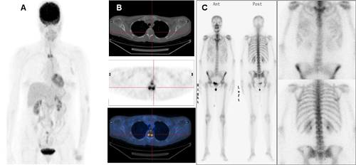
Stage: 18F-FDG-PET/CT detected metastatic disease in 34 of the 254 patients with breast cáncer included in the study, 13.4%. It changed to stage IV in one of the 30 patients in stage I (3.3%), in 18 of the 138 patients in stage II (13%) and in 15 of the186 patients in stage III (17.4%). According to the sub-stages PET/CT changes were recorded in the 4% of the patients with stage IA, none patients in stage IB, in the 8.1% in IIA, 18.7% in IIB, 10.6% in IIIA, 26.3% in IIIB and 25% in stage IIIC. A statistical significance was found in those patients with IIB, IIIA, IIIB y IIIC stages and metastatic findings in the PET-CT (OR: 3.04; IC 95%: 1.2-7.2; p = 0.009) (Figure1).
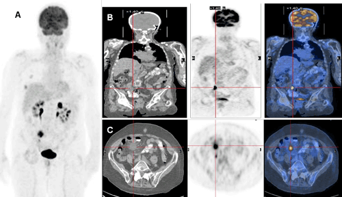
Age: The presence of metastatic disease was seen in 14 of the 49 patients with age above 70 years. (28.6%; OR: 3.7; IC 95%: 1.7- 8; p< 0.05). In the group of patients under 50 years the distance involvement occurred in 11 of 73 (15, 1%) and in the group between 50 and 70 years, 9 of the 132 patients (6.8%). Location: According to the location, the visceral disease was 4.7% in patients with breast cancer who switched to disseminated disease. The most frequently affected was the lung with 9 cases; follow by the liver in 3 cases. PET/CT did not find uptake in other organs, like the brain. The extra-axillary nodal affectation was confirmed in the supraclavicular region [N3] in the 7.9% and metastatic nodal disease was seen in 8 of the 254 patients (8.7%). The sensibility of PET/CT of visceral and lymph node disease was 100% and the specificity 86.2%. Four cases was identified as false positives, three of them in relation to inflammatory disorders [two with lymph node reactive pattern and other with diverticulitis], and one compatible case with benign adrenal gland. Bone involvement was found in 22 of 254 patients (8.7%). The more frequent location was dorsal spine. In this case the sensibility of PET/CT was 95.6%. One case was false negative, new blastic bone lesions were seen in the bone scintigraphy one month later. Also the 7.5% of the bone lesions were classified in the PET/CT as uncertain, requiring bone scintigraphy or follow-up to confirm the diagnosis. Finally, none of these lesions were metastatic disease. In 8 patients (3.1%) visceral /nodal and bone disease occurred simultaneously. Moreover, in three cases PET-CT helped with the diagnosis of two colloid goiters and one ovarian mucinous cystoadenoma. Molecular phenotype: The group with positivity for hormonal receptors and negative Her-2 status globally showed more metastatic disease (15.5%). However, the location analysis, visceral and bone involvement, was prevalent in the positive Her-2 and negative hormonal receptors group, with 7.1% and 10.7% respectively. For lymph node disease, metastatic as well as N3, triple negative tumors were the most frequency. In either cases was found statistical significance (Figure 2). Discussion
The locoregional breast cancer staging 18F-FDG-PET-CT has presented a low sensibility, for the detection of subcentimeter and/or multiples, and also to localize those tumors with low proliferation index or lobulillar histology [8-11]. The size and the accuracy for detection increase in parallel, in Jin Choi et al [9] the sensibility is 100% in breast cancer higher than 2 cm, it means, patients from IIA and IIB stage (7th AJCC edition).
According to molecular phenotype is known that Her-2 expression does not correlate with increased 18F-FDG uptake [9]. This uptake is low in cases of osteoblastic bone lesions [10]. In our study positive Her-2 tumors were associated with significant bone metastasis, almost 30% of the bone lesions, most of them osteoblastic. The confirmation was done by bone scintigraphy and in three cases required bone histological analysis. However, a significant number of breast cancer patients have osteolytic bone lesions, so bone scintigraphy is a technique with risk of false negative. Also other risk of bone scintigraphy is the rate of false positive because of bone injuries. For this reason, the use of PET-CT has shown a higher detection range for osseous metastases than conventional imaging techniques, due to the integration of morphological and functional images [12,13]. Triple negative breast cancer metastases show higher FDG uptake [14]. This relation was checked significantly in our study, more than 95% of triple negative tumors had a high proliferation index (ki67 ≥ 14%). Moreover, this molecular phenotype presented greater lymoph node [metastatic and N3] and visceral involvement at the same time. The higher 18F-FDG uptake in PET-CT was associated with the more aggressive and worse prognosis breast cancer. Sentinel lymph node detection (SLND) has shown a higher sensibility and specificity than PET-CT in breast cancer with locoregional axillary involvement. PET-CT has a similar sensibility as axillar ultrasonography [15]. Although to detect infra/supraclavicular and internal mammary chain lymph nodes PET/CT suggest greater effectiveness compared to other imaging techniques [16-18]. So PET/CT is not the imaging technique of choice for locorregional disease. In newly diagnosis breast cancer patients, the use of PET/CT to initial extension study is recommended starting from IIB stage; in the 18.7% of the patient unknown metastatic disease was discovered. From the therapeutic point of view, the stage changing avoid unnecessary mastectomy more/less lymphadenectomy in this patients group. This also avoids negative psychological impact without benefits in overall survival [19,20]. Other advantage was the improvement of quality of live in these patients because the intensity of chemotherapy was reduced after PET/CT findings due to not being potentially curable. The last advantage was reducing costs by avoiding surgical and radiotherapy treatments and the complications arising. As authors have mentioned above 34 of the 254 patients included [13.4%] were diagnosed of metastasic disease. If researchers excluded the 104 patients with stage I and stage IIB the percentage of patients affected would be almost 18%. Reviewing the other two studies with larger number of patients of the usefulness of the PET/CT in breast cancer, authors observed that the percentage of metastasic disease was 10% of 70 patients [10] and 20.9% of 254 patients [21]. In the latter study they only included patients with II or III stage and the established recommendation for performing the PET-CT was from stage IIB, as in our study. Although the characteristics between the studies were very different. In our population 93.7% of the patients with IIB stage belonged to the subgroup T2N1 versus 58.9 % in the Groheux et al [21], where the metastasis disease was 10.7% for this subgroup. According to the age distribution, the patients above the 70 years old had more diagnostic yield, in 28.6% of these patients was found distant disease. The 26.5% had 76 or more years and only 2.1% has and age between 70 and 76 years. Only in those patients with a good performance status underwent PET/CT, for this reason the incidence of this group could be underestimated. In the other two age groups the majority of the patients met criteria for the PET/CT performance. Only 9 of the 132 patients (6.8%) in the age group between the 50 and 70 years showed metastasic disease. Nowadays this is the group where the breast cancer screening is performance. This data might suggest the effectiveness and adequate monitoring of our patients. However authors cannot forget the patients below the 50 years, 73 in our study, because the breast cancer is the leading cause of death in women between 40 and 80 years in our country, even so the lowest rate of deaths for this cause in Europe [1]. Conclusion
The 18F-FDG-PET/TC showed to be useful in breast cancer metastatic disease detection in patients from IIB stage, in our clinical practice. The incidence of metastasis was higher in those patients above the 70 years and in the HER-2 negative and hormonal positive receptor group. Although the tumours with higher proliferation index (Ki67) and more visceral and lymph nodes disease (N3 and M1) altogether was found in the triple negative breast cancer group.
Ethical approval: All procedures performed in studies involving human participants were in accordance with the ethical standards of the institutional and/or national research committee and with the 1964 Helsinki Declaration and its later amendments or comparable ethical standards. However, this is a retrospective analysis and for this type of study formal consent is not required. Informed consent: Informed consent was obtained from all individual participants included in the study. Open access: This article is distributed under the terms of the Creative Commons Attribution License which permits any use, distribution, and reproduction in any medium, provided the original author(s) and the source are credited. |
References
- Cabanes A, Pérez-Gómez B, Aragonés N, Pollán M, López-Abente G (2009) La situación del cáncer en España 1975-2006. Instituto de Salud Carlos III (Madrid).
- Brasic N, Wisner DJ, Joe BN (2013) Breast MR imaging for extent of disease assessment in patients with newly diagnosed breast cancer. Magn Reson Imaging Clin N Am 21: 519-532.
- Houssami N, Ciatto S, Macaskill P, Lord SJ, Warren RM, et al. (2008) Accuracy and surgical impact of magnetic resonance imaging in breast cancer staging: systematic review and meta-analysis in detection of multifocal and multicentric cancer. J Clin Oncol 26: 3248-3258.
- Goldhirsch A, Wood WC, Coates AS, Gelber RD, Thürlimann B, et al. (2011) Strategies for subtypes--dealing with the diversity of breast cancer: highlights of the St. Gallen International Expert Consensus on the Primary Therapy of Early Breast Cancer 2011. Ann Oncol 22: 1736-1747.
- Goldhirsch A, Winer EP, Coates AS, Gelber RD, Piccart-Gebhart M, et al. (2013) Personalizing the treatment of women with early breast cancer: highlights of the St Gallen International Expert Consensus on the Primary Therapy of Early Breast Cancer 2013. Ann Oncol 24: 2206-2223.
- Bernet L, Piñero A, Vidal-Sicart S, Cano R, Cordero JM, et al. (2010) Updated consensus on selective sentinel node biopsy in breast cancer. Rev Senología Patol Mam 263: 1-11.
- Houssami N, Ciatto S, Turner RM, Cody HS 3rd, Macaskill P (2011) Preoperative ultrasound-guided needle biopsy of axillary nodes in invasive breast cancer: meta-analysis of its accuracy and utility in staging the axilla. Ann Surg 254: 243-251.
- Kumar R, Chauhan A, Zhuang H, Chandra P, Schnall M, et al. (2006) Clinicopathologic factors associated with false negative FDG-PET in primary breast cancer. Breast Cancer Res Treat 98: 267-274.
- Groheux D, Espié M, Giacchetti S, Hindié E (2013) Performance of FDG PET/CT in the clinical management of breast cancer. Radiology 266: 388-405.
- Segaert I, Mottaghy F, Ceyssens S, De Wever W, Stroobants S, et al. (2010) Additional value of PET-CT in staging of clinical stage IIB and III breast cancer. Breast J 16: 617-624.
- Choi YJ, Shin YD, Kang YH, Lee MS, Lee MK, et al. (2012) The Effects of Preoperative (18)F-FDG PET/CT in Breast Cancer Patients in Comparison to the Conventional Imaging Study. J Breast Cancer 15: 441-448.
- Liu T, Cheng T, Xu W, Yan WL, Liu J, et al. (2011) A meta-analysis of 18FDG-PET, MRI and bone scintigraphy for diagnosis of bonemetastases in patients with breast cancer. Skeletal Radiol 40: 523-531.
- Yang HL, Liu T, Wang XM, Xu Y, Deng SM (2011) Diagnosis of bone metastases: a meta-analysis comparing ¹â¸FDG PET, CT, MRI and bone scintigraphy. Eur Radiol 21: 2604-2617.
- Ohara M, Shigematsu H, Tsutani Y, Emi A, Masumoto N, et al. (2013) Role of FDG-PET/CT in evaluating surgical outcomes of operable breast cancer--usefulness for malignant grade of triple-negative breast cancer. Breast 22: 958-963.
- Cooper KL, Harnan S, Meng Y, Ward SE, Fitzgerald P, et al. (2011) Positron emission tomography (PET) for assessment of axillary lymph node status in early breast cancer: a systematic review and meta-analysis. Eur J Surg Oncol 37:187-198
- Aukema TS, Straver ME, Peeters MJ, Russell NS, Gilhuijs KG, et al. (2010) Detection of extra-axillary lymph node involvement with FDG PET/CT in patients with stage II-III breast cancer. Eur J Cancer 46: 3205-3210.
- Danforth DN Jr, Aloj L, Carrasquillo JA, Bacharach SL, Chow C, et al. (2002) The role of 18F-FDG-PET in the local/regional evaluation of women with breast cancer. Breast Cancer Res Treat 75: 135-146.
- Tran A, Pio BS, Khatibi B, Czernin J, Phelps ME, et al. (2005) 18F-FDG PET for staging breast cancer in patients with inner-quadrant versus outer-quadrant tumors: comparison with long-term clinical outcome. J Nucl Med 46: 1455-1459.
- Badwe R, Parmar V, Hawaldar R (2013) Surgical removal of primary tumor and axillary lymph nodes in women with metastatic breast cancer at first presentation: A randomized controlled trial. Paper presented at: 36th Annual San Antonio Breast Cancer Symposium, San Antonio, TX.
- Soran A, Ozmen V, Ozbas S (2013) Early follow up of a randomized trial evaluating resection of the primary breast tumor in women presenting with de novo stage IV breast cancer. Paper presented at: 36th Annual San Antonio Breast Cancer Symposium, San Antonio, TX.
- Groheux D, Hindié E, Delord M, Giacchetti S, Hamy AS, et al. (2012) Prognostic impact of (18)FDG-PET-CT findings in clinical stage III and IIB breast cancer. J Natl Cancer Inst 104: 1879-1887.
Figures at a glance
 |
 |
| Figure 1 | Figure 2 |
Relevant Topics
- Abdominal Radiology
- AI in Radiology
- Breast Imaging
- Cardiovascular Radiology
- Chest Radiology
- Clinical Radiology
- CT Imaging
- Diagnostic Radiology
- Emergency Radiology
- Fluoroscopy Radiology
- General Radiology
- Genitourinary Radiology
- Interventional Radiology Techniques
- Mammography
- Minimal Invasive surgery
- Musculoskeletal Radiology
- Neuroradiology
- Neuroradiology Advances
- Oral and Maxillofacial Radiology
- Radiography
- Radiology Imaging
- Surgical Radiology
- Tele Radiology
- Therapeutic Radiology
Recommended Journals
Article Tools
Article Usage
- Total views: 13731
- [From(publication date):
August-2015 - Aug 29, 2025] - Breakdown by view type
- HTML page views : 9191
- PDF downloads : 4540
